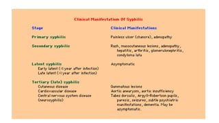
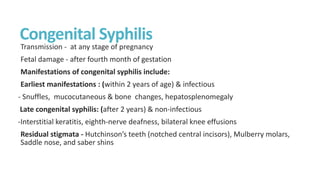
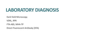
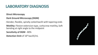
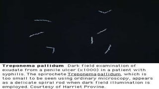
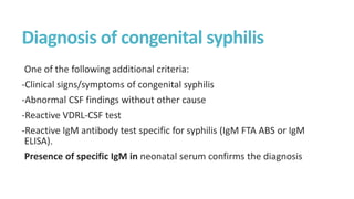
Syphilis is caused by the spirochete Treponema pallidum. It is transmitted sexually or congenitally from mother to fetus. Syphilis increases the risk of HIV transmission. It has various stages including primary, secondary, latent, and tertiary syphilis. Laboratory diagnosis involves direct visualization of T. pallidum via darkfield microscopy or detection of antibodies to T. pallidum via non-treponemal and treponemal tests. Penicillin is the treatment of choice for all stages of syphilis.
This document summarizes bacterial infections including syphilis, gonorrhea, and noma. Syphilis is caused by Treponema pallidum and has primary, secondary, tertiary, and congenital stages. Gonorrhea is caused by Neisseria gonorrhoeae and commonly infects the genital tract, sometimes ascending in women to cause pelvic inflammatory disease. Noma (cancrum oris) is a rapidly progressive infection caused by normal oral flora under conditions of compromised immunity such as malnutrition, affecting the gingiva and soft tissues of the face.
Molluscum contagiosum is a common, harmless skin infection caused by a poxvirus that spreads through direct skin-to-skin contact. It presents as small, flesh-colored bumps with a dimpled center that contain a white, curdy core. While generally asymptomatic, the bumps can occasionally itch or cause a skin rash. Diagnosis is made through visual examination of characteristic lesions, and treatment options range from natural resolution to cryotherapy, curettage, laser surgery, or topical medications depending on severity. Left untreated in healthy individuals, molluscum contagiosum will usually clear up on its own within months without scarring.
This document discusses diagnostic tests for syphilis caused by the bacterium Treponema pallidum. It describes direct detection methods like darkfield microscopy and fluorescent antibody testing to visualize the bacterium in samples. It also covers non-treponemal tests that detect non-specific reagin antibodies like VDRL and RPR, and treponemal tests that detect antibodies specific to T. pallidum like FTA-ABS. The stages of syphilis and clinical manifestations are also briefly outlined.
Cutaneous tuberculosis can occur through either exogenous or endogenous infection by Mycobacterium tuberculosis or M. bovis. Clinical manifestations depend on the site of infection, inoculum type, and host immunity. Diagnosis involves demonstration of acid-fast bacilli in lesions via staining, culture, or PCR. Treatment involves a multi-drug regimen according to WHO guidelines, with surgery sometimes indicated for localized lesions. Drug-resistant tuberculosis requires customized multi-drug treatment regimens.
This document summarizes various skin and soft tissue infections, including their causes, symptoms, and treatments. Erysipelas is a streptococcal infection of the skin that causes a painful, erythematous rash. Impetigo is a contagious superficial infection commonly caused by streptococci or staphylococci in children. Folliculitis is a bacterial infection of hair follicles that causes papules and pustules. Boils are deeper hair follicle infections forming tender, red swellings. Carbuncles are clusters of interconnected boils. Cellulitis is a spreading bacterial skin infection beneath the skin. Necrotizing fasciitis is a severe infection of the fascia requiring aggressive
Pityriasis versicolor is a common fungal infection of the skin caused by Malassezia furfur. It presents with hypo-pigmented or hyper-pigmented macules and patches on the chest and back. Diagnosis can be confirmed with potassium hydroxide examination of skin scrapings or Wood's lamp examination. Treatment involves topical or oral antifungal agents like selenium sulfide, ketoconazole or fluconazole. Recurrence is common so lifestyle changes and prophylactic treatment may help reduce relapse.
This document provides information about histoplasmosis, including its characteristics, pathogenesis, types, clinical presentation, and laboratory diagnosis. It can be summarized as follows:
1. Histoplasmosis is caused by the dimorphic fungus Histoplasma capsulatum, which exists in both a mycelial and yeast form. It is found worldwide in soil contaminated with bird or bat droppings.
2. Infection typically occurs via inhalation of yeast cells into the lungs. It can cause pulmonary or disseminated disease, spreading to organs in immunocompromised individuals.
3. Laboratory diagnosis involves direct examination of samples for yeast cells, culture of the fungus, and serological tests like complement fixation
Pityriasis versicolor is a fungal infection of the skin caused by Malassezia species. It typically presents as hypopigmented or occasionally hyperpigmented macules with fine scaling that often coalesce. Diagnosis is usually clinical but can be confirmed with KOH microscopy showing fungal elements or wood's lamp fluorescence. Treatment involves topical or oral antifungal agents like selenium sulfide or ketoconazole.
Pityriasis rosea is a self-limiting rash of unknown etiology often associated with viral infections. It is characterized by an initial herald patch followed by secondary lesions in a Christmas-tree distribution. Lesions resolve spontaneously over weeks to months with supportive care
This document provides information about Erythema Nodosum Leprosum (ENL), a type 2 reaction that occurs in patients with multibacillary leprosy with a heavy bacterial load. ENL is characterized by red, tender skin nodules and can involve multiple organs. It is caused by an Arthus-type allergic reaction when large numbers of killed leprosy bacteria release antigens. Diagnosis involves clinical presentation of skin and nerve inflammation as well as slit skin smears and histopathology. Treatment focuses on reassurance, continuing multidrug therapy, rest, NSAIDs for mild cases, and oral steroids for severe cases.
Etiology of Leprosy:
A chronic infection caused by Mycobacterium leprae
Acid-fast, rod shaped
Main route of infection:
nasal droplets,
Eating armadillos (south america)
Not very contagious, but close relatives are at high risk of infection
Bulloous disorders (BSDs) are skin conditions characterized by blister formation between the epidermis and dermis layers of the skin. BSDs are mostly autoimmune in nature and can be caused by genetic factors, physical trauma, inflammation, the immune system, or drug reactions. The main types are genetic blistering diseases like epidermolysis bullosa, and immunobullous diseases like pemphigus and pemphigoid which involve antibody-mediated blistering between skin layers. Pemphigus is an intraepidermal immunobullous disease affecting the skin and mucous membranes, while bullous pemphigoid is a subepidermal immunobullous condition commonly affecting
This document provides information on Treponema pallidum, the spirochete bacterium that causes syphilis. It discusses the morphology, cultivation, antigenic structure, and pathogenesis of T. pallidum. It also describes the stages of syphilis infection including primary, secondary, and tertiary syphilis. The document concludes with an overview of laboratory diagnosis of syphilis including microscopy, staining techniques, and various serological tests.
This document provides an overview of a syphilis curriculum, including:
1. The curriculum covers the epidemiology, pathogenesis, clinical manifestations, diagnosis, patient management, and prevention of syphilis.
2. Lessons include the disease epidemiology in the US, the pathogenesis of Treponema pallidum, clinical manifestations across all stages of syphilis, methods for diagnosis including serologic tests and microscopy, and prevention strategies.
3. The goal is for learners to understand all aspects of syphilis including transmission, population trends, microbiology, symptoms, testing, and treatment according to CDC guidelines.
1) Lepra reactions are immunologically inflammatory states that can occur in leprosy patients. There are two main types - Type 1 reactions (downgrading and reversal reactions) and Type 2 reactions (Erythema Nodosum Leprosum or ENL).
2) Type 1 reactions involve the skin and nerves and cause swelling and tenderness. They occur after treatment begins. Type 2 or ENL reactions cause painful swollen skin lesions and other symptoms like fever. They typically occur after treatment in lepromatous patients.
3) Histologically, Type 1 reactions show edema while Type 2 reactions show deep inflammation, necrosis and few bacilli. Both can cause significant morbidity if not properly treated.
This document discusses the normal microbial flora of the skin, intestines, and vagina. It describes the most common bacteria that normally inhabit these areas, including Staphylococcus epidermidis and Streptococcus on the skin. In the intestines, it notes the high numbers of anaerobic bacteria and how the flora changes along the GI tract. It also explains how disruption of the normal flora through factors like antibiotics or medical procedures can allow pathogenic bacteria to colonize.
- A 7 year old female presented with a skin biopsy from her left gluteal region for a clinical diagnosis of granuloma annulare.
- Microscopic examination showed features consistent with granuloma annulare including hyperkeratosis, acanthosis, lymphocytic infiltration, and histiocytes surrounding degenerated collagen extending into the dermis.
- Granuloma annulare is a benign inflammatory dermatosis more common in females that involves skin and subcutaneous tissue, though the etiology is unknown.
Syphilis is a sexually transmitted infection caused by the bacterium Treponema pallidum. It presents in stages, beginning with a painless sore and progressing to a rash and lesions on mucous membranes if left untreated. Without treatment, it can spread throughout the body and cause damage to internal organs, nerves, and blood vessels. Syphilis is treated with antibiotics, most commonly penicillin, with the treatment depending on which stage the infection is in. Congenital syphilis can occur if an infected mother transmits the bacterium to her fetus during pregnancy.
Chlamydiae are small, obligate intracellular parasites that lack the ability to produce their own ATP. There are three medically important species: C. pneumoniae, C. psittaci, and C. trachomatis. C. pneumoniae causes respiratory infections like pneumonia. C. psittaci infects birds and can be transmitted to humans, causing ornithosis or pneumonia. C. trachomatis causes ocular infections like trachoma and inclusion conjunctivitis as well as genital infections like lymphogranuloma venereum and non-gonococcal urethritis. Diagnosis involves microscopy, culture studies, serological tests, and skin tests. Treatment involves sulfon
Non-Gonococcal urethritis. main causative organisms are Chlamydiae, Mycoplasma, Ureaplasma. various other bacteria and viruses can cause this. this powerpoint is made in systemic manner and will be helpful for Postgraduate students.
Cutaneous tuberculosis can present in different clinical forms as a result of Mycobacterium tuberculosis, M. bovis, or BCG vaccine infection of the skin. Lupus vulgaris is the most common form, appearing as painless reddish-brown nodules on the face that slowly enlarge. Scrofuloderma typically affects the neck and chest as an asymptomatic swelling that breaks down into sinuses or ulcers. Diagnosis involves skin biopsy demonstrating tuberculoid granulomas and potentially identifying acid-fast bacilli, along with a positive tuberculin test. Treatment consists of a multi-drug regimen in two phases to reduce the bacterial burden and ensure sterilization.
1. Seminoma is the most common malignant tumor of the testis, occurring most often in men between ages 25-55. Undescended testes are an important risk factor.
2. There are two main types of seminoma: classic seminoma, which accounts for 90% of cases, and spermatocytic seminoma, which comprises 5% of cases.
3. Classic seminoma appears as enlarged, solid testes containing white nodules separated from the testicular tissue. Under the microscope, classic seminoma cells are large with clear cytoplasm and central nuclei, and stain positive for glycogen.
This document discusses superficial fungal infections. It begins by noting that fungal infections affect 20-25% of the world's population. Risk factors include tropical climate, low socioeconomic status, sweating, and immunosuppression. There are three main genera that cause superficial cutaneous fungal infections: Microsporum, Trichophyton, and Epidermophyton. The document then discusses eight specific types of superficial fungal infections in detail, including tinea capitis, tinea barbae, tinea corporis, tinea pedis, tinea cruris, tinea manuum, onychomycosis, and pityriasis versicolor. For each infection, it describes clinical features,
This document discusses laboratory diagnosis of syphilis. It describes various diagnostic methods used including demonstration of Treponemes through dark-field microscopy and direct fluorescent antibody testing. It also discusses serological tests including non-treponemal tests like VDRL and RPR as well as treponemal tests like FTA-ABS. Specimen collection and processing is also outlined. The stages of syphilis and expected serological responses at each stage are provided. Neurosyphilis and congenital syphilis diagnosis are briefly covered.
Cutaneous tuberculosis can present in various forms as a result of hematogenous spread or direct extension from a latent tuberculosis infection. Common types include lupus vulgaris, verrucosa cutis, and scrofuloderma. Diagnosis involves skin biopsy demonstrating tuberculoid granulomas with occasional acid-fast bacilli. Treatment consists of long-term multidrug antitubercular therapy following standard protocols for systemic tuberculosis.
Viral skin diseases are common and include infections caused by herpes simplex virus, varicella zoster virus, and human papillomavirus. Herpes simplex virus causes lesions such as cold sores, genital herpes, and eczema herpeticum. Varicella zoster virus causes chickenpox and shingles. Human papillomavirus causes warts, including common warts, flat warts, plantar warts, and genital warts. These viral infections are generally self-limiting but can be treated with antivirals to reduce symptoms and duration of infection.
1. There are two main types of tests for diagnosing syphilis - direct tests that detect the bacteria Treponema pallidum, and indirect serological tests that detect antibodies produced in response to the infection.
2. Direct tests include dark-field microscopy, direct fluorescent antibody testing, and PCR to identify the bacterium in lesions. Serological tests include non-treponemal tests like VDRL and RPR that detect nonspecific reagin antibodies, and treponemal tests like FTA-ABS, TPHA, and EIA that detect treponema-specific antibodies.
3. Dark-field microscopy examines exudate from lesions under a dark-field microscope to visualize
1. Spirochetes are thin, spiral-shaped bacteria that include Treponema, Borrelia, and Leptospira genera.
2. Treponema pallidum causes syphilis which is transmitted sexually or through direct contact. It has a characteristic spiral shape and causes primary, secondary, latent, and late stages of syphilis with varied clinical manifestations.
3. Laboratory diagnosis of syphilis involves direct visualization of T. pallidum using dark-field microscopy, serological tests like VDRL and RPR that detect non-specific reagin antibodies, and treponemal tests that detect species-specific antibodies.
1. Treponema pallidum is a spirochete bacterium that causes syphilis. It has four subspecies that can cause different diseases.
2. Syphilis progresses through primary, secondary, latent, and tertiary stages. The primary stage involves painless sores called chancres. Secondary syphilis has a widespread rash.
3. Penicillin is the treatment of choice for syphilis. Early syphilis is treated with benzathine penicillin injections or oral antibiotics.
This document summarizes information about spirochetes and Treponema pallidum, the bacterium that causes syphilis. It describes the morphology, culture characteristics, pathogenicity, stages of syphilis (primary, secondary, latent, tertiary), laboratory diagnosis, epidemiology, immunity, prophylaxis, and treatment of T. pallidum. Key points are that T. pallidum is a spiral-shaped bacterium that cannot be grown in culture but causes the sexually transmitted infection syphilis in humans, which progresses through distinct stages if left untreated and can be effectively treated with penicillin.
Treponema pallidum is a spirochete bacterium that causes syphilis and other diseases. It has a thin, helical shape with periplasmic flagella and tightly coiled cells. Syphilis infection progresses through primary, secondary, latent, and tertiary stages and can involve multiple organ systems if left untreated. Diagnosis involves darkfield microscopy, serological tests like VDRL and RPR, and specific treponemal tests since the bacterium cannot be cultured.
bacterial sexually transmitted diseases in the tropicskaluyas934
This document summarizes several bacterial STDs, including:
- Treponema pallidum, which causes syphilis. It is visualized via darkfield microscopy. Primary syphilis presents as a chancre and secondary syphilis has skin and mucous lesions.
- Haemophilus ducreyi causes chancroid presenting as genital ulcers with enlarged lymph nodes.
- Chlamydia trachomatis causes non-gonococcal urethritis and lymphogranuloma venereum.
- Neisseria gonorrhoeae causes gonorrhea presenting as urethral discharge and is diagnosed via gram stain of intracellular diplococci.
A 29-year-old man presented to an STD clinic complaining of multiple papules on his genitals. Examination revealed indurated papules that were painless. Tests determined it was a case of primary syphilis caused by the bacterium Treponema pallidum.
Treponema pallidum is a spirochete bacterium that causes syphilis. It is thin and delicate, about 10 micrometers in length, and actively motile. T. pallidum cannot be grown in culture but can be seen microscopically using dark-field illumination or silver staining. Syphilis has stages including primary, secondary, latent, and tertiary, and can involve multiple organs if untreated. Diagnosis involves microscopic examination of samples or serological tests to detect antibodies. Penicillin is the treatment of choice.
This pptx covers the basics of almost all stds.Use this and for sure you'll get a good score.
It is taken from very hugh yield and authenic sources.
The animations Adds further beauty to the presentation.
The animations itself are self explanatory just download and deliver you'll be very much appreciated for it no need to add further the pptx is well comprehensive.
Syphilis is a sexually transmitted disease caused by the bacterium Treponema pallidum. It is transmitted through direct contact with syphilis sores during sex, from mother to fetus, or rarely through blood transfusion. Syphilis has four stages - primary, secondary, latent, and tertiary. The primary stage involves a painless sore called a chancre, while the secondary stage has diffuse symptoms like rash and fever. Without treatment, later stages can involve serious complications in multiple organ systems. Syphilis is diagnosed through clinical examination, dark-field microscopy of sores, and serological blood tests. Treatment involves antibiotics like penicillin or doxycycline.
This document provides information about the bacteria Spirochaetes. It discusses their morphology, motility, reproduction, pathogenic species, diseases caused, and laboratory diagnosis. Some key points:
- Spirochaetes are elongated, helically coiled, and motile bacteria with endoflagella. They can cause diseases like syphilis, Lyme disease, and leptospirosis.
- Pathogenic species include Treponema pallidum (syphilis), Borrelia burgdorferi (Lyme disease), and Leptospira interrogans (leptospirosis).
- Laboratory diagnosis involves darkfield microscopy, culture, serology, and
Spirochetes are long, helical, motile bacteria that include the genera Treponema, Borrelia, and Leptospira. Treponema pallidum causes syphilis, presenting initially with a chancre then secondary and latent stages before potential progression to tertiary disease. Borrelia burgdorferi causes Lyme disease transmitted by ticks, presenting with a skin lesion and flu-like symptoms. These spirochetes are diagnosed via microscopy, culture, and serology. Penicillin is the treatment of choice for diseases caused by Treponema and Borrelia.
SYPHILIS.pptx bachelors degree in medicineJosphatSiele
Syphilis is caused by the bacterium Treponema pallidum. It is usually transmitted sexually but can also be transmitted congenitally from mother to fetus. There are four stages of syphilis: primary, secondary, latent, and tertiary. Left untreated, syphilis can cause serious long term complications affecting the heart, brain, and other organs. Penicillin remains the treatment of choice and can cure syphilis if administered properly in the early stages of infection. Complications from treatment may include anaphylaxis or Jarisch-Herxheimer reaction.
1. Syphilis is caused by the bacterium Treponema pallidum, which can be transmitted sexually or from mother to fetus.
2. It has four stages: primary, secondary, latent, and tertiary. Primary symptoms include sores, secondary includes rashes, and tertiary can damage organs.
3. Diagnosis involves blood tests to detect antibodies produced against T. pallidum. Treatment is with penicillin, which is usually effective at curing syphilis if administered early enough.
This document summarizes information about Spirochetes bacteria including Treponema pallidum, the causative agent of syphilis. It describes the morphology, stages of infection (primary, secondary, tertiary), methods of diagnosis (microscopy, serological tests), treatment (penicillin), and prevention of syphilis. It also discusses other Spirochetes bacteria such as Borrelia burgdorferi which causes Lyme disease, and Leptospira interrogans which can cause Weil's disease.
This document discusses laboratory diagnosis of syphilis. It begins by describing syphilis stages and transmission routes. It then details the laboratory tests used for syphilis screening and confirmation, including non-treponemal tests that detect reagin antibodies and treponemal tests that detect antibodies to Treponema pallidum antigens. It explains the traditional and reverse screening algorithms and emphasizes the importance of clinical evaluation to properly interpret laboratory results. Follow-up testing intervals are also outlined.
This document discusses syphilis, a sexually transmitted disease caused by the spirochete Treponema pallidum. It defines syphilis and describes the causative organism. It discusses the modes of transmission for acquired and congenital syphilis. The clinical stages of syphilis are outlined including primary, secondary, latent, and tertiary syphilis. The diagnosis, treatment, and prognosis of syphilis are summarized. Congenital syphilis is also described. References are provided.
Immersive Learning That Works: Research Grounding and Paths ForwardLeonel Morgado
We will metaverse into the essence of immersive learning, into its three dimensions and conceptual models. This approach encompasses elements from teaching methodologies to social involvement, through organizational concerns and technologies. Challenging the perception of learning as knowledge transfer, we introduce a 'Uses, Practices & Strategies' model operationalized by the 'Immersive Learning Brain' and ‘Immersion Cube’ frameworks. This approach offers a comprehensive guide through the intricacies of immersive educational experiences and spotlighting research frontiers, along the immersion dimensions of system, narrative, and agency. Our discourse extends to stakeholders beyond the academic sphere, addressing the interests of technologists, instructional designers, and policymakers. We span various contexts, from formal education to organizational transformation to the new horizon of an AI-pervasive society. This keynote aims to unite the iLRN community in a collaborative journey towards a future where immersive learning research and practice coalesce, paving the way for innovative educational research and practice landscapes.
Sexuality - Issues, Attitude and Behaviour - Applied Social Psychology - Psyc...PsychoTech Services
A proprietary approach developed by bringing together the best of learning theories from Psychology, design principles from the world of visualization, and pedagogical methods from over a decade of training experience, that enables you to: Learn better, faster!
SDSS1335+0728: The awakening of a ∼ 106M⊙ black hole⋆Sérgio Sacani
Context. The early-type galaxy SDSS J133519.91+072807.4 (hereafter SDSS1335+0728), which had exhibited no prior optical variations during the preceding two decades, began showing significant nuclear variability in the Zwicky Transient Facility (ZTF) alert stream from December 2019 (as ZTF19acnskyy). This variability behaviour, coupled with the host-galaxy properties, suggests that SDSS1335+0728 hosts a ∼ 106M⊙ black hole (BH) that is currently in the process of ‘turning on’. Aims. We present a multi-wavelength photometric analysis and spectroscopic follow-up performed with the aim of better understanding the origin of the nuclear variations detected in SDSS1335+0728. Methods. We used archival photometry (from WISE, 2MASS, SDSS, GALEX, eROSITA) and spectroscopic data (from SDSS and LAMOST) to study the state of SDSS1335+0728 prior to December 2019, and new observations from Swift, SOAR/Goodman, VLT/X-shooter, and Keck/LRIS taken after its turn-on to characterise its current state. We analysed the variability of SDSS1335+0728 in the X-ray/UV/optical/mid-infrared range, modelled its spectral energy distribution prior to and after December 2019, and studied the evolution of its UV/optical spectra. Results. From our multi-wavelength photometric analysis, we find that: (a) since 2021, the UV flux (from Swift/UVOT observations) is four times brighter than the flux reported by GALEX in 2004; (b) since June 2022, the mid-infrared flux has risen more than two times, and the W1−W2 WISE colour has become redder; and (c) since February 2024, the source has begun showing X-ray emission. From our spectroscopic follow-up, we see that (i) the narrow emission line ratios are now consistent with a more energetic ionising continuum; (ii) broad emission lines are not detected; and (iii) the [OIII] line increased its flux ∼ 3.6 years after the first ZTF alert, which implies a relatively compact narrow-line-emitting region. Conclusions. We conclude that the variations observed in SDSS1335+0728 could be either explained by a ∼ 106M⊙ AGN that is just turning on or by an exotic tidal disruption event (TDE). If the former is true, SDSS1335+0728 is one of the strongest cases of an AGNobserved in the process of activating. If the latter were found to be the case, it would correspond to the longest and faintest TDE ever observed (or another class of still unknown nuclear transient). Future observations of SDSS1335+0728 are crucial to further understand its behaviour. Key words. galaxies: active– accretion, accretion discs– galaxies: individual: SDSS J133519.91+072807.4
When I was asked to give a companion lecture in support of ‘The Philosophy of Science’ (https://shorturl.at/4pUXz) I decided not to walk through the detail of the many methodologies in order of use. Instead, I chose to employ a long standing, and ongoing, scientific development as an exemplar. And so, I chose the ever evolving story of Thermodynamics as a scientific investigation at its best.
Conducted over a period of >200 years, Thermodynamics R&D, and application, benefitted from the highest levels of professionalism, collaboration, and technical thoroughness. New layers of application, methodology, and practice were made possible by the progressive advance of technology. In turn, this has seen measurement and modelling accuracy continually improved at a micro and macro level.
Perhaps most importantly, Thermodynamics rapidly became a primary tool in the advance of applied science/engineering/technology, spanning micro-tech, to aerospace and cosmology. I can think of no better a story to illustrate the breadth of scientific methodologies and applications at their best.
Candidate young stellar objects in the S-cluster: Kinematic analysis of a sub...Sérgio Sacani
Context. The observation of several L-band emission sources in the S cluster has led to a rich discussion of their nature. However, a definitive answer to the classification of the dusty objects requires an explanation for the detection of compact Doppler-shifted Brγ emission. The ionized hydrogen in combination with the observation of mid-infrared L-band continuum emission suggests that most of these sources are embedded in a dusty envelope. These embedded sources are part of the S-cluster, and their relationship to the S-stars is still under debate. To date, the question of the origin of these two populations has been vague, although all explanations favor migration processes for the individual cluster members. Aims. This work revisits the S-cluster and its dusty members orbiting the supermassive black hole SgrA* on bound Keplerian orbits from a kinematic perspective. The aim is to explore the Keplerian parameters for patterns that might imply a nonrandom distribution of the sample. Additionally, various analytical aspects are considered to address the nature of the dusty sources. Methods. Based on the photometric analysis, we estimated the individual H−K and K−L colors for the source sample and compared the results to known cluster members. The classification revealed a noticeable contrast between the S-stars and the dusty sources. To fit the flux-density distribution, we utilized the radiative transfer code HYPERION and implemented a young stellar object Class I model. We obtained the position angle from the Keplerian fit results; additionally, we analyzed the distribution of the inclinations and the longitudes of the ascending node. Results. The colors of the dusty sources suggest a stellar nature consistent with the spectral energy distribution in the near and midinfrared domains. Furthermore, the evaporation timescales of dusty and gaseous clumps in the vicinity of SgrA* are much shorter ( 2yr) than the epochs covered by the observations (≈15yr). In addition to the strong evidence for the stellar classification of the D-sources, we also find a clear disk-like pattern following the arrangements of S-stars proposed in the literature. Furthermore, we find a global intrinsic inclination for all dusty sources of 60 ± 20◦, implying a common formation process. Conclusions. The pattern of the dusty sources manifested in the distribution of the position angles, inclinations, and longitudes of the ascending node strongly suggests two different scenarios: the main-sequence stars and the dusty stellar S-cluster sources share a common formation history or migrated with a similar formation channel in the vicinity of SgrA*. Alternatively, the gravitational influence of SgrA* in combination with a massive perturber, such as a putative intermediate mass black hole in the IRS 13 cluster, forces the dusty objects and S-stars to follow a particular orbital arrangement. Key words. stars: black holes– stars: formation– Galaxy: center– galaxies: star formation
Travis Hills of MN is Making Clean Water Accessible to All Through High Flux ...Travis Hills MN
By harnessing the power of High Flux Vacuum Membrane Distillation, Travis Hills from MN envisions a future where clean and safe drinking water is accessible to all, regardless of geographical location or economic status.
PPT on Alternate Wetting and Drying presented at the three-day 'Training and Validation Workshop on Modules of Climate Smart Agriculture (CSA) Technologies in South Asia' workshop on April 22, 2024.
Discovery of An Apparent Red, High-Velocity Type Ia Supernova at 𝐳 = 2.9 wi...Sérgio Sacani
We present the JWST discovery of SN 2023adsy, a transient object located in a host galaxy JADES-GS
+
53.13485
−
27.82088
with a host spectroscopic redshift of
2.903
±
0.007
. The transient was identified in deep James Webb Space Telescope (JWST)/NIRCam imaging from the JWST Advanced Deep Extragalactic Survey (JADES) program. Photometric and spectroscopic followup with NIRCam and NIRSpec, respectively, confirm the redshift and yield UV-NIR light-curve, NIR color, and spectroscopic information all consistent with a Type Ia classification. Despite its classification as a likely SN Ia, SN 2023adsy is both fairly red (
�
(
�
−
�
)
∼
0.9
) despite a host galaxy with low-extinction and has a high Ca II velocity (
19
,
000
±
2
,
000
km/s) compared to the general population of SNe Ia. While these characteristics are consistent with some Ca-rich SNe Ia, particularly SN 2016hnk, SN 2023adsy is intrinsically brighter than the low-
�
Ca-rich population. Although such an object is too red for any low-
�
cosmological sample, we apply a fiducial standardization approach to SN 2023adsy and find that the SN 2023adsy luminosity distance measurement is in excellent agreement (
≲
1
�
) with
Λ
CDM. Therefore unlike low-
�
Ca-rich SNe Ia, SN 2023adsy is standardizable and gives no indication that SN Ia standardized luminosities change significantly with redshift. A larger sample of distant SNe Ia is required to determine if SN Ia population characteristics at high-
�
truly diverge from their low-
�
counterparts, and to confirm that standardized luminosities nevertheless remain constant with redshift.
Mending Clothing to Support Sustainable Fashion_CIMaR 2024.pdfSelcen Ozturkcan
Ozturkcan, S., Berndt, A., & Angelakis, A. (2024). Mending clothing to support sustainable fashion. Presented at the 31st Annual Conference by the Consortium for International Marketing Research (CIMaR), 10-13 Jun 2024, University of Gävle, Sweden.
2. Syphilis
Caused by Treponema pallidum.
Transmission: sexual; maternal-fetal, and rarely by other means
Syphilis increases the risk of both transmitting and getting infected with HIV.
4. TREPONEMA
Pathogenic species
-T. pallidum subspecies pallidum
-T. pallidum subspecies pertenue
-T. pallidum subspecies endemicum
-T. carateum
Almost identical in their morphology, antigenic structure and in genetic composition
5. TREPONEMA PALLIDUM
Morphology: extremely thin and delicate
with tapering ends
Size: 6–14 μm × 0.2 μm
Spirals: 6–12 spirals at intervals of 1 μm
Motility: flexion extension, translatory,
and corkscrew motility
Endoflagella: About 3–4 flagella - motility
& highly antigenic
6. Microscopy
Dark ground or phase contrast microscope
Staining: Do not take up the ordinary stain.
-Fluorescence staining
-Sliver impregnation methods
Cultivation: Pathogenic treponemes cannot be grown in artificial culture media.
Maintained in rabbit testes. E.g,Nichols strain
Non-pathogenic Treponemes – grow in Smith Noguchi medium under strict anerobic
conditions
7. Antigens
1. Group-specific antigen: Protein antigen
- Present in all treponemes
- Antibodies detected using antigens of Reiter treponemes.
1. Species-specific antigen: Polysaccharide
- Antibodies detected by using specific T. pallidum antigens
1. Non-specific antigen: Heterophile antigen
- Antibody detected using beef heart antigen
8. PATHOGENESIS OF SYPHILIS
Mode of transmission:
-Venereal
-Non-venereal - direct contact, blood transfusion or transplacental
Spread: T. pallidum penetrates through mucosa or abraded skin Enter lymphatics
and blood systemic primary lesion
Incubation period - Variable (9–90 days)
- Inversely proportional to the number of organisms inoculated
9. STAGES OF SYPHILIS
1. Primary
2. Secondary
3. Latent
Early latent
Late latent
4. Late or tertiary
May involve any organ, but main parts are:
Neurosyphilis
Cardiovascular syphilis
Late benign (gumma)
10. CLINICAL MANIFESTATIONS OF SYPHILIS
Primary Syphilis
Incubation period 9-90 days, usually ~21
days.
Develops at site of contact/inoculation.
Primary (or hard) chancre:
-Single painless papule ulcerated &
indurated
-Covered by thick exudate rich in
spirochetes
-Common sites – penis, cervix or labia
-Heals within 4–6 weeks
11. Regional (usually inguinal)
lymphadenopathy
-Painless firm, non-suppurative, and often
bilateral
-May persist for months
If acquired by non-venereal mode -
primary syphilis
-Direct contact → extragenital, usually on
the fingers
- Blood transfusion → primary chancre
does not occur Oral chancres
12. Secondary Syphilis
Develops 4–8 weeks after healing of
primary lesion
Skin and mucous membranes -Usually w
diffuse non-pruritic, indurated rash,
including palms & soles.
13. Secondary Syphilis
Condylomata lata- mucocutaneous papules
coalesce to form large pink to grey lesion in
warm moist intertriginous areas (such as
perianal region, vulva, and scrotum)
Mucous patches (superficial mucosal
erosions)
-30% of secondary syphilis patients develop
mucous patch
-slightly raised, oval area covered by a
grayish white membrane, with a pink base
that does not bleed.
-Highly infectious
14. Secondary Syphilis
May also cause:
Fever, malaise, headache, sore throat, myalgia, arthralgia, generalized
lymphadenopathy
Hepatitis (10%)
Renal: an immune complex type of nephropathy with transient nephrotic syndrome
Iritis or an anterior uveitis
Bone: periostitis
CSF pleocytosis in 10 - 30% (but, symptomatic meningitis is seen in <1%)
15. Latent Syphilis
Absence of clinical manifestations of syphilis with positive serological tests and
normal CSF findings.
Patients are still infectious - bloodstream or in utero
May lead to :
-Persistent lifelong infection (common)
-Development of late syphilis (rare)
-Spontaneous cure
16. Early latent:
• The first year after the resolution of primary or secondary lesions, or
• A reactive serologic test for syphilis in an asymptomatic individual who has had a
negative serologic test within the preceding year.
• Infectious.
Late latent:
Usually not infectious, except for the pregnant woman, who may transmit infection
to her fetus.
Latent Syphilis
17. -1/3 of untreated pts will proceed to tertiary syphilis
•Is the destructive stage of the disease.
•Lesions develop in skin, bone, & visceral organs (any organ).
•Can be crippling and life threatening
•Blindness, deafness, deformity, lack of coordination, paralysis, dementia may occur
•It is usually very slowly progressive, barring certain neurologic syndromes which may
develop suddenly due to endarteritis and thrombosis in the CNS
•Late syphilis is noninfectious.
Late or Tertiary Syphilis
18. Late or Tertiary Syphilis
The main types are:
Gumma (late benign syphilis): Locally destructive granulomatous lesions - bone and
skin
Neurosyphilis:
-Meningeal syphilis (meningitis)
- Meningovascular syphilis (vasculitis of arteries embolic stroke)
-General paresis of insane
-Tabes dorsalis
Cardiovascular syphilis: aneurysm of ascending aorta and aortic regurgitation
19.
20. Congenital Syphilis
Transmission - at any stage of pregnancy
Fetal damage - after fourth month of gestation
Manifestations of congenital syphilis include:
Earliest manifestations : (within 2 years of age) & infectious
- Snuffles, mucocutaneous & bone changes, hepatosplenomegaly
Late congenital syphilis: (after 2 years) & non-infectious
-Interstitial keratitis, eighth-nerve deafness, bilateral knee effusions
Residual stigmata - Hutchinson’s teeth (notched central incisors), Mulberry molars,
Saddle nose, and saber shins
23. LABORATORY DIAGNOSIS OF SYPHILIS
Direct Microscopy
Dark Ground Microscopy (DGM)
-Slender, flexible, spirally coiled bacilli with tapering ends
Motility: Flexion-extension type, corkscrew motility, Soft
bending at right angle to the midpoint
Sensitivity of DGM - 80%
Detection limit of 104 bacilli/mL
24.
25. LABORATORY DIAGNOSIS OF SYPHILIS
Direct Fluorescent Antibody Staining
- Distinct, sharply outlined, apple green
fluorescent bacilli
Sensitivity - 100% when smear made from fresh
lesions are examined
26. LABORATORY DIAGNOSIS OF SYPHILIS
Silver Impregnation Staining
Increase thickness
- Levaditi stain -tissue section
- Fontana stain – exudate smears
Cultivation- maintained by
subcultures in rabbit testes
27. Serology (Antibody Detection)
Non-treponemal tests: Detect non-specific reagin antibody by using cardiolipin
antigen derived from bovine heart
Treponemal tests: Detect species-specific antibody by using T. pallidum specific
antigen
Group-specific tests: Detect group or genus-specific antibody by using Reiter
treponemal strains possessing protein antigen present in all treponemes.
28. Standard Tests for Syphilis
Non-treponemal or Non-specific tests or STS (Standard Tests for Syphilis)
Detect reagin antibody using cardiolipin antigen extracted from beef heart
Cardiolipin antigen - diphosphatidyl glycerol
Reagin antibodies are IgG or rarely IgM type
-Slide flocculation tests - VDRL, RPR, USR, TRUST
-Wassermann test (e.g. of complement fixation test)
-Kahn test (e.g. of tube flocculation test
29. Venereal Disease Research Laboratory
(VDRL)
Widely used, simple and rapid
serological test
VDRL antigen - cardiolipin antigen
to which cholesterol and lecithin
are added
Slide Flocculation test
30. Venereal Disease Research Laboratory
(VDRL)
Qualitative test: Inactivated serum + a drop of VDRL antigen rotated at 180 rpm
for 4 minutes in a VDRL rotator examined under microscope
- Non-reactive: Uniformly distributed fusiform crystals
- Reactive: Medium to large clumps
Quantitative test: Test performed with serial dilutions (1:2, 1:4, 1:8 and so on) of
serum done with 0.9% saline
VDRL-CSF:No preheating of CSF is needed
31. VDRL v/s RPR
VDRL RPR
Results read microscopically - clumps
are smaller
Results read macroscopically - Finely
divided carbon particles coated
cardiolipin antigens are used so that
larger visible clumps are formed
Antigen, once reconstituted, should
be used within 24 hours
EDTA is used as stabilizer; hence RPR
antigen can be stored longer (up to 6
months at 4-100C)
Preheating of serum is required to
remove non specific inhibitors
Preheating of serum is not required as
choline chloride is used to remove
inhibitors
32. VDRL v/s RPR
VDRL RPR
Blood, plasma, serum, and CSF can be
tested
Blood, plasma and serum can be tested
but not CSF
Rotation of slide is done for 4 mins Rotation of card is done for 8 mins
Sensitivity in primary syphilis is 78% Sensitivity in primary syphilis is 86%
It is cheaper; one vial of VDRL antigen
can be used for 250 tests. It is preferred
for field studies and for antenatal
screening
RPR is expensive than VDRL. It is
preferred when sample load is less.
33. Other Reagin Antibody Tests
Unheated Serum Reagin Test (USR) is similar to VDRL except for:
-EDTA - antigen stabilizer daily preparation of antigen is eliminated
-Choline chloride - inhibit the non-specific inhibitors in serum pre-heating of
serum is not needed
Toluidine Red Unheated Serum Test (TRUST)
-Modified RPR test where toluidine red pigment particles
-Does not require microscope for examination
34. Advantages of Non-treponemal Tests
To monitor the response to treatment
- Reagin tests usually become negative 6–18 months after the effective treatment
Neurosyphilis: VDRL detects CSF antibodies
Detectable 7–10 days after the appearance of primary chancre (or 3–5 weeks after
acquiring the infection)
Sensitivity: Varies from 78 to 85% in primary stage, 100% in secondary stage and 95–
98% in latent stage
35. Disadvantages of Non-treponemal
Tests
Biological false-positive (BFP) reactions: Positive non-treponemal tests, with negative
treponemal tests, in absence of syphilis and no technical faults. BFP Antibodies - 1% of
normal sera, IgM type
Conditions - lepromatous leprosy, relapsing fever, malaria, tropical pulmonary
eosinophilia, viral hepatitis, infectious mononucleosis, HIV, pregnancy and IV drug
abusers
Prozone phenomena
Sensitivity of non-treponemal tests is low in late stage
Non-treponemal tests are used as screening tests
36. Treponemal or Specific Tests
T. pallidum Immobilization (TPI) test
-Principle: patient’s antibody and complement to immobilize the live
actively motile T.pallidum (Nichols strain) - observed under dark ground
microscope
37. Treponemal or Specific Tests
Fluorescent Treponemal Antibody-Absorption Test (FTA -ABS)
-Uses killed T.pallidum, indirect fluorescent antibody technique
-Patient’s serum diluted with an extract of nonpathogenic Reiter treponemes to
remove group specific treponemal antibodies layered on a slide previously
coatedwith killed T. pallidum Serum antibodies bound to T. pallidum can be
detected by addition of fluorescent labeled anti-human immunoglobulin
examined under fluorescent microscope
38. Treponemal or Specific Tests
IgM-FTA-ABS test
Advantages: Highly sensitive and specific in all stages of syphilis
-First serological test to be positive following infection
-Detects CSF antibodies
-Disadvantage: False positive in other treponemal diseases (pinta, yaws..) and other
spirochete diseases (Lyme, leptospirosis…)
-Remains reactive for lifein most, despite adequate therapy. Only 15-25 % of those
treated for primay syphilis may turn negative by 2-3 yrs.
39. Treponemal or Specific Tests
T. pallidum Hemagglutination Assay (TPHA)
- Tanned sheep RBCs coated with T.pallidum antigens
-Reactive result: Smooth mat of agglutinated cells in microtiter plate
-Nonreactive result: Compact button in the center of the well
Quantitation - done by serial dilution of patient’s sera
Advantages: Affordable, easy to perform, available as commercial kit and no special
equipment is needed, detects CSF antibodies
- Sensitivity and specificity of TPHA are excellent
40. Treponemal or Specific Tests
Enzyme Immunoassays
-ELISA specific to IgG and IgM, ™
They have excellent sensitivity and
specificity
Western Blot
-Detects IgG and IgM antibodies separately, highly sensitive and specific
Group-specific Test
- Reiter’s protein complement fixation test (RP-CFT)
Molecular Methods - PCR-based techniques
41. Diagnosis of congenital syphilis
Definitive diagnosis:
Demonstration of T. pallidum by DGM of umbilical cord, placenta, nasal
discharge, or skin lesion material
Presumptive diagnosis:
-Infant born to a mother who had syphilis at the time of delivery regardless
of findings in the infant
-Reactive treponemal test in infant
42. Diagnosis of congenital syphilis
One of the following additional criteria:
-Clinical signs/symptoms of congenital syphilis
-Abnormal CSF findings without other cause
-Reactive VDRL-CSF test
-Reactive IgM antibody test specific for syphilis (IgM FTA ABS or IgM
ELISA).
Presence of specific IgM in neonatal serum confirms the diagnosis
43. Syphilis and HIV
Both syphilis and HIV affect each other’s pathogenesis
Problems in the diagnosis of syphilis in HIV infected people are:
-Confusing clinical picture, Lack of serologic response
-Unusually high titers in non-treponemal tests
-Failure of non-treponemal test titers to decline even after treatment
-Disappearance of treponemal test reactivity over time
44. Treatment Syphilis
Penicillin is the drug of choice for all the stages of syphilis
-Primary, secondary, or early latent syphilis: single dose of Penicillin G
-Late latent CVS or benign tertiary stage: penicillin G is given single dose weekly for 3
weeks
-Alternative drug is used in patients with penicillin allergy:
-Primary, secondary, latent, CVS or benign tertiary syphilis—tetracycline
-Neurosyphilis or pregnancy or associated HIV—desensitization to penicillin
45. Evaluation after Treatment
Non-treponemal tests
For primary and secondary syphilis:
-at least fourfold decline in the titer by the third or fourth month and an eightfold
decline in the titer by sixth to eighth month
Latent or late syphilis, or patients with multiple episodes of syphilis:
-gradual decline in titer, low titers may persist for years
47. NON-VENEREAL TREPONEMATOSES
Feature Venereal
Syphilis
Yaws Endemic
Syphilis
Pinta
Agent T. pallidum T. pertenue T. endemicum T.carateum
Mode of
transmission
Sexual,
Transplacental
Blood
Skin-to-skin Household
contacts:
kissing,
sharing
utensils or
insect vector
Skin-to-Skin
Age Adulthood Early
childhood
Early
childhood
Late
childhood
48. NON-VENEREAL TREPONEMATOSES
Feature Venereal
Syphilis
Yaws Endemic
Syphilis
Pinta
Primary lesion Chancre-
painless
non-
indurated
Lymphadenop
athy
Papilloma,
often
ulcerative
Lymphadenop
athy
Rarely seen Non
ulcerating
pruritic
papule
Site of lesion Genital, oral,
anal
Extremities Oral Extremities,
face
49. NON-VENEREAL TREPONEMATOSES
Feature Venereal
Syphilis
Yaws Endemic
Syphilis
Pinta
Secondary
lesions
Skin rashes
Mucosal
patches
Condylomata
lata
Skin lesions-
macular or
papular
Periostitis
Oral mucous
patches
Periostitis,
Lymphadenop
athy
Pintides,
Pigmented &
pruritic
Relapses 25% Common Unknown None
50. NON-VENEREAL TREPONEMATOSES
Feature Venereal
Syphilis
Yaws Endemic
Syphilis
Pinta
Late
complications
Gummas,
CVS and CNS
lesion
Destructive gummas of skin,
bone, cartilage
Destruction of the nose,
maxilla, palate, and pharynx is
termed as gangosa
Non
destructive,
dyschromic
macule