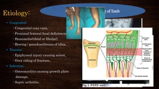
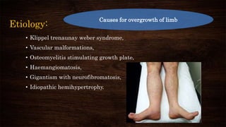
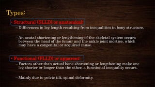
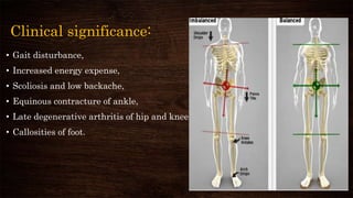
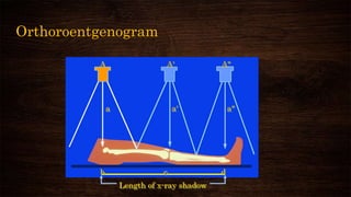
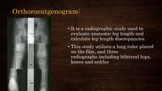
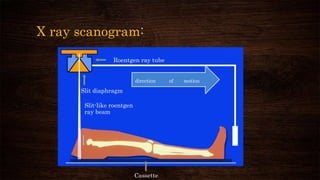
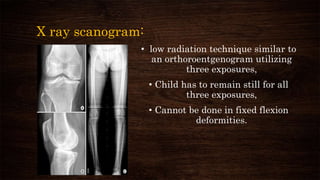
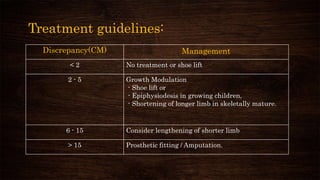
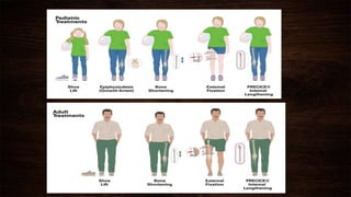
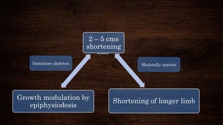
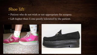
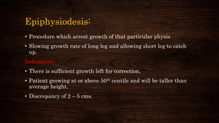
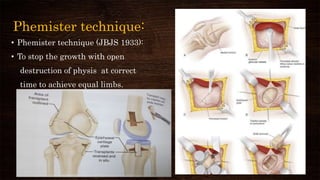
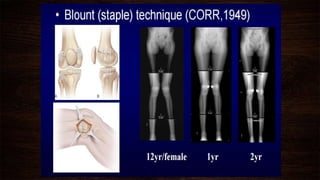
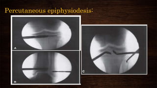
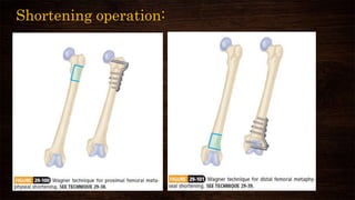
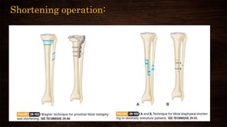
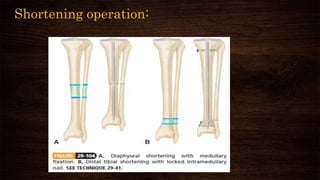
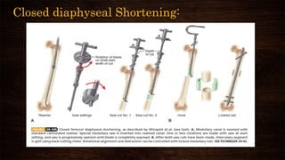
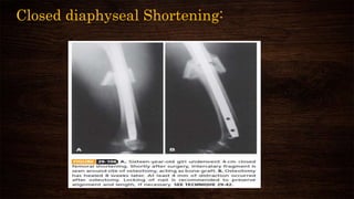
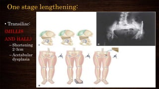
LIMB LENGTH DISCREPANCY
BY
DR. RAMACHANDRA REDDY
DNB RESIDENT
UNDER GUIDANCE OF
PROF. A. DEVADOSS
DR. SATISH DEVADOSS
Definition:
• Limb length discrepancy or anisomelia, is defined as a condition in which
the paired lower extremity have a noticeably unequal length.
• In the lower extremity length discrepancy is not only a cosmetic concern,
but also a functional concern.
• Giles and Taylor and Friberg studied
much larger groups of patients and concluded
that significant limb-length discrepancy causes
low back pain, and that the pain is diminished by
limb equalization.
More than 2.5 cms of LLD causes significant increase in energy
expenditure due to excessive rise and fall of pelvis ,compensatory ankle
movements.
Etiology:
• Congenital:
- Congenital coxa vara,
- Proximal femoral focal deficiencies,
- Hemimelia(tibial or fibular),
- Bowing / pseudoarthrosis of tibia,
• Trauma:
- Epiphyseal injury causing arrest,
- Over riding of fracture,
• Infection:
- Osteomyelitis causing growth plate
damage,
- Septic arthritis,
• Neurologic:
- Cerebral palsy,
- Polio,
- Myelodysplasia,
• Tumours:
- Multiple exostosis,
- Fibrous dysplasia,
• Idiopathic unilateral hypoplasia
Causes for shortening of limb
Etiology:
• Klippel trenaunay weber syndrome,
• Vascular malformations,
• Osteomyelitis stimulating growth plate,
• Haemangiomatosis,
• Gigantism with neurofibromatosis,
• Idiopathic hemihypertrophy.
Causes for overgrowth of limb
Types:
• Structural (SLLD) or anatomical:
– Differences in leg length resulting from inequalities in bony structure.
– An acutal shortening or lengthening of the skeletal system occurs
between the head of the femur and the ankle joint mortise, which
may have a congenital or acquired cause.
• Functional (FLLD) or apparent:
– Factors other than actual bone shortening or lengthening make one
leg shorter or longer than the other, a functional inequality occurs.
– Mainly due to pelvic tilt, spinal deformity.
Classification:
• McCaw and Bates (1991) report the following classification:
– LLD has been classified according to the magnitude of the
inequality, generally expressed in cm or mm, and described as
mild, moderate, or severe.
• Mild Less than 3 cm
• Moderate 3-6 cm
• Severe More than 6 cm
Compensation:
• Shoulder tilt,
• Unequal arm swing,
• Pelvic tilt,
• Scoliosis towards same side,
• On longer side( Knee flexion and pronation of ankle),
• On shorter side(Plantar flexion and supination of
ankle).
Clinical significance:
• Gait disturbance,
• Increased energy expense,
• Scoliosis and low backache,
• Equinous contracture of ankle,
• Late degenerative arthritis of hip and knee,
• Callosities of foot.
MANAGEMENT:
DIFFERS IN 1)IMMATURE GROWING PATIENTS AND,
2) SKELETALLY MATURE PATIENTS.
In children we should have ideas regarding
- Normal growth,
- Techniques for predicting growth and
- Appropriate time for intervention.
Techniques for predicting growth:
• Green and anderson method,
• Moseley’s chart,
• Menelaus method
• Paley’s multiplier method.
• Proximal Femur
– 3mm / year
– 15% leg
• Distal Femur
– 9mm / year
– 37% leg
– 70% of femur
• Growth Cessation
– 14-15 Girls
– 16-17 Boys
• Proximal Tibia
– 6mm / year
– 28%
– 60% tibia
• Distal Tibia
– 3mm / year
– 20%
Menelaus method : relies on chronological age rather
than skeletal age
Green and anderson method:
• Growth remaining method
– uses skeletal age
– requires graph
– estimates growth potential in distal femur and proximal tibia
at various skeletal ages
– separate charts for girls and boys
Moseley chart:
• Straight - Line Graph Method
– uses Green & Anderson data
– applied to a chart
• At least 3 measurements each time
– 1. Length long leg
– 2. Length short leg
– 3. Skeletal age
• Do so 3 times separated by 3-6 months
– accuracy improves with increased plotting
Moseley chart:
Paley multiplier:
• State of the art:
– take LLD for boy or girl
– multiplier for chronological or skeletal age
– predicts LLD at maturity
Paley multiplier:
Paley multiplier:
• Congenital Limb Length Discrepancy
∆m = ∆ x M
– ∆m: Limb discrepancy at skeletal maturity
– ∆: Current Limb-length discrepancy
– M: Multiplier.
• Example:current lld is 4cm in Congenital hemihypertrophy at 10
yrs age
• Using value of 1.310 according to Multiplier chart at age of 10 in
tibia
• 4 x 1.310 = 5.24 cm(lld at maturity)
Developmental LLD Leg-length discrepancy:
• ∆m = ∆ + (IXG)
• I=1 -(S – S’)/(L – L’)
• G=L(M-1)
• G= amount of growth remaining
• I=amount of growth inhibition
• L= current length of long limb
• L’=length of long limb as measured on previous radiographs
• Lm length =length of femur or tibia at skeletal maturity of femur or tibia at skeletal maturity
• M=multiplier
• S= current length of short limb
• S’ =length o f short limb as measured on previous radiographs
• ∆ = current limb-length discrepancy
• ∆m=limb length discrepancy at skeletal maturity
Example:
• Femur length(cm)
right (abnormal) left (normal)
• previous 24 26 at age of 8yrs
• Current 26 29 at age of 10 yrs
• I=1 -(S – S’)/(L – L’)
I =1-(26-24/29-26) = 1-2/3=0.33(amount of growth inhibition)
• G=L(M-1)
G=29(1.310-1)=29 x 0.310=8.99(amount of growth remaining)
• ∆m = ∆ + (IXG)
∆m = 3 +(0.33 x 8.99)=3 + 2.97 = 5.97 cm(lld at skeletal maturity)
Automated software:
•
Clinical Examination:
• Wood block test(coleman’s):
– with the patient standing,
add blocks under the short
leg until the pelvis is level,
then measure the blocks to
determine the discrepancy.
– block testing is considered
the best initial screening
method.
– Add blocks (known height)
until the pelvis is level
Gallezzi’s test: supination and pronation
Supination – femur
Pronation – tibia
Leg-length measurement:
• Apparent length:
– from the Xiphisternum to the
medial malleolus
• True length:
– from the ASIS to the medial
malleolus after squaring
pelvis.
Radiographs (Measure Length Discrepancy)
• Teloroentgenogram
• Orthoroentgenogram
• Scanogram (x-ray/ CT)
Teloroentgenogram
Length of x-ray shadow
• An X-ray photograph taken at a distance of usually six feet with resultant practical
parallelism of the rays and production of shadows of natural size.
Orthoroentgenogram
Length of x-ray shadow
A A A
a a a
b c d
Orthoroentgenogram:
• It is a radiographic study used to
evaluate anatomic leg length and
calculate leg-length discrepancies.
• This study utilises a long ruler placed
on the film, and three
radiographs including bilateral hips,
knees and ankles
X ray scanogram:
Roentgen ray tube
Slit diaphragm
Slit-like roentgen
ray beam
Cassette
motionofdirection
X ray scanogram:
• low radiation technique similar to
an orthoroentgenogram utilizing
three exposures,
• Child has to remain still for all
three exposures,
• Cannot be done in fixed flexion
deformities.
CT scanogram:
• Investigation of choice,
• Software measures
distances
–accurate to 0.2 mm
–legs must be in same
position
–fast
Treatment guidelines:
Discrepancy(CM) Management
< 2 No treatment or shoe lift
2 - 5 Growth Modulation
- Shoe lift or
- Epiphysiodesis in growing children,
- Shortening of longer limb in skeletally mature.
6 - 15 Consider lengthening of shorter limb
> 15 Prosthetic fitting / Amputation.
2 – 5 cms
shortening
Shortening of longer limbGrowth modulation by
epiphysiodesis
Immature skeleton Skeletally mature
Shoe lift:
• Patient who do not wish or not appropriate for surgery.
• Lift higher than 5 cms poorly tolerated by the patient.
Operative management:
• Theoretically lengthening of the short limb is optimal, but it’s
technically difficult and associated with frequent complications,
• For small discrepancies in growing children, epiphysiodesis is
relatively simple procedure with low morbidity and fast recovery,
• In skeletally mature people shortening of longer limb is better
than lengthening as joint stiffness is less.
Epiphysiodesis:
• Procedure which arrest growth of that particular physis
• Slowing growth rate of long leg and allowing short leg to catch
up.
Indications:
• There is sufficient growth left for correction,
• Patient growing at or above 50th centile and will be taller than
average height,
• Discrepancy of 2 – 5 cms.
Disadvantages:
• Normal limb is operated on, instead of pathologic limb,
• Any deformity in pathologic limb cannot be corrected by this
method,
• The final height at maturity may be unacceptably low,
• Body proportions may be cosmetically displeasing.
Techniques:
• Phemister epiphysiodesis (1933),
• Blount’s epiphysiodesis (1949),
• Percutaneous epiphysiodesis by CANALE ET AL,
• Percutaneous trans-epiphyseal screw epiphysiodesis by
METAIZEAU ET AL,
• Tension plate epiphysiodesis.
Phemister technique:
• Phemister technique (JBJS 1933):
• To stop the growth with open
destruction of physis at correct
time to achieve equal limbs.
Percutaneous epiphysiodesis:
Percutaneous trans physeal screw
epiphysiodesis:
PETS(Percutaneous Epiphysiodesis using Trans epiphyseal Screws )
(Metaizeau JP, et al, JPO,1998)
TENSION PLATE EPIPHYSIODESIS
• This technique is largely reserved for hemiepiphysiodesis in angular
corrections,
• It can be used for complete epiphysiodesis if implants are used on both sides
of the physis.
• This technique also has the advantage of potential growth resumption with
implant removal; however, restoration of normal growth often is
unpredictable after implant removal, and careful timing of epiphysiodesis is
still important
TENSION PLATE EPIPHYSIODESIS
• Most of these plating systems are nonlocking, which allows some
degree of screw divergence within the plate as the physis
continues to grow.
• It is likely that growth arrest does not occur until maximal screw
divergence is reached.
• Therefore, it is advisable to place the screws in a divergent
fashion at the time of implantation to allow growth arrest to
occur as quickly as possible.
TENSION PLATE EPIPHYSIODESIS
Problems of Epiphysiodesis
• Under correction of growth or angulation
• Overcorrection growth or angulation
• Asymmetric growth arrest
• Nerve injury, infection
• Implant failures.
Limb Shortening operation:
• WAGNER outlined the approach to limb shortening,
• WINQUIST deviced closed technique for diaphyseal shortening,
INDICATIONS:
• Skeletally mature patient,
• Tibia 2-3cms, Femur 4-5cms can be removed without affecting muscle
function, (Discrepancy less than 5cms),
• Patient height more than 50th percentile.
Femoral shortening is preferred
Femoral shortening:
• Upto 5 cms tolerable,
• Only one bone is involved and is
protected by muscles around the
thigh,
• Delayed and non union are less
common,
• Muscles regain strength and
tension quickly.
Tibial shortening:
• Upto 3 cms tolerable,
• Has to deal with two bones,
• Chances of neurovascular bundle
injury is higher, (Fasciotomy is
required)
• Recovery of muscle takes longer
time.
Shortening operation:
Closed diaphyseal Shortening:
Limb lengthening operation:
Indications:
• Shortening >6 cms,
• nearing skeletal maturity where epiphyseal arrest or shortening of bone of
long limb would not produce satisfactory equalization,
• When discrepancy is more in a single bone due to trauma/ infection,
Pre requisites:
• Neighbouring joints should be free with good ROM,
• Absence of scarring of skin or soft tissue,
• Bone should be normal,(Fracture if any should be united).
Limb lengthening operation:
• Can be performed on both children and adults with limb length
discrepancies ( >6cms) and angular deformities due to birth
defects, injuries or diseases.
• The success depends largely on
- patients and families commitment in maintaining external
fixator,
- Efforts in physiotheraphy and
- Patience.
Limb lengthening:
• Not advisable in --
• Patients who are unable to participate in frequent follow-up or who do not
have the support to care for the fixator properly and to undergo vigorous
physical therapy are best treated by means other than lengthening,
Limb lengthening
Acute
Gradual
Acute lengthening:
• When performing acute lengthening, cut the bone, spread the two sections
apart, and insert a graft and internal fixation is done to maintain the
length.
• Surrounding muscles, nerves and blood vessels do not tolerate a lot of
stretching.
• So acute lengthening can only achieve limited increase is acheived. For
example, forearm bones (radius or ulna) and foot bones (metatarsals) are
lengthened by this method when only a small gain in length is needed.
One stage lengthening:
• Transiliac:
(MILLIS
AND HALL)
– Shortening
2-3cm
– Acetabular
dysplasia
Gradual lengthening - Distraction Osteogenesis:
• Principle:
– 1) Corticotomy: preserve endosteal & periosteal blood supply in
metaphyseal region,
– 2) Ilizarov Ring fixator or unilateral LRS
– 3) Latency period: 7-14 days
– 4) Proper rate & Rhythm: 0.25mm x4 / day
– 5) Encourage Joint motion
Limb lengthening operation:
• Devices for gradual lengthening
1)External fixators:
– Unilateral fixator (Orthofix / LRS)
– Circular ring fixator (Ilizarov, Taylor spatial frame )
2)Intramedullary lengthening device
– PRECISE – Approved in USA,
– ISKD(inter medullary skeletal kinetic device),
– Fitbone.
Distraction Epiphysiolysis
• Chondrodiastasis (Gelbke,1951, De Bastiani,1986)
• Separation of the epiphyseal plate
• Immature patient
• Risk of septic arthritis
• Painful stiffness of the joint
• Premature closure of the physis
Four phases of Gradual limb lenghtening:
• Preparation:
– Consultation, X- rays of the limbs to build a custom- build external fixator,
psychological evaluation
• Surgery:
– External fixator is attached to the bones
• Lengthening:
– Fixator is lengthened about 1 mm every day for new bone growth.
• Strengthening:
– For proper alignment and consolidation of new bone, removal of external
fixator, PT rehabilitation.
Gradual lengthening :
• By Orthofix: Instruments.
C D unit
Conical pins
Clamp
Rail
schanz pin
Tibia Lengthening:
(DEBASTIANI ET AL) Orthofix lengthening devices
Femur
lengthening:
• Ilizarov Technique (Instruments):
Rings 120 -220mm
Tensioners
Wires - olive and plain Connecting rods
Tibial lengthening:
Femoral lengthening:
Complications:
• Muscle contractures
• Joint subluxations
• Neurological or vascular insult
• Premature or delayed consolidation
• Re- fracture
• Pin- site infections
• Psychological stress
Intramedullary lengthening devices:
Advantages:
• No pin tract infection and soft-tissue transfixation,
• To maintain mechanical alignment and stability during lengthening
and consolidation, and
• To improve patient comfort and tolerance.
Types of intramedullary devices:
• Lengthening may be initiated by
- controlled rotation, ambulation, and weight bearing
ISKD(Intramedullary Skeletal Kinetic Device; Orthofix,
McKinney, Tex);
- An implanted electrically activated motorized drive (FITBONE;
Wittenstein Igersheim, Germany).
- An Magnetically controlled distractors using an external remote
(PRECICE; Ellipse Tech., Irvine, USA).
ISKD:
ISKD:
Fitbone:
External remote for distraction of
precice nail:
- It takes 7 mins for 1mm
distraction,
-So three times a day patient uses
this remote for 2.5 mins for
accurate lengthening,
- Approved by FDA for use in US.
For LLD more than
15cms
Prosthetic fitting
Amputation.
Prosthetic fitting
• Significant discrepancies, deformed functionally useless
feet
• Discrepancies greater than 15-20cm and femoral length
less than 50%
• Fibular hemimelia with unstable ankle
• PFFD: A/K prosthesis or BK prosthesis with Van Nes
rotationplasty
Amputation:
• Significant length discrepancy or loss of sensation in foot,
• Poor underlying bone quality for lengthening,
• Dysfunctional/ painful limb.
Clinical case:
14 years old girl with idiopathis shortening of rt
lower limb -- LLD of 20 cms
Pre operative After femoral
lengthening
Pre op CT scanogram Immediate post op 2 months 4 months
Thank you…

Limb length discrepancy

  • 1.
    LIMB LENGTH DISCREPANCY BY DR.RAMACHANDRA REDDY DNB RESIDENT UNDER GUIDANCE OF PROF. A. DEVADOSS DR. SATISH DEVADOSS
  • 2.
    Definition: • Limb lengthdiscrepancy or anisomelia, is defined as a condition in which the paired lower extremity have a noticeably unequal length. • In the lower extremity length discrepancy is not only a cosmetic concern, but also a functional concern. • Giles and Taylor and Friberg studied much larger groups of patients and concluded that significant limb-length discrepancy causes low back pain, and that the pain is diminished by limb equalization.
  • 3.
    More than 2.5cms of LLD causes significant increase in energy expenditure due to excessive rise and fall of pelvis ,compensatory ankle movements.
  • 4.
    Etiology: • Congenital: - Congenitalcoxa vara, - Proximal femoral focal deficiencies, - Hemimelia(tibial or fibular), - Bowing / pseudoarthrosis of tibia, • Trauma: - Epiphyseal injury causing arrest, - Over riding of fracture, • Infection: - Osteomyelitis causing growth plate damage, - Septic arthritis, • Neurologic: - Cerebral palsy, - Polio, - Myelodysplasia, • Tumours: - Multiple exostosis, - Fibrous dysplasia, • Idiopathic unilateral hypoplasia Causes for shortening of limb
  • 5.
    Etiology: • Klippel trenaunayweber syndrome, • Vascular malformations, • Osteomyelitis stimulating growth plate, • Haemangiomatosis, • Gigantism with neurofibromatosis, • Idiopathic hemihypertrophy. Causes for overgrowth of limb
  • 6.
    Types: • Structural (SLLD)or anatomical: – Differences in leg length resulting from inequalities in bony structure. – An acutal shortening or lengthening of the skeletal system occurs between the head of the femur and the ankle joint mortise, which may have a congenital or acquired cause. • Functional (FLLD) or apparent: – Factors other than actual bone shortening or lengthening make one leg shorter or longer than the other, a functional inequality occurs. – Mainly due to pelvic tilt, spinal deformity.
  • 7.
    Classification: • McCaw andBates (1991) report the following classification: – LLD has been classified according to the magnitude of the inequality, generally expressed in cm or mm, and described as mild, moderate, or severe. • Mild Less than 3 cm • Moderate 3-6 cm • Severe More than 6 cm
  • 8.
    Compensation: • Shoulder tilt, •Unequal arm swing, • Pelvic tilt, • Scoliosis towards same side, • On longer side( Knee flexion and pronation of ankle), • On shorter side(Plantar flexion and supination of ankle).
  • 9.
    Clinical significance: • Gaitdisturbance, • Increased energy expense, • Scoliosis and low backache, • Equinous contracture of ankle, • Late degenerative arthritis of hip and knee, • Callosities of foot.
  • 10.
    MANAGEMENT: DIFFERS IN 1)IMMATUREGROWING PATIENTS AND, 2) SKELETALLY MATURE PATIENTS. In children we should have ideas regarding - Normal growth, - Techniques for predicting growth and - Appropriate time for intervention.
  • 11.
    Techniques for predictinggrowth: • Green and anderson method, • Moseley’s chart, • Menelaus method • Paley’s multiplier method.
  • 12.
    • Proximal Femur –3mm / year – 15% leg • Distal Femur – 9mm / year – 37% leg – 70% of femur • Growth Cessation – 14-15 Girls – 16-17 Boys • Proximal Tibia – 6mm / year – 28% – 60% tibia • Distal Tibia – 3mm / year – 20% Menelaus method : relies on chronological age rather than skeletal age
  • 13.
    Green and andersonmethod: • Growth remaining method – uses skeletal age – requires graph – estimates growth potential in distal femur and proximal tibia at various skeletal ages – separate charts for girls and boys
  • 14.
    Moseley chart: • Straight- Line Graph Method – uses Green & Anderson data – applied to a chart • At least 3 measurements each time – 1. Length long leg – 2. Length short leg – 3. Skeletal age • Do so 3 times separated by 3-6 months – accuracy improves with increased plotting
  • 15.
  • 16.
    Paley multiplier: • Stateof the art: – take LLD for boy or girl – multiplier for chronological or skeletal age – predicts LLD at maturity
  • 17.
  • 18.
    Paley multiplier: • CongenitalLimb Length Discrepancy ∆m = ∆ x M – ∆m: Limb discrepancy at skeletal maturity – ∆: Current Limb-length discrepancy – M: Multiplier. • Example:current lld is 4cm in Congenital hemihypertrophy at 10 yrs age • Using value of 1.310 according to Multiplier chart at age of 10 in tibia • 4 x 1.310 = 5.24 cm(lld at maturity)
  • 19.
    Developmental LLD Leg-lengthdiscrepancy: • ∆m = ∆ + (IXG) • I=1 -(S – S’)/(L – L’) • G=L(M-1) • G= amount of growth remaining • I=amount of growth inhibition • L= current length of long limb • L’=length of long limb as measured on previous radiographs • Lm length =length of femur or tibia at skeletal maturity of femur or tibia at skeletal maturity • M=multiplier • S= current length of short limb • S’ =length o f short limb as measured on previous radiographs • ∆ = current limb-length discrepancy • ∆m=limb length discrepancy at skeletal maturity
  • 20.
    Example: • Femur length(cm) right(abnormal) left (normal) • previous 24 26 at age of 8yrs • Current 26 29 at age of 10 yrs • I=1 -(S – S’)/(L – L’) I =1-(26-24/29-26) = 1-2/3=0.33(amount of growth inhibition) • G=L(M-1) G=29(1.310-1)=29 x 0.310=8.99(amount of growth remaining) • ∆m = ∆ + (IXG) ∆m = 3 +(0.33 x 8.99)=3 + 2.97 = 5.97 cm(lld at skeletal maturity)
  • 21.
  • 22.
    Clinical Examination: • Woodblock test(coleman’s): – with the patient standing, add blocks under the short leg until the pelvis is level, then measure the blocks to determine the discrepancy. – block testing is considered the best initial screening method. – Add blocks (known height) until the pelvis is level
  • 23.
    Gallezzi’s test: supinationand pronation Supination – femur Pronation – tibia
  • 24.
    Leg-length measurement: • Apparentlength: – from the Xiphisternum to the medial malleolus • True length: – from the ASIS to the medial malleolus after squaring pelvis.
  • 25.
    Radiographs (Measure LengthDiscrepancy) • Teloroentgenogram • Orthoroentgenogram • Scanogram (x-ray/ CT)
  • 26.
    Teloroentgenogram Length of x-rayshadow • An X-ray photograph taken at a distance of usually six feet with resultant practical parallelism of the rays and production of shadows of natural size.
  • 27.
    Orthoroentgenogram Length of x-rayshadow A A A a a a b c d
  • 28.
    Orthoroentgenogram: • It isa radiographic study used to evaluate anatomic leg length and calculate leg-length discrepancies. • This study utilises a long ruler placed on the film, and three radiographs including bilateral hips, knees and ankles
  • 29.
    X ray scanogram: Roentgenray tube Slit diaphragm Slit-like roentgen ray beam Cassette motionofdirection
  • 30.
    X ray scanogram: •low radiation technique similar to an orthoroentgenogram utilizing three exposures, • Child has to remain still for all three exposures, • Cannot be done in fixed flexion deformities.
  • 31.
    CT scanogram: • Investigationof choice, • Software measures distances –accurate to 0.2 mm –legs must be in same position –fast
  • 32.
    Treatment guidelines: Discrepancy(CM) Management <2 No treatment or shoe lift 2 - 5 Growth Modulation - Shoe lift or - Epiphysiodesis in growing children, - Shortening of longer limb in skeletally mature. 6 - 15 Consider lengthening of shorter limb > 15 Prosthetic fitting / Amputation.
  • 34.
    2 – 5cms shortening Shortening of longer limbGrowth modulation by epiphysiodesis Immature skeleton Skeletally mature
  • 35.
    Shoe lift: • Patientwho do not wish or not appropriate for surgery. • Lift higher than 5 cms poorly tolerated by the patient.
  • 36.
    Operative management: • Theoreticallylengthening of the short limb is optimal, but it’s technically difficult and associated with frequent complications, • For small discrepancies in growing children, epiphysiodesis is relatively simple procedure with low morbidity and fast recovery, • In skeletally mature people shortening of longer limb is better than lengthening as joint stiffness is less.
  • 37.
    Epiphysiodesis: • Procedure whicharrest growth of that particular physis • Slowing growth rate of long leg and allowing short leg to catch up. Indications: • There is sufficient growth left for correction, • Patient growing at or above 50th centile and will be taller than average height, • Discrepancy of 2 – 5 cms.
  • 38.
    Disadvantages: • Normal limbis operated on, instead of pathologic limb, • Any deformity in pathologic limb cannot be corrected by this method, • The final height at maturity may be unacceptably low, • Body proportions may be cosmetically displeasing.
  • 39.
    Techniques: • Phemister epiphysiodesis(1933), • Blount’s epiphysiodesis (1949), • Percutaneous epiphysiodesis by CANALE ET AL, • Percutaneous trans-epiphyseal screw epiphysiodesis by METAIZEAU ET AL, • Tension plate epiphysiodesis.
  • 40.
    Phemister technique: • Phemistertechnique (JBJS 1933): • To stop the growth with open destruction of physis at correct time to achieve equal limbs.
  • 42.
  • 43.
    Percutaneous trans physealscrew epiphysiodesis: PETS(Percutaneous Epiphysiodesis using Trans epiphyseal Screws ) (Metaizeau JP, et al, JPO,1998)
  • 44.
    TENSION PLATE EPIPHYSIODESIS •This technique is largely reserved for hemiepiphysiodesis in angular corrections, • It can be used for complete epiphysiodesis if implants are used on both sides of the physis. • This technique also has the advantage of potential growth resumption with implant removal; however, restoration of normal growth often is unpredictable after implant removal, and careful timing of epiphysiodesis is still important
  • 45.
    TENSION PLATE EPIPHYSIODESIS •Most of these plating systems are nonlocking, which allows some degree of screw divergence within the plate as the physis continues to grow. • It is likely that growth arrest does not occur until maximal screw divergence is reached. • Therefore, it is advisable to place the screws in a divergent fashion at the time of implantation to allow growth arrest to occur as quickly as possible.
  • 46.
  • 47.
    Problems of Epiphysiodesis •Under correction of growth or angulation • Overcorrection growth or angulation • Asymmetric growth arrest • Nerve injury, infection • Implant failures.
  • 48.
    Limb Shortening operation: •WAGNER outlined the approach to limb shortening, • WINQUIST deviced closed technique for diaphyseal shortening, INDICATIONS: • Skeletally mature patient, • Tibia 2-3cms, Femur 4-5cms can be removed without affecting muscle function, (Discrepancy less than 5cms), • Patient height more than 50th percentile.
  • 49.
    Femoral shortening ispreferred Femoral shortening: • Upto 5 cms tolerable, • Only one bone is involved and is protected by muscles around the thigh, • Delayed and non union are less common, • Muscles regain strength and tension quickly. Tibial shortening: • Upto 3 cms tolerable, • Has to deal with two bones, • Chances of neurovascular bundle injury is higher, (Fasciotomy is required) • Recovery of muscle takes longer time.
  • 50.
  • 51.
  • 52.
  • 53.
  • 54.
  • 55.
    Limb lengthening operation: Indications: •Shortening >6 cms, • nearing skeletal maturity where epiphyseal arrest or shortening of bone of long limb would not produce satisfactory equalization, • When discrepancy is more in a single bone due to trauma/ infection, Pre requisites: • Neighbouring joints should be free with good ROM, • Absence of scarring of skin or soft tissue, • Bone should be normal,(Fracture if any should be united).
  • 56.
    Limb lengthening operation: •Can be performed on both children and adults with limb length discrepancies ( >6cms) and angular deformities due to birth defects, injuries or diseases. • The success depends largely on - patients and families commitment in maintaining external fixator, - Efforts in physiotheraphy and - Patience.
  • 57.
    Limb lengthening: • Notadvisable in -- • Patients who are unable to participate in frequent follow-up or who do not have the support to care for the fixator properly and to undergo vigorous physical therapy are best treated by means other than lengthening, Limb lengthening Acute Gradual
  • 58.
    Acute lengthening: • Whenperforming acute lengthening, cut the bone, spread the two sections apart, and insert a graft and internal fixation is done to maintain the length. • Surrounding muscles, nerves and blood vessels do not tolerate a lot of stretching. • So acute lengthening can only achieve limited increase is acheived. For example, forearm bones (radius or ulna) and foot bones (metatarsals) are lengthened by this method when only a small gain in length is needed.
  • 59.
    One stage lengthening: •Transiliac: (MILLIS AND HALL) – Shortening 2-3cm – Acetabular dysplasia
  • 60.
    Gradual lengthening -Distraction Osteogenesis: • Principle: – 1) Corticotomy: preserve endosteal & periosteal blood supply in metaphyseal region, – 2) Ilizarov Ring fixator or unilateral LRS – 3) Latency period: 7-14 days – 4) Proper rate & Rhythm: 0.25mm x4 / day – 5) Encourage Joint motion
  • 61.
    Limb lengthening operation: •Devices for gradual lengthening 1)External fixators: – Unilateral fixator (Orthofix / LRS) – Circular ring fixator (Ilizarov, Taylor spatial frame ) 2)Intramedullary lengthening device – PRECISE – Approved in USA, – ISKD(inter medullary skeletal kinetic device), – Fitbone.
  • 62.
    Distraction Epiphysiolysis • Chondrodiastasis(Gelbke,1951, De Bastiani,1986) • Separation of the epiphyseal plate • Immature patient • Risk of septic arthritis • Painful stiffness of the joint • Premature closure of the physis
  • 63.
    Four phases ofGradual limb lenghtening: • Preparation: – Consultation, X- rays of the limbs to build a custom- build external fixator, psychological evaluation • Surgery: – External fixator is attached to the bones • Lengthening: – Fixator is lengthened about 1 mm every day for new bone growth. • Strengthening: – For proper alignment and consolidation of new bone, removal of external fixator, PT rehabilitation.
  • 64.
    Gradual lengthening : •By Orthofix: Instruments. C D unit Conical pins Clamp Rail schanz pin
  • 65.
    Tibia Lengthening: (DEBASTIANI ETAL) Orthofix lengthening devices
  • 66.
  • 67.
    • Ilizarov Technique(Instruments): Rings 120 -220mm Tensioners Wires - olive and plain Connecting rods
  • 68.
  • 69.
  • 70.
    Complications: • Muscle contractures •Joint subluxations • Neurological or vascular insult • Premature or delayed consolidation • Re- fracture • Pin- site infections • Psychological stress
  • 71.
    Intramedullary lengthening devices: Advantages: •No pin tract infection and soft-tissue transfixation, • To maintain mechanical alignment and stability during lengthening and consolidation, and • To improve patient comfort and tolerance.
  • 72.
    Types of intramedullarydevices: • Lengthening may be initiated by - controlled rotation, ambulation, and weight bearing ISKD(Intramedullary Skeletal Kinetic Device; Orthofix, McKinney, Tex); - An implanted electrically activated motorized drive (FITBONE; Wittenstein Igersheim, Germany). - An Magnetically controlled distractors using an external remote (PRECICE; Ellipse Tech., Irvine, USA).
  • 73.
  • 74.
  • 75.
  • 76.
    External remote fordistraction of precice nail: - It takes 7 mins for 1mm distraction, -So three times a day patient uses this remote for 2.5 mins for accurate lengthening, - Approved by FDA for use in US.
  • 77.
    For LLD morethan 15cms Prosthetic fitting Amputation.
  • 78.
    Prosthetic fitting • Significantdiscrepancies, deformed functionally useless feet • Discrepancies greater than 15-20cm and femoral length less than 50% • Fibular hemimelia with unstable ankle • PFFD: A/K prosthesis or BK prosthesis with Van Nes rotationplasty
  • 79.
    Amputation: • Significant lengthdiscrepancy or loss of sensation in foot, • Poor underlying bone quality for lengthening, • Dysfunctional/ painful limb.
  • 80.
    Clinical case: 14 yearsold girl with idiopathis shortening of rt lower limb -- LLD of 20 cms Pre operative After femoral lengthening
  • 82.
    Pre op CTscanogram Immediate post op 2 months 4 months
  • 83.