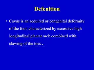
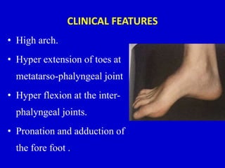
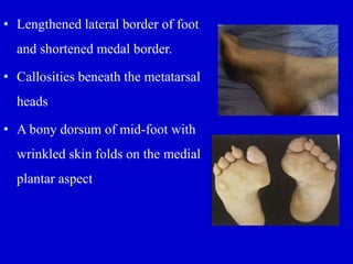
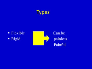
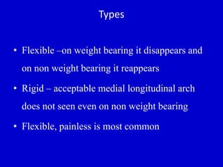
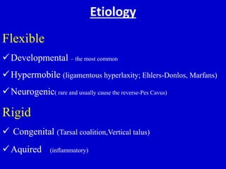
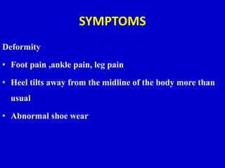
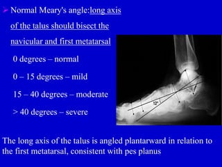
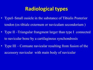
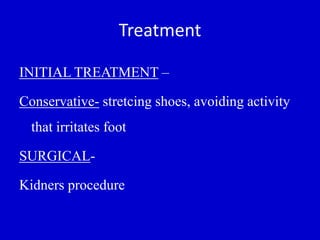
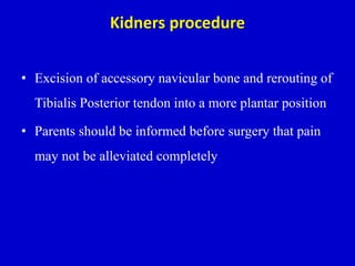
Pes Cavus and Pes Planus
Moderator:PROF.DR.K.PRAKASAM
M.S.Ortho,D.Ortho,DSc(HON)
Director&HOD
Presentor:Dr.Thouseef A Majeed
ANATOMY OF THE ARCHES OF FOOT
A) Two longitudinal arches
– Medial longitudinal arch
– Lateral longitudinal arch
B) Transverse arch
• Anterior transverse arch
• Posterior transverse arch
USE OF THE ARCHED FOOT
 Supports body weight in upright posture
 Acts as a lever to propel the body forwards in walking,
running and jumping
 Acts as a shock absorber
 Concavity of the arches protects the soft tissues of the sole
against pressure
Medial longitudinal arch
• Higher than lateral
• Composed of – Calcaneous
- Talus
- Navicular
- 3 cuneiform
- 3 metatarsals
• Talar head is key stone of this arch
• Tibialis anterior attached to – 1st metatarsal,medial cuneiform –
strength for this arch.
• Peroneus longus tendon – pass laterally to this arch providing
support
Lateral longitudinal Arch
• Flatter than medial longitudinal arch.
• Rests on the ground during standing.
• It is made up of – calcaneous, cuboid, 2 lateral
metatarsals.
Transverse arch
• Runs from side to side
• It is formed by – cuboid,
cuneiforms, bases of
metatarsals
• Medial and lateral parts
of longitudinal arch act as
pillars
• Tendons of fibularis
longus and tibialis
posterior
Integrity of bony arches
• Maintained by passive factors and dynamic
supports
Passive factors
• Shape of the united bones
• Four successive layers of fibrous
tissue – bowstring the longitudinal
arch
– Plantar aponeurosis
– Long plantar ligament
– Plantar calcaneocuboid (short
plantar) ligament
– Plantar calcaneonavicular
(spring) ligament
Dynamic supports
• Active bracing action of intrinsic muscles of foot
• Active and tonic contraction of muscles with long
tendons extending in to foot
– Flexor hallusis and digitorum longus – longitudinal arch
– Fibularis longus and tibialis posterior – transverse arch
• Plantar ligaments and plantar aponeurosis bear
greatest stress and important in maintaining arches
MECHANISM OF ARCH SUPPORT
SHAPE OF BONES
• Bones are wedge-shaped with the thin edge lying inferiorly
• This applies particularly to the bone occupying the center of
the arch“keystone”
MECHANISM OF ARCH SUPPORT
SUSPENDING THE ARCH FROM ABOVE
• Medial longtitudinal arch: Tibialis anterior, Tibialis
posterior, medial ligament of ankle joint
• Lateral longtitudinal arch: Peroneus longus, Peroneus
brevis
• Transverse arch: Peroneus longus
MECHANISM OF ARCH SUPPORT
SUSPENDING THE ARCH FROM ABOVE
PES CAVUS
Synonyms for Cavus Foot
• Schaffer Foot
• Lotus Flower Foot
• Bolt Foot
• Claw Foot
• Vault Foot
• Hollow Foot
• Anterior Equinus
• Pes Cavo Varus
• Contracted Foot
• Talipes (Pes)
Arcuatus
• Talipes Plantaris
Defenition
• Cavus is an acquired or congenital deformity
of the foot ,characterized by excessive high
longitudinal plantar arch combined with
clawing of the toes .
Etiology
Neurological Causes
• Charcot Marie Tooth disease
• Friedrich’s Ataxia
• Roussy-Levy syndrome
• Poliomyelitis
• Cerebral Palsy
Congenital
– Spina Bifida
– Talipes Equinovarus
– Myelodysplasia
– Clubfoot
Iatrogenic
– Post surgery or trauma
– Peroneal nerve injury
Etiology
Infection
– Syphillis
– Poliomyelitis
Idiopathic
– Most common
Development of the deformity
• The intrinsic musculature
normally flexes the
metatarsophalyngeal joint
and extends the
interphalyngeal joint.
• When the long flexor contracts on the straight digit it slings
up the heads of the metatarsals and prevents the drop of the
forefoot on the hind foot
• In the absence of lumbricals ,the long flexor pulls the toes
into flexion and no longer supports the metatarsal head.
• So the forefoot drops and the lax structures in the sole
contracts and forms claw foot.
• Dropping of fore foot on the hind foot followed by a
contracture of the plantar fascia and clawing of the
toes
CLINICAL FEATURES
• High arch.
• Hyper extension of toes at
metatarso-phalyngeal joint
• Hyper flexion at the inter-
phalyngeal joints.
• Pronation and adduction of
the fore foot .
• Lengthened lateral border of foot
and shortened medal border.
• Callosities beneath the metatarsal
heads
• A bony dorsum of mid-foot with
wrinkled skin folds on the medial
plantar aspect
Radiographic findings –pes cavus
Standing weight bearing Antero –posterior and Lateral views
X Rays taken to
• Demonstrate the apex of the deformity
• Talo calcaneal ankle
• Calcaneal pitch
• Degree of plantar flexion of the great toe
• Asess the contribution of cavus by hind foot,midfoot and
fore foot
DEGREES OF PES CAVUS
• 5 degrees
First degree pes cavus
• Child is clumsy with repeated falls
• Foot appears normal
• Deformity appears when foot is relaxed
• Child catches his toes against low objects such as edges of
carpet.
• Mild extensor weakness
Treatment of first degree pes cavus
• Daily manipulation –supinating fore foot and everting heel
• Anterior arch bar in shoes
• If not corrected then Girdle stone tendon transfer
operation.
• Through an incision on each toe
extending distally from metatarso-
phalyngeal joint .
• Long and short toe flexors are brought
to lateral aspect of proximal phalynx
and sutured to the extensor expansion.
Second degree pes cavus
• Flexion of the fore foot
• Plantar fascia is felt to be tense and contracted
• Clawing of great toe .
• Great toe clawing can be corrected by upward pressure on
the ball of great toe.
Treatment of second degree Pes cavus
• A shoe fitted with a metatarsal bar may give temporary
relief.
• Stiendlers Procedure : Plantar fascia release
• Jones Procedure:The Extensor hallucis longus tendon is
divided at its insertion and passed though the neck of first
metatarsal + Interphalyngeal joint fusion.
Third degree pes cavus
• The arches of foot is markedly raised.
• All toes are clawed .
• Tendocalcaneus may begin to appear contracted.
• Painfull callosities are seen.
• Deformities are rigid and cannot be corrected by finger
pressure under Ist metatarsal head
Treatment of third degree Pes cavus
• Stiendlers procedure +Muscle sliding
operation.
• Japas ‘ V‘osteotomy of tarsus : Apex of V is
proximal and highest point of cavus
• Dwayers Calcaneal Ostetomy
Fourth degree pes cavus
• In addition to cavus and claw toes
• Adduction at tarsometatarsal joints resulting in varus
deformity.
• Rigid and painful foot
• Walking becomes painful and difficult.
Fifth degree-pes cavus
• Seen on paralytic conditions.(poliomyelitis)
• Whole foot is contracted into rigid equino varus with high
arch.
• Tender callosities.
• The patient is very disabled .
Treatment of fourth and fifth degree Pes cavus
• Dunns triple arthrodesis
• Lambrinudis arthrodesis
(triple arthrodesis :subtalar+calcneo cuboid +talo
navicular joint fusion)
• Cols Anterior tarsal wedge osteotomy
PES-PLANUS
Synonyms
• Pes planovalgus
• Flat feet
• Fallen arches
• Pronation of feet
Definition
• Absence of normal medial longitudinal arch
• Instep of the foot collapses and comes in
contact with the ground.
• In some individuals, this arch never develops
Other abnormalities
• Heel valgus
• Mild subluxation of subtalar joint(talus tilts medially
and plantarwards)
• Eversion of the calcaneus at the subtalar joint
• Lateral angulation of midtarsal joints (Talo Calcaneal
,Calcaneo Cuboid)
• Supination of forefeet
• Flat feet are a common condition.
• In infants and toddlers, the longitudinal arch is not
developed and flat feet are normal.
• The arch develops in childhood
• By adulthood (12-13yrs), most people have
developed normal arches
Types
 Flexible Can be
 Rigid painless
Painful
Types
• Flexible –on weight bearing it disappears and
on non weight bearing it reappears
• Rigid – acceptable medial longitudinal arch
does not seen even on non weight bearing
• Flexible, painless is most common
Etiology
Flexible
Developmental – the most common
Hypermobile (ligamentous hyperlaxity; Ehlers-Donlos, Marfans)
Neurogenic( rare and usually cause the reverse-Pes Cavus)
Rigid
 Congenital (Tarsal coalition,Vertical talus)
Aquired )inflammatory)
SYMPTOMS
Deformity
• Foot pain ,ankle pain, leg pain
• Heel tilts away from the midline of the body more than
usual
• Abnormal shoe wear
FLAT FEET CAN produce
• Tendonitis. posterior tibial tendon and it can either fail,
rupture, stretch or just hurt. This condition is called
POSTERIOR TIBIAL DYSFUNCTION (PTD OR TPD) .
• Arthritis.
• Plantar fasciitis
• Bunions & Hammertoes
• Corns and callosities
Radiography
• Asymptomatic flatfoot radiological evaluation unnecessary
• First Antero posterior and Lateral views of the foot should
be taken to evaluate severity of deformity
• Antero-posterior ankle to rule out valgus at the distal end of
tibia
• Special view - 45 degree eversion oblique for accessory
navicular bone
Radiography
• AP standing view is to asses heel valgus , Talocalcaneal
angle more than 35 degree is associated with incresed heel
valgus
• CT scan accurately defines anatomy of subtalar joint ,
allows surgical plannig if it is involved.
Meary’s Angle
• Most common angle to indicate
flat foot
• Intersects at apex of the
deformity
• Meary’s angle - between long
axis of talus and long axis of
first metatarsal on a standing
lateral X ray
 Normal Meary's angle:long axis
of the talus should bisect the
navicular and first metatarsal
0 degrees – normal
0 – 15 degrees – mild
15 – 40 degrees – moderate
> 40 degrees – severe
The long axis of the talus is angled plantarward in relation to
the first metatarsal, consistent with pes planus
Treatment
0-3 years old:
 No treatment unless very strong family hx of persistent
flatfeet
 Orthotic shoes with thomas heels ,medial heel wedges and
navicular pads
 Convince the parents.
Treatment
3-9 years
• Conservative management
• No surgery
• Custom orthosis inserted with leather ,cork,
propylene .
Treatment
• 10-14 yrs
• No symptom- No treatment
• Symptomatic – conservative management
initially
• Surgical
Surgical treatment
Indications
1.pain
2.failure to respond to orthotic control
3.Ulceration or callus under the head of the plantiflexed talus
4.Excessive shoe wear
Surgical treatment
• The surgeon , patient, and parents must be willing to
exchange loss of eversion and inversion of the foot
for relief of pain and disability .
Surgical treatment
• Arthrodesis for relieving painful flat foot have been
most successful when the subtalar joint is involved .
• Although midtarsal arthtrodesis without inclusion of
the subtalar joint has gained popularity
Surgeries
• Durham flatfoot plasty
• Posterior calcaneal displacement osteotomy
• Anterior calcaneal lengthening – distraction
wedge osteotomy
• Triple atrhrodesis (triplane)
Durham plasty for pes planus
A, Incision.
B, Elevation of posterior tibial
tendon.
C, Elevation of osteo-periosteal
flap from proximal to distal.
D, Arthrodesis of navicular–first
cuneiform joint.
E, Extent of arthrodesis resection
through midfoot.
F, Internal fixation of navicular–
first cuneiform joint.
•
pull the posterior tibial tendon taut
into its prepared bed on the plantar
surface of the waist of the
navicular, and tie the suture
dorsally
Calcaneal osteotomy (Dilwyn-
Evana,Mosca)
• Lengthening of lateral
column of the foot by
inserting a tibial bone graft
and calcaneocuboidal
fusion
Posterior calcaneal displacement
osteotomy(koutsgiannis)
• Symptomatic patients with excessive heel valgus , a
calcaneal osteotomy is intended to displace the
posterior part of the calcaneum medially , to restore
normal Weight bearing alignment
Triple Arthrodesis
Joints fused are:
• Subtalar joint
• Calcaneo cuboid joint
• Talo navicular joint
AGE
• Usually done after the age of 12
• Triple arthrodesis tend to have a high (50%) failure rate in
children under 10 years of age;
• contra-indicated in young children (less than 10-12 yrs)
because the procedure limits foot growth
Complications
• Nonunion
• Degenerative joint disease
• Avascular necrosis
• Lateral instability
• Stiff foot
Accessory navicular bone
• It is a most common accessory bone in the foot
• Listed as a cause of flat foot
Pathoanatomy
• Abnormal insertion of Tibialis Posterior into
accessory navicular bone believe to cause the flat
foot
Clinical presntation
• Often incidental, many patients are asymptomatic
• Pain
• Prominence of medial aspect of foot
• On attempted inversion of the foot against resistance
, Tibialis posterior tendon is inserted into the bump
and this maneuver produces pain
Radiography
• Special view - 45 degree eversion oblique for
accessory navicular bone
• Antero-Posterior view and Lateral weight bearing
views of the foot should be taken to evaluate other
deformities
Radiological types
• TypeI–Small ossicle in the substance of Tibialis Posterior
tendon (os tibiale externum or naviculam secondorium )
• Type II –Triangular frangment larger than type I connected
to navicular bone by a cartilaginous synchondrosis
• Type III – Cornuate navicular resulting from fusion of the
accessory navicular with main body of navicular
Treatment
INITIAL TREATMENT –
Conservative- stretcing shoes, avoiding activity
that irritates foot
SURGICAL-
Kidners procedure
Kidners procedure
• Excision of accessory navicular bone and rerouting of
Tibialis Posterior tendon into a more plantar position
• Parents should be informed before surgery that pain
may not be alleviated completely
“Our feet are no more alike than our
faces”
THANK YOU

Flat foot and Cavus foot

  • 1.
    Pes Cavus andPes Planus Moderator:PROF.DR.K.PRAKASAM M.S.Ortho,D.Ortho,DSc(HON) Director&HOD Presentor:Dr.Thouseef A Majeed
  • 2.
    ANATOMY OF THEARCHES OF FOOT A) Two longitudinal arches – Medial longitudinal arch – Lateral longitudinal arch B) Transverse arch • Anterior transverse arch • Posterior transverse arch
  • 3.
    USE OF THEARCHED FOOT  Supports body weight in upright posture  Acts as a lever to propel the body forwards in walking, running and jumping  Acts as a shock absorber  Concavity of the arches protects the soft tissues of the sole against pressure
  • 4.
    Medial longitudinal arch •Higher than lateral • Composed of – Calcaneous - Talus - Navicular - 3 cuneiform - 3 metatarsals • Talar head is key stone of this arch
  • 5.
    • Tibialis anteriorattached to – 1st metatarsal,medial cuneiform – strength for this arch. • Peroneus longus tendon – pass laterally to this arch providing support
  • 6.
    Lateral longitudinal Arch •Flatter than medial longitudinal arch. • Rests on the ground during standing. • It is made up of – calcaneous, cuboid, 2 lateral metatarsals.
  • 7.
    Transverse arch • Runsfrom side to side • It is formed by – cuboid, cuneiforms, bases of metatarsals • Medial and lateral parts of longitudinal arch act as pillars • Tendons of fibularis longus and tibialis posterior
  • 8.
    Integrity of bonyarches • Maintained by passive factors and dynamic supports
  • 9.
    Passive factors • Shapeof the united bones • Four successive layers of fibrous tissue – bowstring the longitudinal arch – Plantar aponeurosis – Long plantar ligament – Plantar calcaneocuboid (short plantar) ligament – Plantar calcaneonavicular (spring) ligament
  • 10.
    Dynamic supports • Activebracing action of intrinsic muscles of foot • Active and tonic contraction of muscles with long tendons extending in to foot – Flexor hallusis and digitorum longus – longitudinal arch – Fibularis longus and tibialis posterior – transverse arch • Plantar ligaments and plantar aponeurosis bear greatest stress and important in maintaining arches
  • 11.
    MECHANISM OF ARCHSUPPORT SHAPE OF BONES • Bones are wedge-shaped with the thin edge lying inferiorly • This applies particularly to the bone occupying the center of the arch“keystone”
  • 12.
    MECHANISM OF ARCHSUPPORT SUSPENDING THE ARCH FROM ABOVE • Medial longtitudinal arch: Tibialis anterior, Tibialis posterior, medial ligament of ankle joint • Lateral longtitudinal arch: Peroneus longus, Peroneus brevis • Transverse arch: Peroneus longus
  • 13.
    MECHANISM OF ARCHSUPPORT SUSPENDING THE ARCH FROM ABOVE
  • 14.
  • 15.
    Synonyms for CavusFoot • Schaffer Foot • Lotus Flower Foot • Bolt Foot • Claw Foot • Vault Foot • Hollow Foot • Anterior Equinus • Pes Cavo Varus • Contracted Foot • Talipes (Pes) Arcuatus • Talipes Plantaris
  • 16.
    Defenition • Cavus isan acquired or congenital deformity of the foot ,characterized by excessive high longitudinal plantar arch combined with clawing of the toes .
  • 17.
    Etiology Neurological Causes • CharcotMarie Tooth disease • Friedrich’s Ataxia • Roussy-Levy syndrome • Poliomyelitis • Cerebral Palsy
  • 18.
    Congenital – Spina Bifida –Talipes Equinovarus – Myelodysplasia – Clubfoot Iatrogenic – Post surgery or trauma – Peroneal nerve injury
  • 19.
  • 20.
    Development of thedeformity • The intrinsic musculature normally flexes the metatarsophalyngeal joint and extends the interphalyngeal joint.
  • 21.
    • When thelong flexor contracts on the straight digit it slings up the heads of the metatarsals and prevents the drop of the forefoot on the hind foot • In the absence of lumbricals ,the long flexor pulls the toes into flexion and no longer supports the metatarsal head.
  • 22.
    • So theforefoot drops and the lax structures in the sole contracts and forms claw foot. • Dropping of fore foot on the hind foot followed by a contracture of the plantar fascia and clawing of the toes
  • 23.
    CLINICAL FEATURES • Higharch. • Hyper extension of toes at metatarso-phalyngeal joint • Hyper flexion at the inter- phalyngeal joints. • Pronation and adduction of the fore foot .
  • 24.
    • Lengthened lateralborder of foot and shortened medal border. • Callosities beneath the metatarsal heads • A bony dorsum of mid-foot with wrinkled skin folds on the medial plantar aspect
  • 25.
    Radiographic findings –pescavus Standing weight bearing Antero –posterior and Lateral views X Rays taken to • Demonstrate the apex of the deformity • Talo calcaneal ankle • Calcaneal pitch • Degree of plantar flexion of the great toe • Asess the contribution of cavus by hind foot,midfoot and fore foot
  • 26.
    DEGREES OF PESCAVUS • 5 degrees First degree pes cavus • Child is clumsy with repeated falls • Foot appears normal • Deformity appears when foot is relaxed • Child catches his toes against low objects such as edges of carpet. • Mild extensor weakness
  • 27.
    Treatment of firstdegree pes cavus • Daily manipulation –supinating fore foot and everting heel • Anterior arch bar in shoes • If not corrected then Girdle stone tendon transfer operation.
  • 28.
    • Through anincision on each toe extending distally from metatarso- phalyngeal joint . • Long and short toe flexors are brought to lateral aspect of proximal phalynx and sutured to the extensor expansion.
  • 29.
    Second degree pescavus • Flexion of the fore foot • Plantar fascia is felt to be tense and contracted • Clawing of great toe . • Great toe clawing can be corrected by upward pressure on the ball of great toe.
  • 30.
    Treatment of seconddegree Pes cavus • A shoe fitted with a metatarsal bar may give temporary relief. • Stiendlers Procedure : Plantar fascia release • Jones Procedure:The Extensor hallucis longus tendon is divided at its insertion and passed though the neck of first metatarsal + Interphalyngeal joint fusion.
  • 32.
    Third degree pescavus • The arches of foot is markedly raised. • All toes are clawed . • Tendocalcaneus may begin to appear contracted. • Painfull callosities are seen. • Deformities are rigid and cannot be corrected by finger pressure under Ist metatarsal head
  • 33.
    Treatment of thirddegree Pes cavus • Stiendlers procedure +Muscle sliding operation. • Japas ‘ V‘osteotomy of tarsus : Apex of V is proximal and highest point of cavus • Dwayers Calcaneal Ostetomy
  • 35.
    Fourth degree pescavus • In addition to cavus and claw toes • Adduction at tarsometatarsal joints resulting in varus deformity. • Rigid and painful foot • Walking becomes painful and difficult.
  • 36.
    Fifth degree-pes cavus •Seen on paralytic conditions.(poliomyelitis) • Whole foot is contracted into rigid equino varus with high arch. • Tender callosities. • The patient is very disabled .
  • 37.
    Treatment of fourthand fifth degree Pes cavus • Dunns triple arthrodesis • Lambrinudis arthrodesis (triple arthrodesis :subtalar+calcneo cuboid +talo navicular joint fusion) • Cols Anterior tarsal wedge osteotomy
  • 39.
  • 40.
    Synonyms • Pes planovalgus •Flat feet • Fallen arches • Pronation of feet
  • 41.
    Definition • Absence ofnormal medial longitudinal arch • Instep of the foot collapses and comes in contact with the ground. • In some individuals, this arch never develops
  • 42.
    Other abnormalities • Heelvalgus • Mild subluxation of subtalar joint(talus tilts medially and plantarwards) • Eversion of the calcaneus at the subtalar joint • Lateral angulation of midtarsal joints (Talo Calcaneal ,Calcaneo Cuboid) • Supination of forefeet
  • 43.
    • Flat feetare a common condition. • In infants and toddlers, the longitudinal arch is not developed and flat feet are normal. • The arch develops in childhood • By adulthood (12-13yrs), most people have developed normal arches
  • 44.
    Types  Flexible Canbe  Rigid painless Painful
  • 45.
    Types • Flexible –onweight bearing it disappears and on non weight bearing it reappears • Rigid – acceptable medial longitudinal arch does not seen even on non weight bearing • Flexible, painless is most common
  • 46.
    Etiology Flexible Developmental – themost common Hypermobile (ligamentous hyperlaxity; Ehlers-Donlos, Marfans) Neurogenic( rare and usually cause the reverse-Pes Cavus) Rigid  Congenital (Tarsal coalition,Vertical talus) Aquired )inflammatory)
  • 47.
    SYMPTOMS Deformity • Foot pain,ankle pain, leg pain • Heel tilts away from the midline of the body more than usual • Abnormal shoe wear
  • 48.
    FLAT FEET CANproduce • Tendonitis. posterior tibial tendon and it can either fail, rupture, stretch or just hurt. This condition is called POSTERIOR TIBIAL DYSFUNCTION (PTD OR TPD) . • Arthritis. • Plantar fasciitis • Bunions & Hammertoes • Corns and callosities
  • 49.
    Radiography • Asymptomatic flatfootradiological evaluation unnecessary • First Antero posterior and Lateral views of the foot should be taken to evaluate severity of deformity • Antero-posterior ankle to rule out valgus at the distal end of tibia • Special view - 45 degree eversion oblique for accessory navicular bone
  • 50.
    Radiography • AP standingview is to asses heel valgus , Talocalcaneal angle more than 35 degree is associated with incresed heel valgus • CT scan accurately defines anatomy of subtalar joint , allows surgical plannig if it is involved.
  • 51.
    Meary’s Angle • Mostcommon angle to indicate flat foot • Intersects at apex of the deformity • Meary’s angle - between long axis of talus and long axis of first metatarsal on a standing lateral X ray
  • 52.
     Normal Meary'sangle:long axis of the talus should bisect the navicular and first metatarsal 0 degrees – normal 0 – 15 degrees – mild 15 – 40 degrees – moderate > 40 degrees – severe The long axis of the talus is angled plantarward in relation to the first metatarsal, consistent with pes planus
  • 53.
    Treatment 0-3 years old: No treatment unless very strong family hx of persistent flatfeet  Orthotic shoes with thomas heels ,medial heel wedges and navicular pads  Convince the parents.
  • 54.
    Treatment 3-9 years • Conservativemanagement • No surgery • Custom orthosis inserted with leather ,cork, propylene .
  • 55.
    Treatment • 10-14 yrs •No symptom- No treatment • Symptomatic – conservative management initially • Surgical
  • 56.
    Surgical treatment Indications 1.pain 2.failure torespond to orthotic control 3.Ulceration or callus under the head of the plantiflexed talus 4.Excessive shoe wear
  • 57.
    Surgical treatment • Thesurgeon , patient, and parents must be willing to exchange loss of eversion and inversion of the foot for relief of pain and disability .
  • 58.
    Surgical treatment • Arthrodesisfor relieving painful flat foot have been most successful when the subtalar joint is involved . • Although midtarsal arthtrodesis without inclusion of the subtalar joint has gained popularity
  • 59.
    Surgeries • Durham flatfootplasty • Posterior calcaneal displacement osteotomy • Anterior calcaneal lengthening – distraction wedge osteotomy • Triple atrhrodesis (triplane)
  • 60.
    Durham plasty forpes planus A, Incision. B, Elevation of posterior tibial tendon. C, Elevation of osteo-periosteal flap from proximal to distal. D, Arthrodesis of navicular–first cuneiform joint. E, Extent of arthrodesis resection through midfoot. F, Internal fixation of navicular– first cuneiform joint.
  • 61.
    • pull the posteriortibial tendon taut into its prepared bed on the plantar surface of the waist of the navicular, and tie the suture dorsally
  • 62.
    Calcaneal osteotomy (Dilwyn- Evana,Mosca) •Lengthening of lateral column of the foot by inserting a tibial bone graft and calcaneocuboidal fusion
  • 63.
    Posterior calcaneal displacement osteotomy(koutsgiannis) •Symptomatic patients with excessive heel valgus , a calcaneal osteotomy is intended to displace the posterior part of the calcaneum medially , to restore normal Weight bearing alignment
  • 64.
    Triple Arthrodesis Joints fusedare: • Subtalar joint • Calcaneo cuboid joint • Talo navicular joint
  • 65.
    AGE • Usually doneafter the age of 12 • Triple arthrodesis tend to have a high (50%) failure rate in children under 10 years of age; • contra-indicated in young children (less than 10-12 yrs) because the procedure limits foot growth
  • 66.
    Complications • Nonunion • Degenerativejoint disease • Avascular necrosis • Lateral instability • Stiff foot
  • 67.
    Accessory navicular bone •It is a most common accessory bone in the foot • Listed as a cause of flat foot
  • 68.
    Pathoanatomy • Abnormal insertionof Tibialis Posterior into accessory navicular bone believe to cause the flat foot
  • 69.
    Clinical presntation • Oftenincidental, many patients are asymptomatic • Pain • Prominence of medial aspect of foot • On attempted inversion of the foot against resistance , Tibialis posterior tendon is inserted into the bump and this maneuver produces pain
  • 70.
    Radiography • Special view- 45 degree eversion oblique for accessory navicular bone • Antero-Posterior view and Lateral weight bearing views of the foot should be taken to evaluate other deformities
  • 72.
    Radiological types • TypeI–Smallossicle in the substance of Tibialis Posterior tendon (os tibiale externum or naviculam secondorium ) • Type II –Triangular frangment larger than type I connected to navicular bone by a cartilaginous synchondrosis • Type III – Cornuate navicular resulting from fusion of the accessory navicular with main body of navicular
  • 73.
    Treatment INITIAL TREATMENT – Conservative-stretcing shoes, avoiding activity that irritates foot SURGICAL- Kidners procedure
  • 74.
    Kidners procedure • Excisionof accessory navicular bone and rerouting of Tibialis Posterior tendon into a more plantar position • Parents should be informed before surgery that pain may not be alleviated completely
  • 75.
    “Our feet areno more alike than our faces” THANK YOU