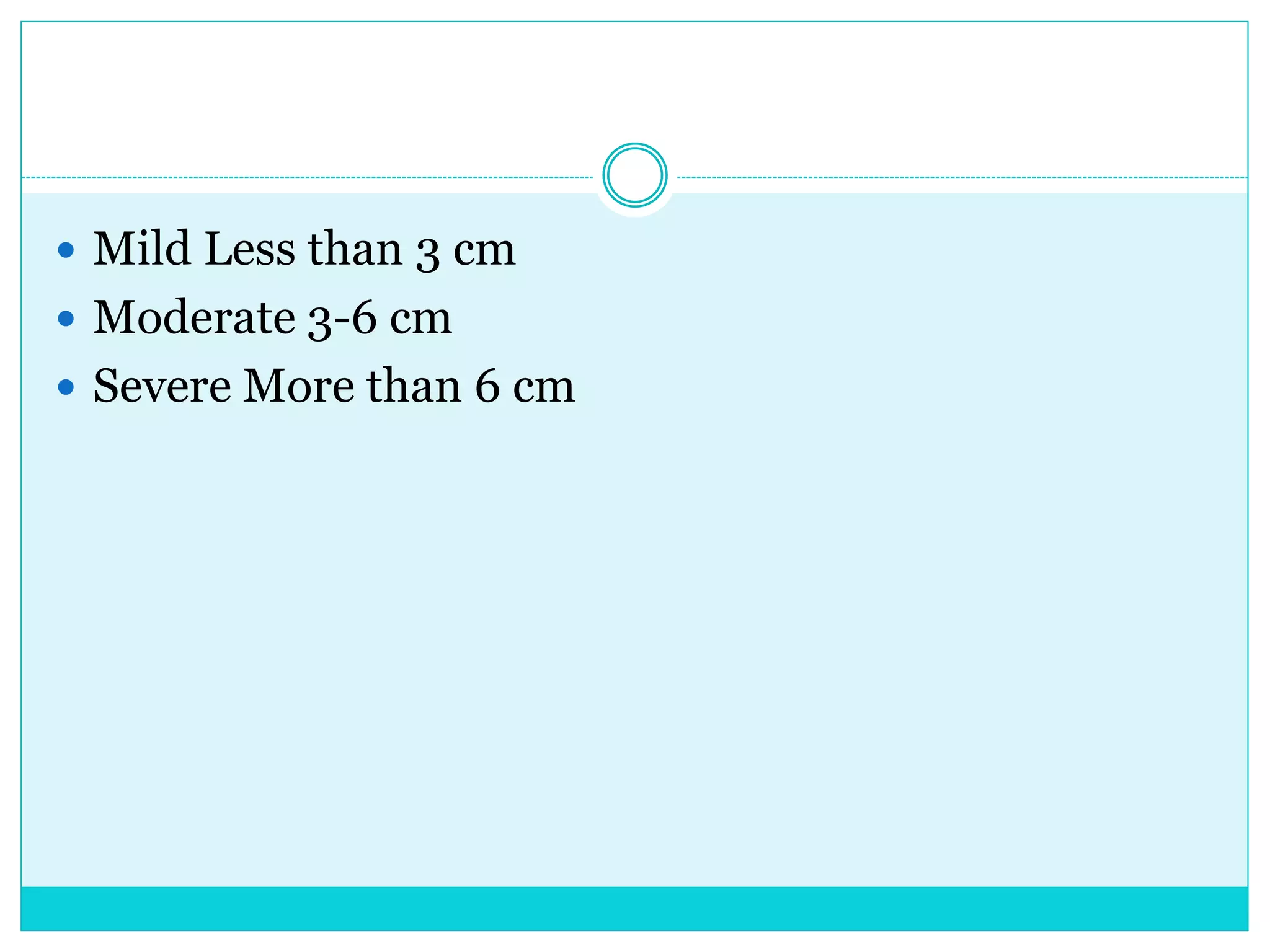
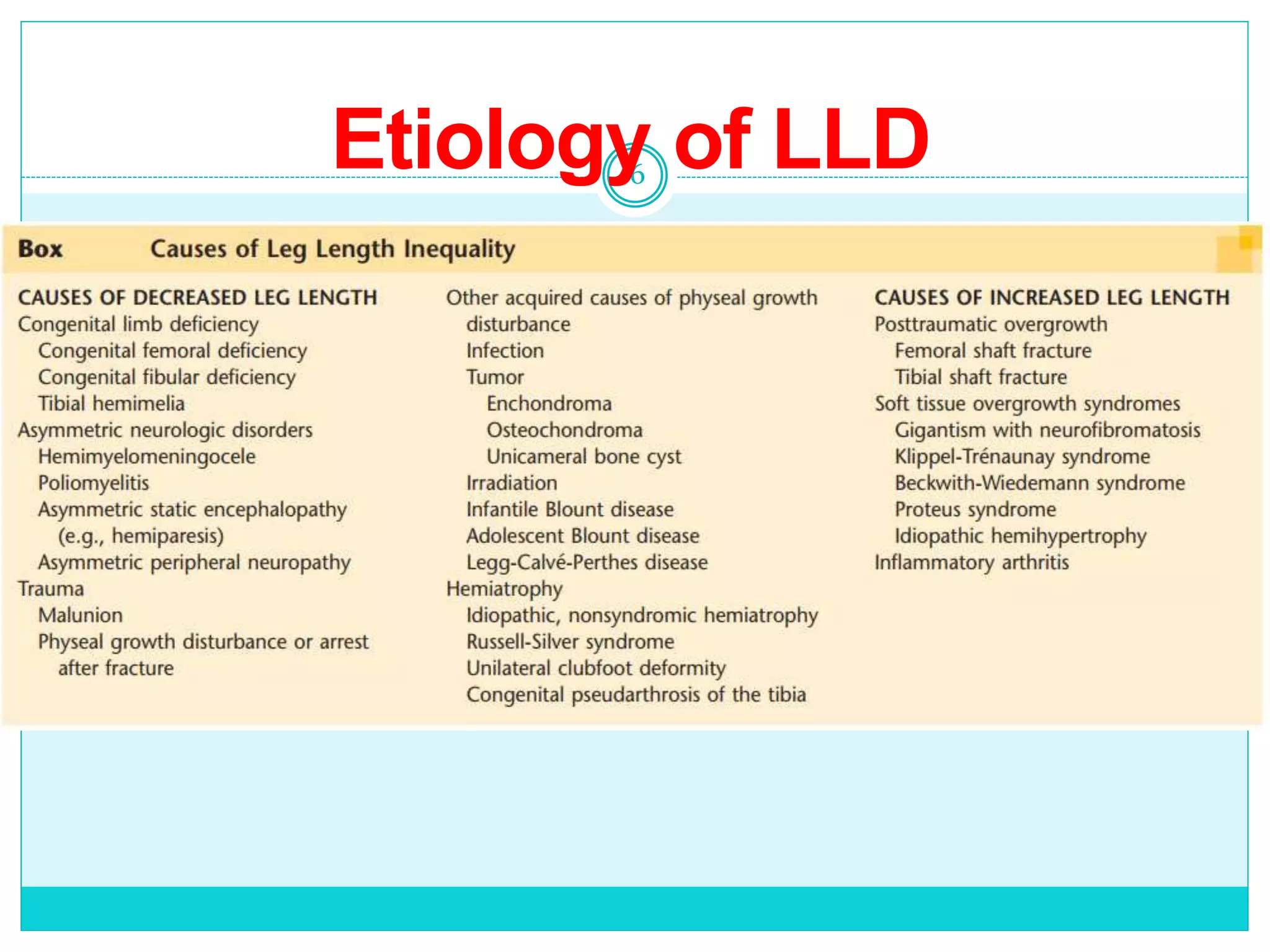
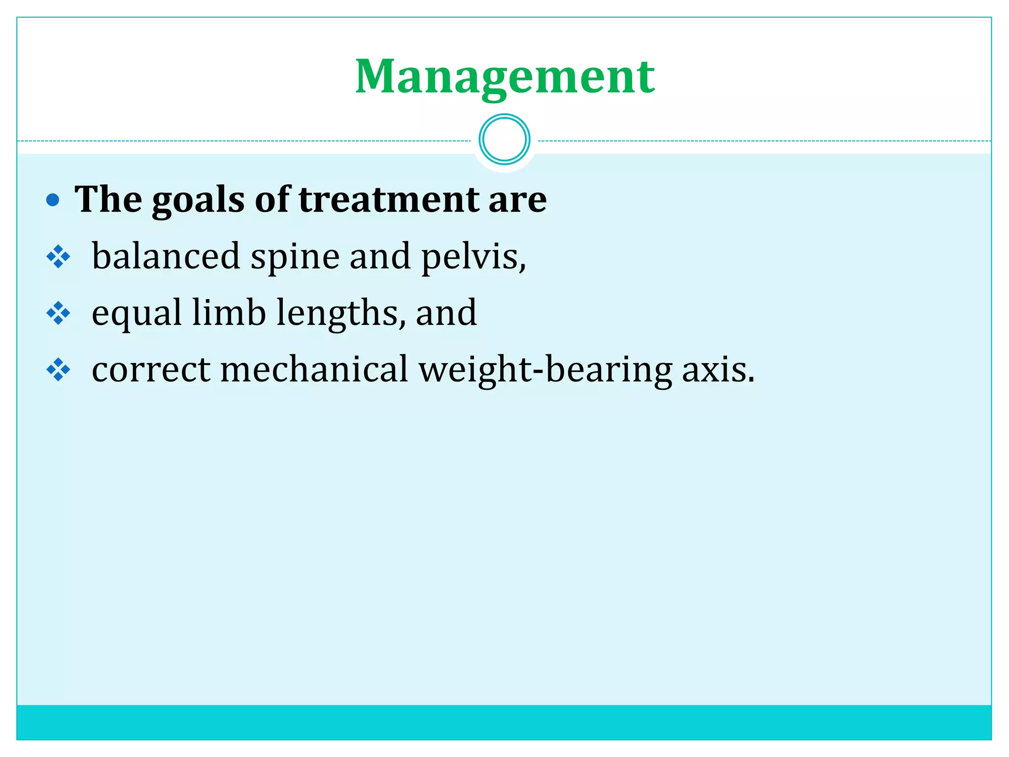
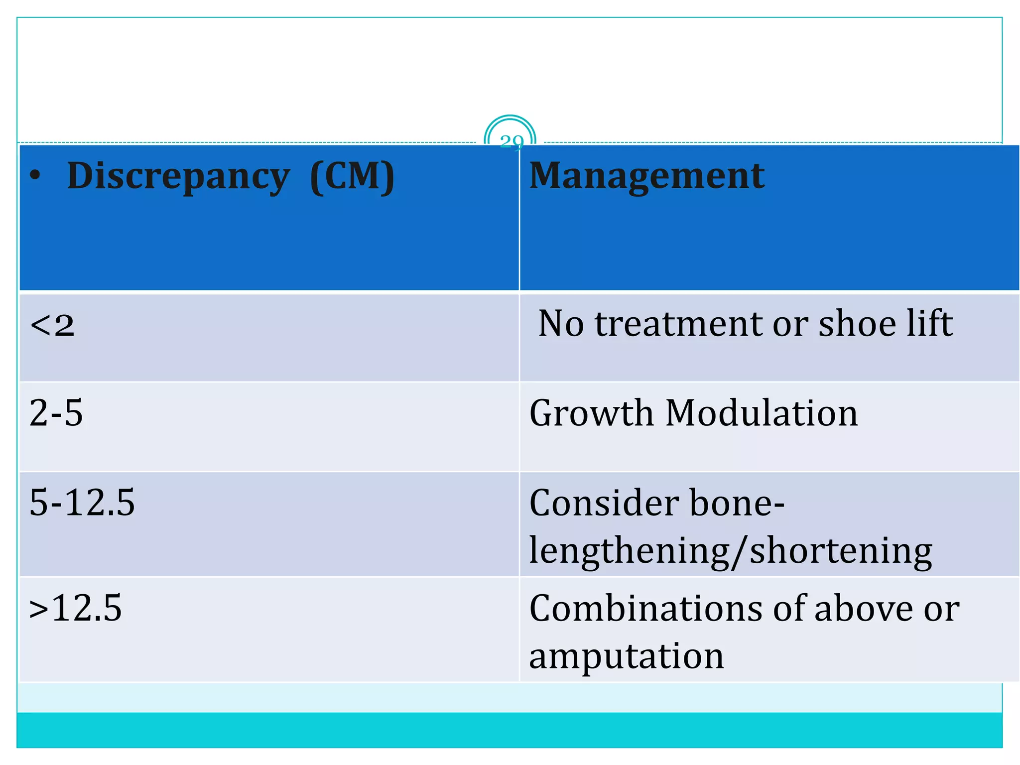
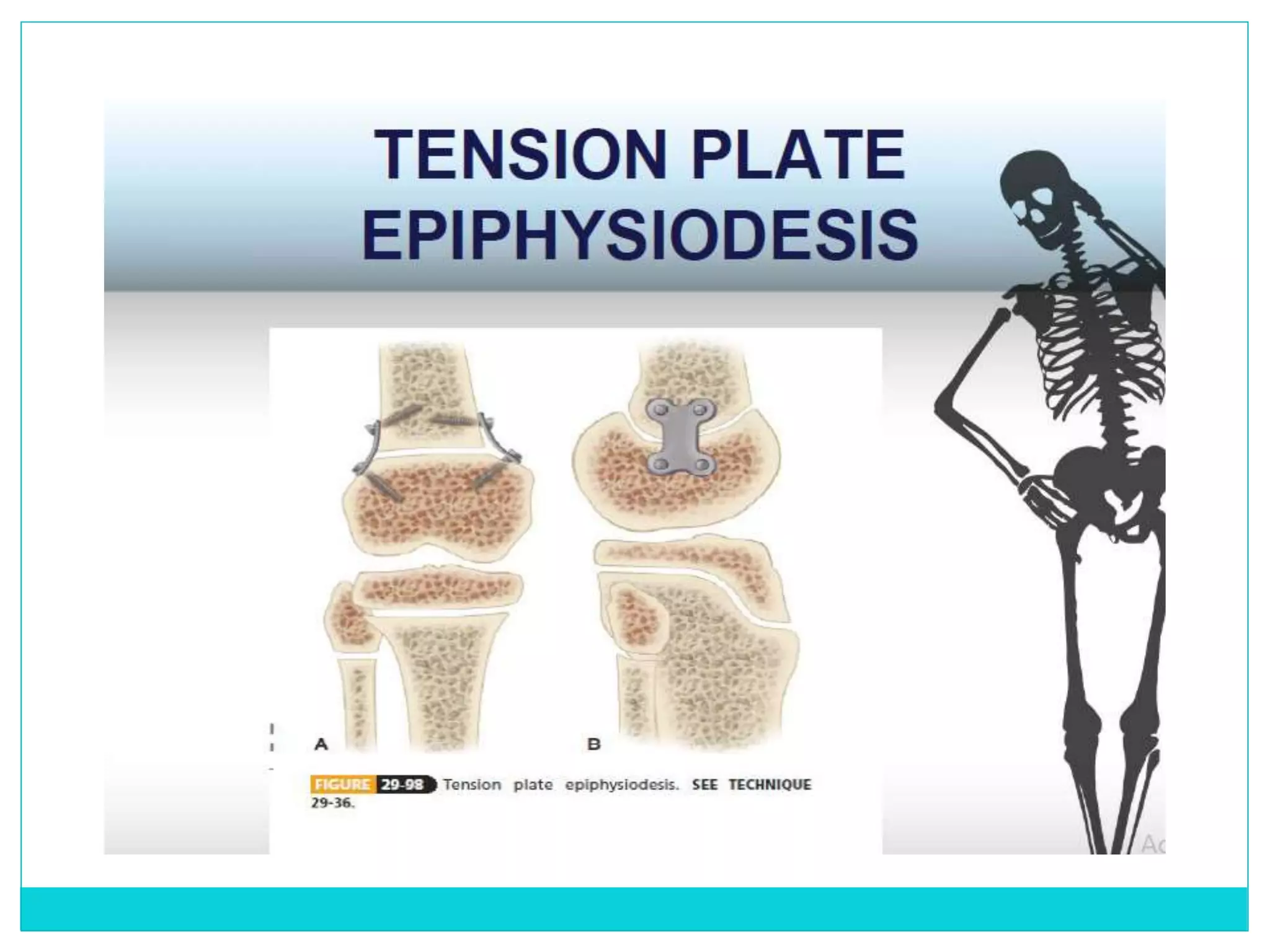
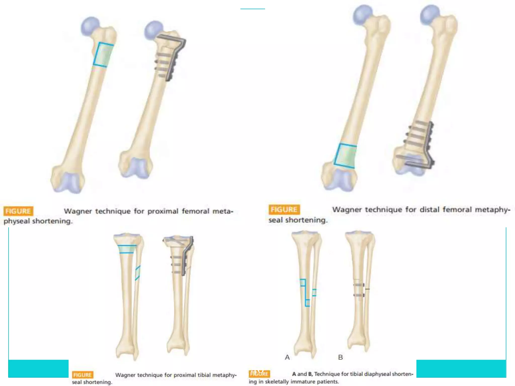
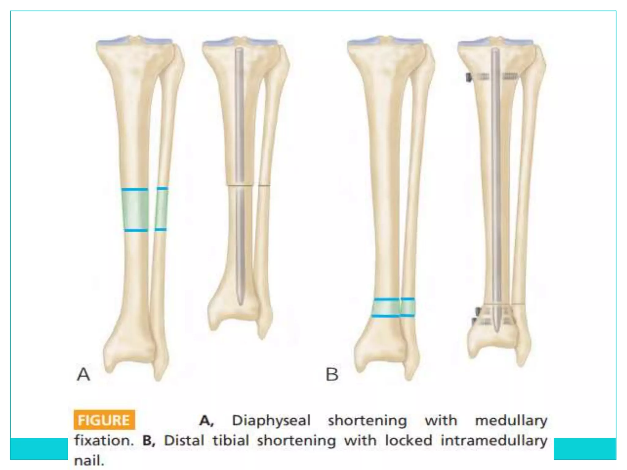
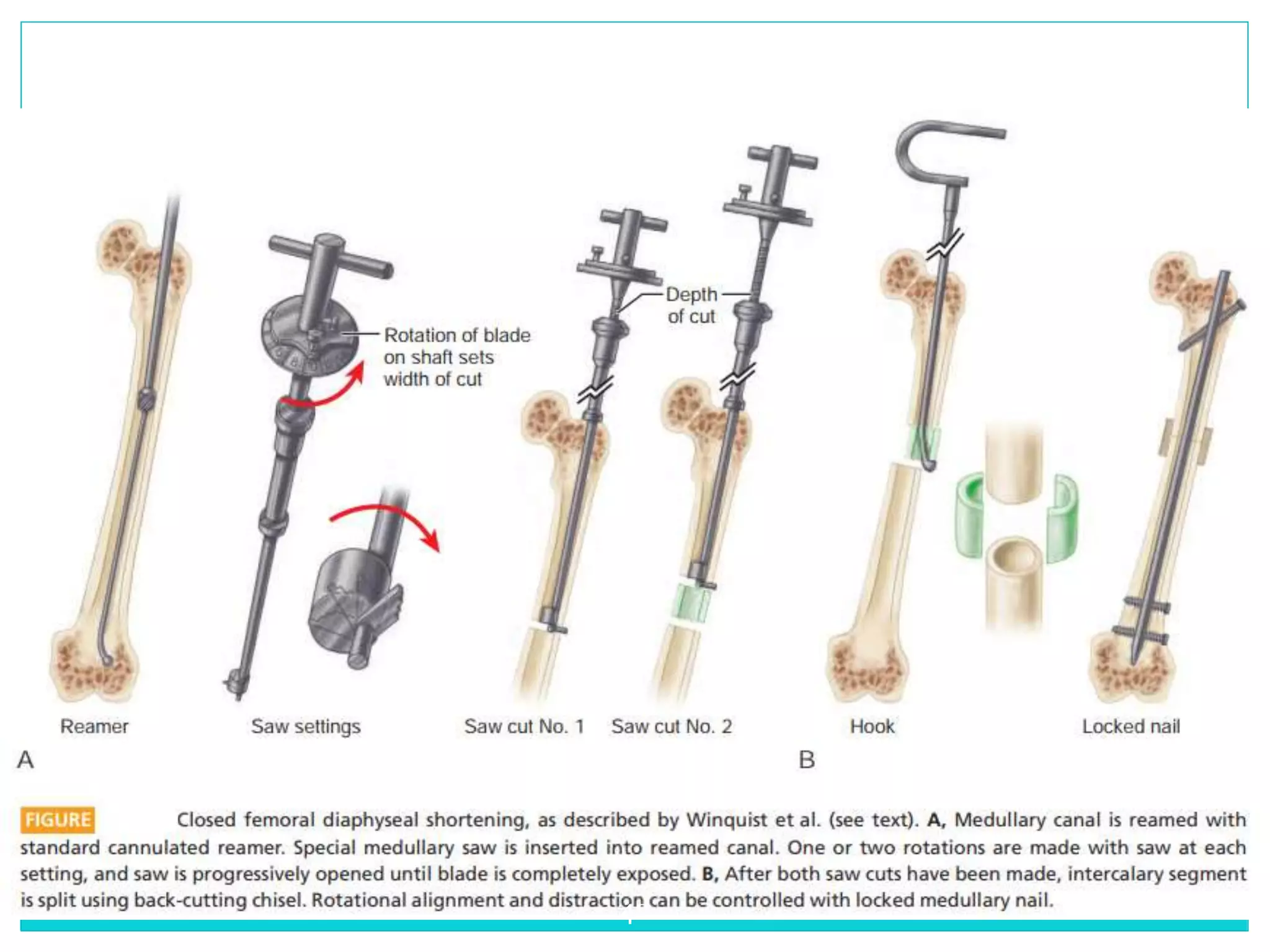
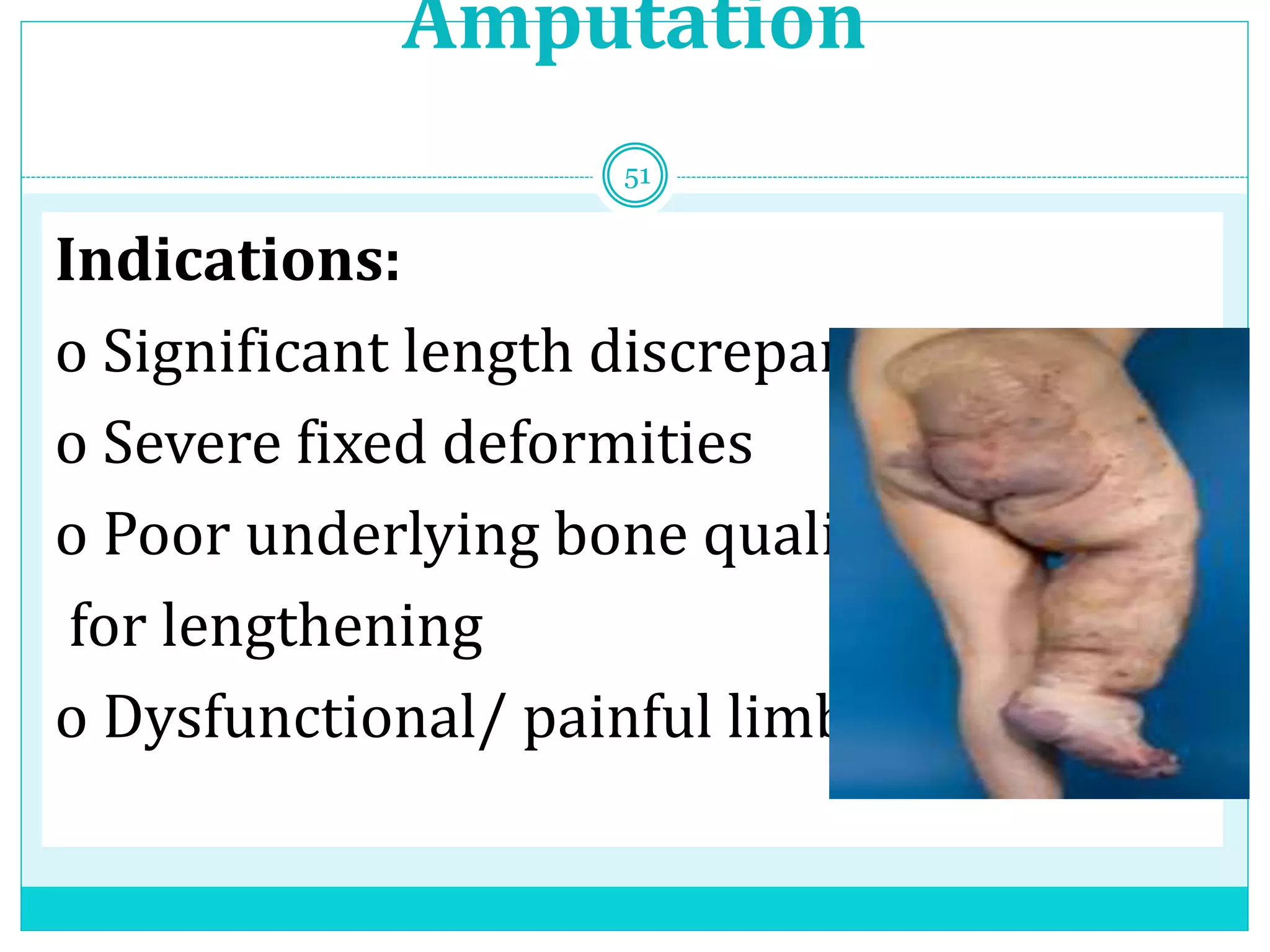
3. Introduction
Limb length discrepancy is defined as a condition in
which the paired lower extremity limbs have a noticeably
unequal length
LLD of 0.5 to 2.0 cm are common in the normal,
asymptomatic population
LLD of more than 2.5 cm is significant
4. Classification
Structural (SLLD) or anatomical:
Differences in leg length resulting from inequalities
in bony structure.
Functional (FLLD) or apparent:
factors other than actual bone shortening or
lengthening make one leg shorter or longer than the
other, a functional inequality occurs
5. Mild Less than 3 cm
Moderate 3-6 cm
Severe More than 6 cm
7. Mechanisms of compensation
specific compensation
Pronation in the ankle of the longer leg
Plantar flexion in the shorter leg
Knee and hip extension of the shorter or flexion in the longer leg
Uncompensated sacral based unleveling &/ scoliosis
8. Effects
On spine
Low back pain & late
degenerative disease
Scoliosis
On hip
Pelvic obliquity
OA
On long bones
Greater incidence of stress
fractures
Tibia
metatarsals and femur
8
10. Evaluation
History:
– Congenital or acquired
– Trauma / infection
– Progressive / static
– Onset and mode of deformity
– Any Syndrome associated features
10
11. Examination:
Gait
Lower limb;
o Determine which segment is short
o Is it too long or too short
o Exclude fixed deformities(Hip,knee and foot)
o Muscle wasting & neuro-vascular status
Spine;
o Scoliosis
11
13. – Wood block test
best initial screening method
13
14.
15. Imaging
Teleoroentgenogram
• Single exposure of both leg on a long film
• Reveals angular deformity
• Error of magnification
• Best technique for small children
•
16. Orthoroentgenogram
• Expos each joints individually
• Avoiding errors of magnification
• Multiple exposure
• Cannot assess whole shape of leg
17. Scanography
• Moving film cassette
• Avoid magnification
• Cannot assess whole shape of leg
18. CT scan
Advantages of CT
• lower radiation exposure
• greater accuracy,
• less susceptibility to error if the patient is poorly
positioned,
• specifically indicated when the patient has a knee
flexion contracture or is in a circular external fixator
19. Prediction of Discrepancy
There are various methods of
assessment of patient and predicting the
LLD
Growth remaining method — by Green & Anderson
Arithmetic method — by White Menelaus
Straight line graph method — by Moseley
Multiplier method — by Paley et al.
23. Paley multiplier
Take LLD for boy or girl
Multiplier for chronological or skeletal age
Predicts LLD at maturity
Automated Software
24.
25. Congenital Limb Length Discrepancy
Δm = Δ x M
– Δ:Current Limb-length discrepancy
– Δm: Limb discrepancy at skeletal maturity
– M: multiplier
• Example: If current LLD is 4cm in Congenital
hemihypertrophy of 10 years old boy
• Using value of 1.310 according to Multiplier chart at
age of 10
Δm = 4 x 1.310 = 5.24 cm(LLD at maturity)
26. Developmental Leg-length discrepancy
• Δm = Δ + (IXG)
• I=1 -(S – S’)/(L – L’)
• G=L(M-1)
Δm=limb length discrepancy at skeletal maturity
Δ = current limb-length discrepancy
G= amount of growth remaining
I=amount of growth inhibition
L= current length of long limb
L’=length of long limb as measured on previous radiographs
Lm length =length of femur or tibia at skeletal maturity
M=multiplier
S= current length of short limb
S’ =length o f short limb as measured on previous radiographs
27. Cont..
Lm=L x M
• L ε = Lm – G ε
• M ε =Lm/Lε
– Lm= length of femur or tibia at skeletal maturity
– L= current length of long limb
– M=multiplier
– Lε =desired length of bone to undergo epiphysiodesis at time
of epiphysiodesis
– ε=desired correction following epiphysiodesis
– Gε=amount of femoral or tibial growth remaining at age of
epiphysiodesis(G ε= ε/0.71 for femur and ε/0.57 for tibia)
– Mε=multiplier at age of epiphysiodesis
28. Management
The goals of treatment are
balanced spine and pelvis,
equal limb lengths, and
correct mechanical weight-bearing axis.
29. • Discrepancy (CM) Management
<2 No treatment or shoe lift
2-5 Growth Modulation
5-12.5 Consider bone-
lengthening/shortening
>12.5 Combinations of above or
amputation
29
30. Orthotic Management
(Shoe lift)
• Patient who do not wish or are not
appropriate for surgery.
• Lift higher than 5 cm poorly tolerated
30
31. Prosthetic fitting
Significant discrepancies,
Deformed functionally useless feet
Discrepancies greater than 15-20cm
Femoral length less than 50%
Fibular hemimelia with unstable ankle
31
46. Tibia Lengthening
Principle:
– It is based on the principle of distraction
osteogenesis.
– A bone that has been cut during surgery are
gradually pulled apart (distraction)
– Leads to new bone formation (osteogenesis) at the
site of lengthening
LLD of more than 2.5 cm is significant, with an increased likelihood of knee, hip, and lumbar spine pain
An acutal shortening or lengthening of the skeletal system occurs between the head of the femur and the ankle joint
mortise, which may have a congenital or acquired cause.
– Unilateral asymmetry of the lower extremity without any
concomitant shortening of the osseous components of the
lower limb
LLD has been classified according to the magnitude of the inequality, generally expressed in cm or mm,
Etiology ;due to undergrowth or over growth
Most common inequality is seen in ;Perthes disease ,Slipped capital femoral epiphysis,Cerebral palsy.
While the most severe discrepancy is found in,; PFFD (proximal focal femoral deficiency) ,Enchondromatosis,Poliomyelitis ,Multiple infective epiphyseal damage
**BWS manifest with omphalocele (exomphalos), macroglossia, and macrosomia (gigantism) .
***Blount disease is a pathologic varus deformity that results from disruption of normal cartilage growth at the medial aspect of the proximal tibial physis
The child compensates better than the adult by walking on the toes of shot leg
Adult seldom compensate that manner tend to heel - toe gait: vaulting gait & excessive pelvic motion and tilt
Knee and hip extension of the shorter or flexion in the longer leg
*****If the leg is left uncompensated, the anterior and posterior iliac spine on the side of the short leg can be lower which may result in a sacral base unleveling and/or scoliosis.
Increased muscle activity in several muscle groups
***Pelvic obliquity• Relatively uncovered hip of long leg and increased coverage of the hip of the short leg.• A longer leg might be a predisposing factor in Osteoarthritis (OA).• With length increasing, femoral head contact/ weight bearing area is decreasing.• Combined with an increased tone in hip abductors caused by elongated distance between origin andinsertion and an increased ground reaction force puts the longer leg at risk.
*****centra l edge angle:
• Decreased in CE angle on the long leg side • decreased in the load bearing area• causes late degenerative arthritis ** #of tibia, metatarsals and femur (consistent with thegreater forces emitted through the longer leg)
**Wood block test:– with the patient standing, add blocks under the short leg until thepelvis is level, then measure the blocks to determine thediscrepancy.– block testing is considered the best initial screening method.– Add blocks (known height) until the pelvis is level
Galeazzi (Allis) test **FIGURE :Apparent limb length inequality in a patient with hemiparesis. A, Clinical examination of the patient supine, with the hips flexed, suggests limb length inequality, with the hemiparetic side shorter than the unaffected side. This apparent shortening is caused by adduction of the hemiparetic leg.
Plain radiography
Teloroentgenogram
Orthoroentgenogram
Scanogram
CT
Teleoroentgenography. Teleoroentgenography is the simplest whole-leg radiographic technique for the assessment of leg length inequality. A radiograph of the entire lower extremity is obtained with the patient supine on the radiography table, with a long film and radiographic ruler beneath the patient. This technique is also the most susceptible to magnification error because a single exposure is made from a midpoint on the patient’s lower extremities. It is useful, however, if the patient is unlikely or unwilling to hold still for multiple-exposure techniques.
********An X-ray photograph taken at a distance of usually six feet with resultant practical parallelism of the rays and production of shadows of natural size.
taken with a single exposure at a distance of 2 m centered on the knee joint
Recent one has magnificafion facror of 5%
Orthoroentgenography. Orthoroentgenography is a radiographic technique described by Green and colleagues in 1946. The purpose is to minimize measurement error resulting from magnification by making three separate exposures of the lower extremities centered over the hips, knees, and ankles. One long film with a radiographic ruler is obtained, similar to in the teleoroentgenogram. In patients with significant leg length inequality, separate exposures for each leg may be made, with the x-ray beam centered over each joint. It is important that the patient not move between exposures (Green and colleagues ensured this immobility by strapping the patient to the table after positioning) and that the limbs be aligned neutrally. Salehand Milne described the use of this technique in a weight bearing position so that angular deformities, mechanical axis deviations, and limb length can be assessed simultaneously in that position. Weight bearing is not important to the accurate measurement of leg length inequality.
Scanography. A, A 14 × 17-inch film and a ruler are placed under the patient. Three (or six) exposures are made, centered
over the hips, knees, and ankles. The film is advanced under the joint to be radiographed and is exposed sequentially. B, Scanogram.
A smaller film is used than in orthoroentgenography, thus making storage and handling easier. Only a portion of the bone segment is
visible on the radiograph
the x-ray source and the film are both adjusted to reduce parallax error
• Software measures distances– accurate to 0.2mm– legs must be in same position– fast
It was developed to help predict the timing of epiphysiodesis and not to describe growth.
it only be used when skeletal and chronologic age are within 1 year off each other and clinical used leg-length differences as determined by blocks and not radiographic length
measurements. This method is best suited for those patients during the last few years of growth whose skeletal age correlates well with their chronologic age. The results of this technique for timing epiphysiodesis have found that 80% of patients were within a ½ in. when compared with the 90% obtained by using the Green and Anderson technique
At least 3 measurements each time
Length of long leg
Length of short leg
Skeletal age
Do so 3 times separated by 3-6 months
accuracy improves with increased plotting.
Example
Femur length(cm)
right (abnormal) left (normal)
• previous 24 26 at age of 8yrs
• Current 26 29 at age of 10 yrs
• I=1 -(S – S’)/(L – L’)
I =1-(26-24/29-26) = 1-2/3=0.33(amount of growth inhibition)
• G=L(M-1)
G=29(1.310-1)=29 x 0.310=8.99(amount of growth remaining)
• Δm = Δ + (IXG)
Δm = 3 +(0.33 x 8.99)=3 + 2.97 = 5.97 cm(LLD at skeletal maturity)
Time of Epiphysiodesis for the above pt
Lm = L(29) X M(1.31) = 37.99(length at maturity)
• Lε = Lm(37.99 ) – Gε(3/0.71) =37.99-4.22 = 33.77 (desired length of bone to undergo epiphysiodesis at time of epiphysiodesis)
• Mε=Lm(37.99)/Lε(33 .77) =1.125(multiplier at age of epiphysiodesis)
****child is compensating for leg length inequality by toe-walking and other strategies such as vaulting, circumducting, or increased flexion of the long leg, we consider not only a lift but an orthosis as well. Lifts greater than 8 cm are not easy for patients to manage and may cause them to fall over or sprain their ankles. The addition of an ankle-foot orthosis can be helpful in such circumstances ****Lifs of 5 to 10 cm are unsightly and unstable, however, and may require additional uprights or an ankle-foot orthosis to help support the ankle.
, monolateral fxator, DeBastiani’s Orthofx (McKinney,TX), the Ilizarov device, and the Taylor Spatial Frame (Smithand Nephew, Memphis, TN), all of which have undergonenumerous modifcations. Te original Wagner device is adjustable in only two planes, and the Hoffman modifcation isadjustable in one additional plane. DeBastiani’s device (Orthofx) has modular components that allow certain simple angularcorrections. Te Ilizarov device is extremely modular and canbe adapted with extensions and hinges to lengthen and correctangular and translational deformities simultaneously. Rotational deformities can be corrected either at the time of fxatorapplication or later by applying outriggers to the rings.
***
**• The reconstruction system with three screws in each segment• The telescopic device with three screws in each segment.• The Garches device with three screws in each segment.