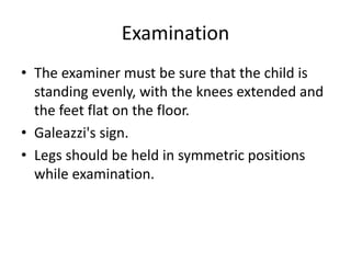
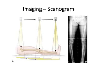
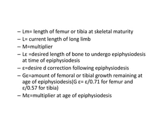
Limb Length Discrepancies
Dr. Sairamakrishnan S
• Limb length discrepancy or anisomelia, is
defined as a condition in which the paired
limbs have a noticeably unequal length.
• Asymptomatic lengthening of <2cm has been
found in upto 50% of normal population
• Any LLD of more than 2.5 cms is significant
and needs to be corrected.
Causes – Shortening
• Congenital limb deficiency
– Congenital femoral deficiency
– Congenital fibular deficiency
– Tibial hemimelia
• Asymmetric neurologic disorders
– Hemimyelomeningocele
– Poliomyelitis
– Asymmetric static encephalopathy
– Asymmetric peripheral neuropathy
• Trauma
– Malunion
– Physeal growth disturbance or arrest after fracture
• Other acquired causes of physeal growth
disturbance
– Infection
– Tumor
– Irradiation
– Blount's disease
– Legg-Calvé-Perthes disease
Causes - Lengthening
• Post-traumatic overgrowth
– Femoral shaft fracture
– Tibial shaft fracture
• Soft tissue overgrowth syndromes
– Gigantism with neurofibromatosis
– Klippel-Trénaunay syndrome
– Beckwith-Wiedemann syndrome
– Proteus syndrome
– Idiopathic hemihypertrophy
• Inflammatory arthritis
Classification
• Congenital
• Developmental
• Acquired
Classification
• Structural
• Functional
Classification – McCaw and Bates
• Mild – <3 cm
• Moderate – 3-6 cm
• Severe – >6 cm
Evaluation
• History
• Exmination
• Imaging
History
• Congenital or developmental.
• Onset and mode of deformity
• Family history
• Fractures/injury
Examination
• The examiner must be sure that the child is
standing evenly, with the knees extended and
the feet flat on the floor.
• Galeazzi's sign.
• Legs should be held in symmetric positions
while examination.
Thigh – leg technique
Imaging - Telerongentography
Imaging – Orthorongentography
Imaging – Scanogram
Imaging – CT scan
• Software measures distances
– accurate to 0.2 mm
– legs must be in same position
– fast
Determining skeletal age
Greulich- Pyle Atlas
– Xray non dominant hand
– less accurate
– improved accuracy by focusing on hand bones
rather than carpal bones
Tanner- Whitehouse Atlas
– more refined
– 20 landmarks graded L Hand
– more accurate
Prediction of shortening
• Green & Anderson tables
– Growth remaining method
– Uses skeletal age
– Requires graph
– Estimates growth potential in distal femur and
proximal tibia at various skeletal ages
– Separate charts for girls and boys
• Moseley charts
– Straight - Line Graph Method
– At least 3 measurements each time
• Length long leg
• Length short leg
• Skeletal age
– Do so atleast 3 times separated by 3-6 months
• Menelaus Method
• Paley multiplier
– State of the art
– multiplier for chronological or skeletal age
– predicts LLD at maturity
Congenital Limb Length Discrepancy
• Δm = Δ x M
– Δ:Current Limb-length discrepancy
– Δm: Limb discrepancy at skeletal maturity
– M:multiplier
Developmental LLD Leg-length discrepancy
• Δm = Δ + (IxG)
• I=1 -(S – S’)/(L – L’)
• G=L(M-1)
– G= amount of growth remaining
– I=amount of growth inhibition
– L= current length of long limb
– L’=length of long limb as measured on previous
radiograph
– S= current length of short limb
– S’ =length o f short limb as measured on previous
radiographs
Time of Epiphysiodesis
• Lm=L x M
• L ε = Lm – G ε
• M ε =Lm/Lε
– Lm= length of femur or tibia at skeletal maturity
– L= current length of long limb
– M=multiplier
– Lε =desired length of bone to undergo epiphysiodesis
at time of epiphysiodesis
– ε=desire d correction following epiphysiodesis
– Gε=amount of femoral or tibial growth remaining at
age of epiphysiodesis(G ε= ε/0.71 for femur and
ε/0.57 for tibia)
– Mε=multiplier at age of epiphysiodesis
Management
• <2 - No treatment or shoe lift
• 2-5 - Growth Modulation(Shoe lift /
Epiphysiodesis / Shortening)
• 5-12.5 - Consider bone-lengthening
• >12.5 - Combinations of above or amputation
Amputation
• Significant length discrepancy
• Poor underlying bone quality for lengthening
• Dysfunctional/ painful limb

Limb length discrepancies

  • 1.
  • 2.
    • Limb lengthdiscrepancy or anisomelia, is defined as a condition in which the paired limbs have a noticeably unequal length.
  • 3.
    • Asymptomatic lengtheningof <2cm has been found in upto 50% of normal population • Any LLD of more than 2.5 cms is significant and needs to be corrected.
  • 4.
    Causes – Shortening •Congenital limb deficiency – Congenital femoral deficiency – Congenital fibular deficiency – Tibial hemimelia • Asymmetric neurologic disorders – Hemimyelomeningocele – Poliomyelitis – Asymmetric static encephalopathy – Asymmetric peripheral neuropathy
  • 5.
    • Trauma – Malunion –Physeal growth disturbance or arrest after fracture • Other acquired causes of physeal growth disturbance – Infection – Tumor – Irradiation – Blount's disease – Legg-Calvé-Perthes disease
  • 6.
    Causes - Lengthening •Post-traumatic overgrowth – Femoral shaft fracture – Tibial shaft fracture • Soft tissue overgrowth syndromes – Gigantism with neurofibromatosis – Klippel-Trénaunay syndrome – Beckwith-Wiedemann syndrome – Proteus syndrome – Idiopathic hemihypertrophy • Inflammatory arthritis
  • 7.
  • 8.
  • 9.
    Classification – McCawand Bates • Mild – <3 cm • Moderate – 3-6 cm • Severe – >6 cm
  • 10.
  • 11.
    History • Congenital ordevelopmental. • Onset and mode of deformity • Family history • Fractures/injury
  • 12.
    Examination • The examinermust be sure that the child is standing evenly, with the knees extended and the feet flat on the floor. • Galeazzi's sign. • Legs should be held in symmetric positions while examination.
  • 15.
    Thigh – legtechnique
  • 16.
  • 17.
  • 18.
  • 19.
    Imaging – CTscan • Software measures distances – accurate to 0.2 mm – legs must be in same position – fast
  • 20.
    Determining skeletal age Greulich-Pyle Atlas – Xray non dominant hand – less accurate – improved accuracy by focusing on hand bones rather than carpal bones
  • 21.
    Tanner- Whitehouse Atlas –more refined – 20 landmarks graded L Hand – more accurate
  • 22.
    Prediction of shortening •Green & Anderson tables – Growth remaining method – Uses skeletal age – Requires graph – Estimates growth potential in distal femur and proximal tibia at various skeletal ages – Separate charts for girls and boys
  • 23.
    • Moseley charts –Straight - Line Graph Method – At least 3 measurements each time • Length long leg • Length short leg • Skeletal age – Do so atleast 3 times separated by 3-6 months
  • 25.
  • 26.
    • Paley multiplier –State of the art – multiplier for chronological or skeletal age – predicts LLD at maturity
  • 27.
    Congenital Limb LengthDiscrepancy • Δm = Δ x M – Δ:Current Limb-length discrepancy – Δm: Limb discrepancy at skeletal maturity – M:multiplier
  • 28.
    Developmental LLD Leg-lengthdiscrepancy • Δm = Δ + (IxG) • I=1 -(S – S’)/(L – L’) • G=L(M-1)
  • 29.
    – G= amountof growth remaining – I=amount of growth inhibition – L= current length of long limb – L’=length of long limb as measured on previous radiograph – S= current length of short limb – S’ =length o f short limb as measured on previous radiographs
  • 30.
    Time of Epiphysiodesis •Lm=L x M • L ε = Lm – G ε • M ε =Lm/Lε
  • 31.
    – Lm= lengthof femur or tibia at skeletal maturity – L= current length of long limb – M=multiplier – Lε =desired length of bone to undergo epiphysiodesis at time of epiphysiodesis – ε=desire d correction following epiphysiodesis – Gε=amount of femoral or tibial growth remaining at age of epiphysiodesis(G ε= ε/0.71 for femur and ε/0.57 for tibia) – Mε=multiplier at age of epiphysiodesis
  • 32.
    Management • <2 -No treatment or shoe lift • 2-5 - Growth Modulation(Shoe lift / Epiphysiodesis / Shortening) • 5-12.5 - Consider bone-lengthening • >12.5 - Combinations of above or amputation
  • 33.
    Amputation • Significant lengthdiscrepancy • Poor underlying bone quality for lengthening • Dysfunctional/ painful limb