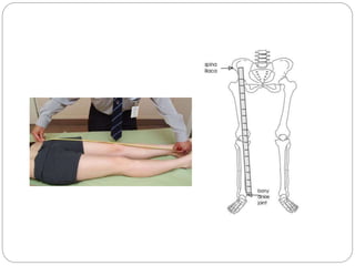
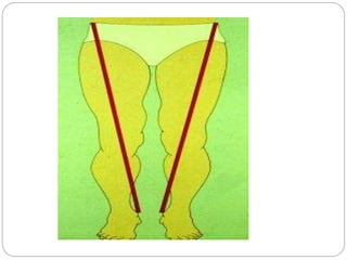
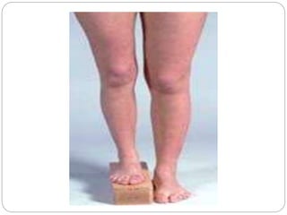
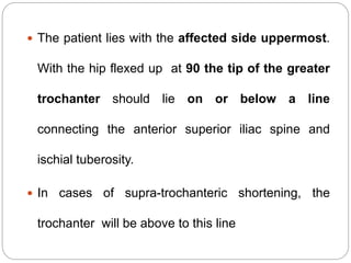
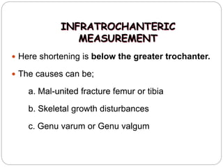
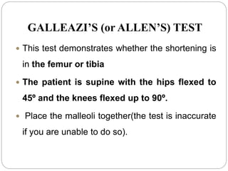
LIMB LENGTH
MEASUREMENT
BY: Dr Sneha Bhalala
Limb length measurement aims to
measure length of individual limbs
(usually lower limbs) with the help of inch
tape.
In clinical practice, exact length of each
lower limb is relatively unimportant, but
important is the difference in length which
exists between the two lower limbs.
 Differences between the lengths of the upper
and/or lower arms and the upper and/or lower
legs are called limb length discrepancies
(LLD).
 Except in extreme cases, arm length differences
cause little or no problem in how the arms
function.
Incidence
 A limb length difference may simply be a mild variation
between the two sides of the body.
 This is not unusual in the general population.
 For example, one study reported that 32 percent of 600
military recruits had a 1/5 inch to a 3/5 inch difference
between the lengths of their legs. This is a normal variation.
 Greater differences may need treatment because a
significant difference can affect a patient's well-being and
quality of life.
Classification of leg length discrepancy (LLD):
 Structural (SLLD) or anatomical or true : Differences in
leg length resulting from inequalities in bony structure.
 Functional (FLLD) or apparent: Unilateral asymmetry of
the lower extremity without any concomitant shortening of
the osseous components of the lower limb.
Etiological factors :
• Idiopathic developmental abnormalities
• Fracture
• Trauma to the epiphysis ( endplate ) prior to skeletal
maturity
• Degenerative disorders
• Legg-calvé- Perthes disease
• Cancer or neoplastic changes
• Infections
• Functional:
o Shortening of soft tissues
o Joint contractures
o Ligamentous laxity
o Axial malalignments
o Foot biomechanics (such as excessive ankle
 Legg-Calvé-Perthes disease (LCPD) is avascular
necrosis of the proximal femoral head resulting from
compromise of the tenuous(weak) blood supply to this
area. LCPD usually occurs in children aged 4-10 years.
The disease has an insidious onset and may occur after an
injury to the hip
Causes
A. Previous Injury to a Bone in the Leg or
Growth plate injury :
1. Trauma (fracture)
2. Infection
1. Trauma
 A broken leg bone may lead to a limb length
discrepancy if it heals in a shortened position.
 This is more likely if the bone was broken in many
pieces.
 It also is more likely if skin and muscle tissue
around the bone were severely injured and exposed,
as in an open fracture.
 Broken bones in children sometimes grow faster for several
years after healing, causing the injured bone to become longer.
 A break in a child's bone through the growth center near the
end of the bone may cause slower growth, resulting in a
shorter leg.
2. Bone Infection:
 Bone infections that occur in children while they are
growing may cause a significant limb length discrepancy.
 This is especially true if the infection happens in infancy.
 Inflammation of joints during growth may cause unequal
leg length. One example is juvenile arthritis.
B. Bone Diseases (Dysplasias) :
 Bone diseases may cause limb length discrepancy, as
well.
C. Neurologic conditions
 Asymmetric paralysis
1. Poliomyelitis
2. Cerebral palsy
3. Hemiplegia
D. Mass induced growth
1. Tumor
2. Post-fracture hyper-vascularity
3. Neurofibromatosis
Fibrous dysplasia:
 Scar-like (fibrous) tissue
develops in place of
normal bone. As the bone
grows, the softer, fibrous
tissue expands, causes
weakening the bone.
Fibrous dysplasia can
cause the affected bone to
deform and become
susceptible to fracture.
Skeletal dysplasia
poliomyelitis
Neurofibromatosis
E. Idiopathic
 Unilateral Hemi hypertrophy (one side too big)
or hemi atrophy (one side too small) are rare
limb length discrepancy conditions.
 In these conditions, the arm and leg on one
side of the body are either longer or shorter
than the arm and leg on the other side of the
body.
 There may also be a difference between the two
sides of the face. Sometimes no cause can be
found. This is known as an "idiopathic"
difference.
F. Long femoral component after total
hip replacement
• Anatomical deformities:
1. Coxa vara or coxa valga
2. Genu varum or genu valgum
3. Congenital or acquired dislocation of hip
4. Congenital talipus equino varus
 Sometimes the cause of limb length discrepancy is unknown,
particularly in cases involving under development of the inner or
outer side of the leg, or partial overgrowth of one side of the body.
 These conditions are usually present at birth, but the leg length
difference may be too small to be detected.
 As the child grows, the limb length discrepancy
increases and becomes more noticeable.
 In underdevelopment, one of the two bones tibia and
fibula is abnormally short.
 There also may be related foot or knee problems.
Associated problems :
Inequality in leg length is commonly associated with
compensatory gait abnormalities and may lead to
degenerative arthritis of the lower extremity and lumbar
spine.
Patients with leg-length discrepancy (LLD) can also have
angular and torsional deformities as well as soft tissue
contractures of the ipsilateral or contralateral extremity
that may influence their functional leg lengths.
 For instance, flexion contractures around the knee and hip
can cause apparent shortening of the leg, while abduction
contractures of the hip and equinus deformity of the ankle
tend to functionally lengthen the affected extremity.
Examination
The most accurate method to identify leg (limb) length
inequality (discrepancy) is through radiography.
Radiography: Limitations are an inherent inaccuracy in
patients with hip or knee flexion contracture.
However, radiography has to be performed by a
specialist, takes more time and is costly. It should only
be used when accuracy is critical.
Computed Tomography (CT-scan):
 It has no greater accuracy compared to the standard
radiography.
Scan method ( x-ray)
 Not available without professional medical
equipment. This methods involves scanning the pelvis
and legs and with the use of computer technology, the
images are enlarged to measure the distance
between reference points (again, usually the pelvis
and ankle joints), with very high accuracy.
 Some health professionals stress that the more
traditional methods of measuring such as the
apparent and true methods are accurate enough
without the need for a scan, which is expensive and
time consuming and can even expose you to
unnecessary radiation.
 It is therefore recommended that people try the
easier and harmless methods first as clinical
evidence suggests these are highly accurate when
carried out correctly.
Direct methods:
 Measuring limb length with a tape measure between 2
defined points. If you choose for this method, keep
following possible errors in mind:
• Always use the mean of at least 2 or 3 measures
• If possible, compare measures between 2 or more
clinicians
• Iliac asymmetries may mask or accentuate a limb length
inequality
• Unilateral deviations in the long axis of the lower limb (eg.
Genu varum,…) may mask or accentuate a limb length
Indirect methods:
 Palpation of bony landmarks, most commonly the iliac
crests or anterior superior iliac spines, in stand. These
methods consist in detecting if bony landmarks are at
(horizontal) level or if limb length inequality is present.
 Palpation and visual estimation of the iliac crest (or
ASIS) in combination with the use of blocks or book
pages of known thickness ,under the shorter limb to
adjust the level of the iliac crests (or ASIS), appears to
be the best (most accurate and precise) clinical method
Types
Limb length measurement is of two
types-
1. True limb length measurement
2. Apparent limb length measurement
TRUE LIMB LENGTH MEASUREMENT
True length is the measurement of lower
limb to diagnose true shortening or true
discrepancy.
Present when there is a decrease in
distance between the upper surface of
head of femur & lower surface of
calcaneum when compared on both the
For clinical purpose, two fixed bony points are taken
for reference;
ASIS (anterior superior iliac spine) because
it lies proximal & lateral to upper surface of head of
femur
&
Medial malleolus (or lateral malleolus)
because its tip corresponds to calcaneum
Direct method
1. Position of patient – supine lying
 Identify ASIS on both sides & draw an imaginary line
connecting both.
 Prior to measuring, place the normal limb in a similar position
to that of affected limb (squaring of pelvis).
 Place one end of the inch tape over ASIS of affected side &
take the tape through the thigh till medial border of patella &
then along the medial border of leg till the tip of medial
malleolus.
 Note the reading of tape & repeat for other limb.
 Compare reading & note if there is any difference in
measurements
 The difference indicates shortening.
 The medial malleolus should present no problem as
it is easily palpated subcutaneously but the ASIS can
sometimes be difficult to palpate in obese patients.
 It is easy to introduce an error of a cm or so in leg
length measurements if you do not measure from
equivalent positions bilaterally.
Squaring of pelvis
 It is a technique to place lower limbs
in such a position that both ASIS are at
same level and in a straight horizontal
line.
Adduction or Abduction deformity:
 If both ASIS are at the same level and the pelvis is square
there is no adduction or abduction deformity.
 If the ASIS on the affected side is higher, an adduction
deformity is present.
 Similarly if the ASIS on the affected side is lower than the
normal side, an abduction deformity is present.
 An abduction contracture of the hip causes a functional leg
length difference.
 The pelvis dips towards the affected side, the normal leg
appears shortened and the affected leg lengthened.
 The opposite occurs with an adduction deformity.
 The affected leg appears shortened and the patient attempts
to compensate with elevating the ASIS on the affected side
to bring his or her legs into parallel alignment.
Flexion contracture of the knee
 Unable to place the legs straight because of the fixed
flexion of the knee. You must place the other leg in the
same position. One could flex the unaffected knee over a
bolster to the same degree and then measure leg lengths
 Valgus knee -
 Measure component parts of the leg.
 This approximates to a true leg length
 Measure from the ASIS to the tibial tuberosity and then
from the tibial tuberosity to the medial malleolus.
Indirect method
 Procedure of the indirect measurement method: the use of
blocks :
Position of patient – standing with both knees fully extended
with feet 10 cm apart and equal weight on both feet.
 The clinician places his/her hands on a bilateral anatomical
structure: posterior superior iliac spine, anterior superior iliac
spine or iliac crest on left and right.
 Identify ASIS on both sides; ASIS on the side of shortened
limb will be at lower level.
Now the clinician visually assesses if there is a length
inequality, and if so, Place wooden blocks of varying
thickness under the foot of shortened limb until both the
ASIS are at the same level and equal length is reached.
Total height of wooden blocks equals the difference
in length.
Variables reported by literature are: pelvic asymmetry,
incorrect positioning of feet, obesity, joint contractures,
scoliosis and inaccurate measurement.
 After measuring the true length, if there is any
shortening, it is now to find out the site of
shortening – whether it is supratrochanteric
or infratrochanteric.
SUPRATROCHANTERIC
MEASUREMENT
Here the shortening is above greater
trochanter.
Causes to supratrochanteric shortening
are-
1. Coxa vara or coxa valga
2. Congenital or acquired hip dislocation
3. Fracture neck femur
4. Hip arthritis
Congenital /Acquired hip dislocation
Fracture neck femur
Hip arthritis
METHODS OF MEASUREMENT
1.BRYANT’S TRIANGLE
2.SHOEMAKER’S LINE
3.CHIENE’S LINE
4.NELATON’S LINE
1. BRYANT’S TRIANGLE
This triangle is constructed on both sides as follows;
 Line 1 – Drawn from ASIS to greater trochanter.
 Line 2 – Imaginary line drawn from ASIS to couch
perpendicularly
 Line 3 – horizontal line drawn from greater
trochanter to line 2 above
Measure & compare with other side.
 Identify the ASIS with your thumb and the tip of the
greater trochanter with your main finger and the base of
the triangle with your index finger.
 Bryant’s triangle is drawn on both sides & length of line is
measured & compared.
 Difference in length of 3rd line indicates the upward shift of
trochanter & confirms supratrochanteric shortening.
 Difference in length of 1st & 2nd line indicates anterior or
posterior shift of greater trochanter.
 This test is not useful in bilateral hip disease.
Causes :
 Shortening above the trochanter may be caused by -
- Destruction of the femoral head or acetabulum or both
- Dislocated hip
- Coxa -vara deformity
- Mal -united inter-trochanteric fracture.
2. SHOEMAKER’S LINE
 Supine position:
 Line joined by connecting two points greater trochanter & ASIS
when extended up on both sides
1. May cross above the umbilicus in the midline – Normal
2. May cross below the umbilicus away from midline – opposite
unilateral supratrochanteric shortening
3. May cross in midline below the umbilicus – bilateral
supratrochanteric shortening
 Not useful in case of bilateral disease.
3. CHIENE’S LINE
Supine position:
The lines joining two ASIS & two greater
trochanter are usually parallel.
This is disturbed if one of the greater
trochanter is shifted upwards.
Useful in unilateral disease only.
4. NELATON’S LINE
In supine lying, when a line is drawn from ischial
tuberosity to the ASIS, the greater trochanter just
touches this line.
If greater trochanter is above the line then
supratrochanteric shortening is confirmed.
Useful in unilateral as well as bilateral hip disease.
 The patient lies with the affected side uppermost.
With the hip flexed up at 90 the tip of the greater
trochanter should lie on or below a line
connecting the anterior superior iliac spine and
ischial tuberosity.
 In cases of supra-trochanteric shortening, the
trochanter will be above to this line
INFRATROCHANTERIC
MEASUREMENT
 Here shortening is below the greater trochanter.
 The causes can be;
a. Mal-united fracture femur or tibia
b. Skeletal growth disturbances
c. Genu varum or Genu valgum
METHODS OF MEASUREMENT
 GALLEZI’S (OR ALLEN’S) TEST
 INDIVIDUAL BONE LENGTH or
segmental MEASUREMENT
GALLEAZI’S (or ALLEN’S) TEST
 This test demonstrates whether the shortening is
in the femur or tibia
 The patient is supine with the hips flexed to
45º and the knees flexed up to 90º.
 Place the malleoli together(the test is inaccurate
if you are unable to do so).
 The examiner assesses the position of both knees
from the end of the bed and from the side
 In Case of femoral shortening, both thighs are at
level but the knee is slightly lower than normal one.
The leg is also slightly behind the normal leg.
 In case of tibial shortening, both legs are at level
but thigh is not at level & knee appears forwards
2. INDIVIDUAL BONE LENGTH
MEASUREMENT
 Length of femur can be measured from
ASIS to the medial joint line of knee &
tibial measurement can be done from
medial joint line of knee to the medial
malleolus.
 Measure on both sides & compare.
COMPLICATIONS DUE TO TRUE
DISCREPANCY
 Compensatory scoliosis
 Low back pain
 Gait abnormalities with secondary injuries
 Over stretching/contracture of muscles
APPARENT LIMB LENGTH
MEASUREMENT
 Apparent measurement helps in assessing the extent
of natural compensation developed for concealing the
natural deformity/disparity.
 Apparent limb length discrepancy (LLD) may be due to:
- Suprapelvic obliquity : Scoliosis
- Intrapelvic obliquity : Pelvic fracture
- Infrapelvic obliquity : Hip contracture
- Knee contracture
-Ankle contracture
METHOD OF MEASUREMENT
Position of patient – Supine lying.
Arrange the lower limbs parallel to each another; for
that bring the unaffected limb near to the affected limb &
align the trunk in line with the lower limbs. (Apparent
length is measured by positioning both the lower limbs
parallel to each other & without correcting any of the
existing deformities. In bilateral affections, apparent
measurement is not of much significance.)
 With inch tape measure the distance between
xiphisternum (or umbilicus) to the tip of medial
malleolus & note the reading.
 Repeat it for the other limb & if difference exists then it
indicates apparent discrepancy.
 Shortening in one limb is usually compensated by --
- Tilting the pelvis down (ASIS dips at a lower level)
- Equinus position of the foot
- Flexing the opposite lower limb at the hip and knee
 If the true shortening is equal to apparent shortening
it indicates there is no compensation.
 If the true shortening is more than the apparent one it
indicates that part of the shortening has been
compensated for.
 If the true shortening is less than the apparent
shortening it would suggest a fixed adduction
TREATMENT OF LIMB LENGTH
DISCREPANCY
0 – 2 cm: no treatment
Difference of less than 3cm are usually accepted by
the patients & can be corrected by shoe raise of the
shortened limb.
Difference in length between 4 to 15cm are corrected
by surgery; Surgical intervention
 Epiphysiodesis of long leg (Epiphysiodesis is a
pediatric surgical procedure in which the epiphyseal
(growth) plate of a bone is fused either temporarily or
permanently to delay growth of a long bone. This
procedure is often used to slow or halt the growth of a
morphologically normal leg to allow a shorter leg to
grow to a matching length)
 Long leg shortening or short leg lengthening
 Any difference in length of more than 15cm are
usually difficult to be corrected by surgical methods
also.
Foot orthosis
Ilizarov’s tech -
References
 Canale (1998) Campbell's Orthopaedics, Mosby,
p. 986
Hoppenfeld (1976) Spine and Extremities, p. 165
Review: Limb Length Inequality: Clinical
Implications for Assessment and Intervention
(2003) Rebecca J. Brady PT, John B. Dean
PT;ATC, T. Marc Skinner, PT;ATC, Michael T.
Gross, PT;PhD.Level of evidence: 1
Review: Leg Length Discrepancy (2001) – Burke
Gurney
Measurement of limb-length inequality.
Comparison of clinical methods with
 Changes in Pain and Disability Secondary to Shoe Lift
Intervention in Subjects With Limb Length Inequality and
Chronic Low Back Pain - A Preliminary Report – Yvonne
M. Golightly et al (2007) – Journal of orthopedics and
sports physical therapy E.B. LEVEL (2)
Conservative Correction of Leg-lengts discrepancies of
10mm of less for the relief of chronic low back pain. –
Ruth Defrin, Sarit Ben Benyamin et al. (2005) Arch Phys
Med Rehabil. E.B. LEVEL (2)
Erector Spinae and Quadratus Lomborum Muscle
endurance tests and supine leg-length alignment
asymmetry: and observational study – Gary A. Knuston
and Edward Owens (OCT 2005) Journal of Manipulative
and Physiological Therapeutics. E.B. LEVEL (2)
The accuracy of the palpation meter (PALM) for
measuring pelvic crest difference and leg length
discrepancy. – Matthew R. Petrone, Jennifer Guin et al
(JUN 2003) – Journal Orthop Sports Phys Ther VOL 33
(n°6) E.B. LEVEL (2)
lld- leg length.ppt

lld- leg length.ppt

  • 1.
  • 2.
    Limb length measurementaims to measure length of individual limbs (usually lower limbs) with the help of inch tape. In clinical practice, exact length of each lower limb is relatively unimportant, but important is the difference in length which exists between the two lower limbs.
  • 3.
     Differences betweenthe lengths of the upper and/or lower arms and the upper and/or lower legs are called limb length discrepancies (LLD).  Except in extreme cases, arm length differences cause little or no problem in how the arms function.
  • 4.
    Incidence  A limblength difference may simply be a mild variation between the two sides of the body.  This is not unusual in the general population.  For example, one study reported that 32 percent of 600 military recruits had a 1/5 inch to a 3/5 inch difference between the lengths of their legs. This is a normal variation.  Greater differences may need treatment because a significant difference can affect a patient's well-being and quality of life.
  • 5.
    Classification of leglength discrepancy (LLD):  Structural (SLLD) or anatomical or true : Differences in leg length resulting from inequalities in bony structure.  Functional (FLLD) or apparent: Unilateral asymmetry of the lower extremity without any concomitant shortening of the osseous components of the lower limb.
  • 6.
    Etiological factors : •Idiopathic developmental abnormalities • Fracture • Trauma to the epiphysis ( endplate ) prior to skeletal maturity • Degenerative disorders • Legg-calvé- Perthes disease • Cancer or neoplastic changes • Infections • Functional: o Shortening of soft tissues o Joint contractures o Ligamentous laxity o Axial malalignments o Foot biomechanics (such as excessive ankle
  • 7.
     Legg-Calvé-Perthes disease(LCPD) is avascular necrosis of the proximal femoral head resulting from compromise of the tenuous(weak) blood supply to this area. LCPD usually occurs in children aged 4-10 years. The disease has an insidious onset and may occur after an injury to the hip
  • 8.
    Causes A. Previous Injuryto a Bone in the Leg or Growth plate injury : 1. Trauma (fracture) 2. Infection 1. Trauma  A broken leg bone may lead to a limb length discrepancy if it heals in a shortened position.  This is more likely if the bone was broken in many pieces.  It also is more likely if skin and muscle tissue around the bone were severely injured and exposed, as in an open fracture.
  • 9.
     Broken bonesin children sometimes grow faster for several years after healing, causing the injured bone to become longer.  A break in a child's bone through the growth center near the end of the bone may cause slower growth, resulting in a shorter leg.
  • 10.
    2. Bone Infection: Bone infections that occur in children while they are growing may cause a significant limb length discrepancy.  This is especially true if the infection happens in infancy.  Inflammation of joints during growth may cause unequal leg length. One example is juvenile arthritis.
  • 11.
    B. Bone Diseases(Dysplasias) :  Bone diseases may cause limb length discrepancy, as well. C. Neurologic conditions  Asymmetric paralysis 1. Poliomyelitis 2. Cerebral palsy 3. Hemiplegia D. Mass induced growth 1. Tumor 2. Post-fracture hyper-vascularity 3. Neurofibromatosis
  • 12.
    Fibrous dysplasia:  Scar-like(fibrous) tissue develops in place of normal bone. As the bone grows, the softer, fibrous tissue expands, causes weakening the bone. Fibrous dysplasia can cause the affected bone to deform and become susceptible to fracture.
  • 13.
  • 14.
  • 15.
  • 16.
    E. Idiopathic  UnilateralHemi hypertrophy (one side too big) or hemi atrophy (one side too small) are rare limb length discrepancy conditions.  In these conditions, the arm and leg on one side of the body are either longer or shorter than the arm and leg on the other side of the body.  There may also be a difference between the two sides of the face. Sometimes no cause can be found. This is known as an "idiopathic" difference. F. Long femoral component after total hip replacement
  • 17.
    • Anatomical deformities: 1.Coxa vara or coxa valga 2. Genu varum or genu valgum 3. Congenital or acquired dislocation of hip 4. Congenital talipus equino varus
  • 18.
     Sometimes thecause of limb length discrepancy is unknown, particularly in cases involving under development of the inner or outer side of the leg, or partial overgrowth of one side of the body.  These conditions are usually present at birth, but the leg length difference may be too small to be detected.
  • 19.
     As thechild grows, the limb length discrepancy increases and becomes more noticeable.  In underdevelopment, one of the two bones tibia and fibula is abnormally short.  There also may be related foot or knee problems.
  • 20.
    Associated problems : Inequalityin leg length is commonly associated with compensatory gait abnormalities and may lead to degenerative arthritis of the lower extremity and lumbar spine.
  • 21.
    Patients with leg-lengthdiscrepancy (LLD) can also have angular and torsional deformities as well as soft tissue contractures of the ipsilateral or contralateral extremity that may influence their functional leg lengths.  For instance, flexion contractures around the knee and hip can cause apparent shortening of the leg, while abduction contractures of the hip and equinus deformity of the ankle tend to functionally lengthen the affected extremity.
  • 23.
    Examination The most accuratemethod to identify leg (limb) length inequality (discrepancy) is through radiography. Radiography: Limitations are an inherent inaccuracy in patients with hip or knee flexion contracture. However, radiography has to be performed by a specialist, takes more time and is costly. It should only be used when accuracy is critical.
  • 24.
    Computed Tomography (CT-scan): It has no greater accuracy compared to the standard radiography.
  • 25.
    Scan method (x-ray)  Not available without professional medical equipment. This methods involves scanning the pelvis and legs and with the use of computer technology, the images are enlarged to measure the distance between reference points (again, usually the pelvis and ankle joints), with very high accuracy.
  • 27.
     Some healthprofessionals stress that the more traditional methods of measuring such as the apparent and true methods are accurate enough without the need for a scan, which is expensive and time consuming and can even expose you to unnecessary radiation.  It is therefore recommended that people try the easier and harmless methods first as clinical evidence suggests these are highly accurate when carried out correctly.
  • 28.
    Direct methods:  Measuringlimb length with a tape measure between 2 defined points. If you choose for this method, keep following possible errors in mind: • Always use the mean of at least 2 or 3 measures • If possible, compare measures between 2 or more clinicians • Iliac asymmetries may mask or accentuate a limb length inequality • Unilateral deviations in the long axis of the lower limb (eg. Genu varum,…) may mask or accentuate a limb length
  • 29.
    Indirect methods:  Palpationof bony landmarks, most commonly the iliac crests or anterior superior iliac spines, in stand. These methods consist in detecting if bony landmarks are at (horizontal) level or if limb length inequality is present.  Palpation and visual estimation of the iliac crest (or ASIS) in combination with the use of blocks or book pages of known thickness ,under the shorter limb to adjust the level of the iliac crests (or ASIS), appears to be the best (most accurate and precise) clinical method
  • 30.
    Types Limb length measurementis of two types- 1. True limb length measurement 2. Apparent limb length measurement
  • 31.
    TRUE LIMB LENGTHMEASUREMENT True length is the measurement of lower limb to diagnose true shortening or true discrepancy. Present when there is a decrease in distance between the upper surface of head of femur & lower surface of calcaneum when compared on both the
  • 32.
    For clinical purpose,two fixed bony points are taken for reference; ASIS (anterior superior iliac spine) because it lies proximal & lateral to upper surface of head of femur & Medial malleolus (or lateral malleolus) because its tip corresponds to calcaneum
  • 33.
    Direct method 1. Positionof patient – supine lying  Identify ASIS on both sides & draw an imaginary line connecting both.  Prior to measuring, place the normal limb in a similar position to that of affected limb (squaring of pelvis).  Place one end of the inch tape over ASIS of affected side & take the tape through the thigh till medial border of patella & then along the medial border of leg till the tip of medial malleolus.  Note the reading of tape & repeat for other limb.  Compare reading & note if there is any difference in measurements  The difference indicates shortening.
  • 34.
     The medialmalleolus should present no problem as it is easily palpated subcutaneously but the ASIS can sometimes be difficult to palpate in obese patients.  It is easy to introduce an error of a cm or so in leg length measurements if you do not measure from equivalent positions bilaterally.
  • 35.
    Squaring of pelvis It is a technique to place lower limbs in such a position that both ASIS are at same level and in a straight horizontal line.
  • 36.
    Adduction or Abductiondeformity:  If both ASIS are at the same level and the pelvis is square there is no adduction or abduction deformity.  If the ASIS on the affected side is higher, an adduction deformity is present.  Similarly if the ASIS on the affected side is lower than the normal side, an abduction deformity is present.
  • 37.
     An abductioncontracture of the hip causes a functional leg length difference.  The pelvis dips towards the affected side, the normal leg appears shortened and the affected leg lengthened.  The opposite occurs with an adduction deformity.  The affected leg appears shortened and the patient attempts to compensate with elevating the ASIS on the affected side to bring his or her legs into parallel alignment.
  • 38.
    Flexion contracture ofthe knee  Unable to place the legs straight because of the fixed flexion of the knee. You must place the other leg in the same position. One could flex the unaffected knee over a bolster to the same degree and then measure leg lengths  Valgus knee -  Measure component parts of the leg.  This approximates to a true leg length  Measure from the ASIS to the tibial tuberosity and then from the tibial tuberosity to the medial malleolus.
  • 41.
    Indirect method  Procedureof the indirect measurement method: the use of blocks : Position of patient – standing with both knees fully extended with feet 10 cm apart and equal weight on both feet.  The clinician places his/her hands on a bilateral anatomical structure: posterior superior iliac spine, anterior superior iliac spine or iliac crest on left and right.  Identify ASIS on both sides; ASIS on the side of shortened limb will be at lower level.
  • 42.
    Now the clinicianvisually assesses if there is a length inequality, and if so, Place wooden blocks of varying thickness under the foot of shortened limb until both the ASIS are at the same level and equal length is reached. Total height of wooden blocks equals the difference in length. Variables reported by literature are: pelvic asymmetry, incorrect positioning of feet, obesity, joint contractures, scoliosis and inaccurate measurement.
  • 44.
     After measuringthe true length, if there is any shortening, it is now to find out the site of shortening – whether it is supratrochanteric or infratrochanteric.
  • 45.
    SUPRATROCHANTERIC MEASUREMENT Here the shorteningis above greater trochanter. Causes to supratrochanteric shortening are- 1. Coxa vara or coxa valga 2. Congenital or acquired hip dislocation 3. Fracture neck femur 4. Hip arthritis
  • 48.
  • 50.
  • 51.
  • 52.
    METHODS OF MEASUREMENT 1.BRYANT’STRIANGLE 2.SHOEMAKER’S LINE 3.CHIENE’S LINE 4.NELATON’S LINE
  • 53.
    1. BRYANT’S TRIANGLE Thistriangle is constructed on both sides as follows;  Line 1 – Drawn from ASIS to greater trochanter.  Line 2 – Imaginary line drawn from ASIS to couch perpendicularly  Line 3 – horizontal line drawn from greater trochanter to line 2 above Measure & compare with other side.
  • 54.
     Identify theASIS with your thumb and the tip of the greater trochanter with your main finger and the base of the triangle with your index finger.
  • 57.
     Bryant’s triangleis drawn on both sides & length of line is measured & compared.  Difference in length of 3rd line indicates the upward shift of trochanter & confirms supratrochanteric shortening.  Difference in length of 1st & 2nd line indicates anterior or posterior shift of greater trochanter.  This test is not useful in bilateral hip disease.
  • 58.
    Causes :  Shorteningabove the trochanter may be caused by - - Destruction of the femoral head or acetabulum or both - Dislocated hip - Coxa -vara deformity - Mal -united inter-trochanteric fracture.
  • 59.
    2. SHOEMAKER’S LINE Supine position:  Line joined by connecting two points greater trochanter & ASIS when extended up on both sides 1. May cross above the umbilicus in the midline – Normal 2. May cross below the umbilicus away from midline – opposite unilateral supratrochanteric shortening 3. May cross in midline below the umbilicus – bilateral supratrochanteric shortening  Not useful in case of bilateral disease.
  • 61.
    3. CHIENE’S LINE Supineposition: The lines joining two ASIS & two greater trochanter are usually parallel. This is disturbed if one of the greater trochanter is shifted upwards. Useful in unilateral disease only.
  • 62.
    4. NELATON’S LINE Insupine lying, when a line is drawn from ischial tuberosity to the ASIS, the greater trochanter just touches this line. If greater trochanter is above the line then supratrochanteric shortening is confirmed. Useful in unilateral as well as bilateral hip disease.
  • 64.
     The patientlies with the affected side uppermost. With the hip flexed up at 90 the tip of the greater trochanter should lie on or below a line connecting the anterior superior iliac spine and ischial tuberosity.  In cases of supra-trochanteric shortening, the trochanter will be above to this line
  • 65.
    INFRATROCHANTERIC MEASUREMENT  Here shorteningis below the greater trochanter.  The causes can be; a. Mal-united fracture femur or tibia b. Skeletal growth disturbances c. Genu varum or Genu valgum
  • 66.
    METHODS OF MEASUREMENT GALLEZI’S (OR ALLEN’S) TEST  INDIVIDUAL BONE LENGTH or segmental MEASUREMENT
  • 67.
    GALLEAZI’S (or ALLEN’S)TEST  This test demonstrates whether the shortening is in the femur or tibia  The patient is supine with the hips flexed to 45º and the knees flexed up to 90º.  Place the malleoli together(the test is inaccurate if you are unable to do so).
  • 68.
     The examinerassesses the position of both knees from the end of the bed and from the side  In Case of femoral shortening, both thighs are at level but the knee is slightly lower than normal one. The leg is also slightly behind the normal leg.  In case of tibial shortening, both legs are at level but thigh is not at level & knee appears forwards
  • 71.
    2. INDIVIDUAL BONELENGTH MEASUREMENT  Length of femur can be measured from ASIS to the medial joint line of knee & tibial measurement can be done from medial joint line of knee to the medial malleolus.  Measure on both sides & compare.
  • 72.
    COMPLICATIONS DUE TOTRUE DISCREPANCY  Compensatory scoliosis  Low back pain  Gait abnormalities with secondary injuries  Over stretching/contracture of muscles
  • 73.
    APPARENT LIMB LENGTH MEASUREMENT Apparent measurement helps in assessing the extent of natural compensation developed for concealing the natural deformity/disparity.  Apparent limb length discrepancy (LLD) may be due to: - Suprapelvic obliquity : Scoliosis - Intrapelvic obliquity : Pelvic fracture - Infrapelvic obliquity : Hip contracture - Knee contracture -Ankle contracture
  • 74.
    METHOD OF MEASUREMENT Positionof patient – Supine lying. Arrange the lower limbs parallel to each another; for that bring the unaffected limb near to the affected limb & align the trunk in line with the lower limbs. (Apparent length is measured by positioning both the lower limbs parallel to each other & without correcting any of the existing deformities. In bilateral affections, apparent measurement is not of much significance.)
  • 75.
     With inchtape measure the distance between xiphisternum (or umbilicus) to the tip of medial malleolus & note the reading.  Repeat it for the other limb & if difference exists then it indicates apparent discrepancy.
  • 76.
     Shortening inone limb is usually compensated by -- - Tilting the pelvis down (ASIS dips at a lower level) - Equinus position of the foot - Flexing the opposite lower limb at the hip and knee
  • 77.
     If thetrue shortening is equal to apparent shortening it indicates there is no compensation.  If the true shortening is more than the apparent one it indicates that part of the shortening has been compensated for.  If the true shortening is less than the apparent shortening it would suggest a fixed adduction
  • 78.
    TREATMENT OF LIMBLENGTH DISCREPANCY 0 – 2 cm: no treatment Difference of less than 3cm are usually accepted by the patients & can be corrected by shoe raise of the shortened limb. Difference in length between 4 to 15cm are corrected by surgery; Surgical intervention
  • 79.
     Epiphysiodesis oflong leg (Epiphysiodesis is a pediatric surgical procedure in which the epiphyseal (growth) plate of a bone is fused either temporarily or permanently to delay growth of a long bone. This procedure is often used to slow or halt the growth of a morphologically normal leg to allow a shorter leg to grow to a matching length)  Long leg shortening or short leg lengthening  Any difference in length of more than 15cm are usually difficult to be corrected by surgical methods also.
  • 80.
  • 82.
  • 83.
    References  Canale (1998)Campbell's Orthopaedics, Mosby, p. 986 Hoppenfeld (1976) Spine and Extremities, p. 165 Review: Limb Length Inequality: Clinical Implications for Assessment and Intervention (2003) Rebecca J. Brady PT, John B. Dean PT;ATC, T. Marc Skinner, PT;ATC, Michael T. Gross, PT;PhD.Level of evidence: 1 Review: Leg Length Discrepancy (2001) – Burke Gurney Measurement of limb-length inequality. Comparison of clinical methods with
  • 84.
     Changes inPain and Disability Secondary to Shoe Lift Intervention in Subjects With Limb Length Inequality and Chronic Low Back Pain - A Preliminary Report – Yvonne M. Golightly et al (2007) – Journal of orthopedics and sports physical therapy E.B. LEVEL (2) Conservative Correction of Leg-lengts discrepancies of 10mm of less for the relief of chronic low back pain. – Ruth Defrin, Sarit Ben Benyamin et al. (2005) Arch Phys Med Rehabil. E.B. LEVEL (2) Erector Spinae and Quadratus Lomborum Muscle endurance tests and supine leg-length alignment asymmetry: and observational study – Gary A. Knuston and Edward Owens (OCT 2005) Journal of Manipulative and Physiological Therapeutics. E.B. LEVEL (2) The accuracy of the palpation meter (PALM) for measuring pelvic crest difference and leg length discrepancy. – Matthew R. Petrone, Jennifer Guin et al (JUN 2003) – Journal Orthop Sports Phys Ther VOL 33 (n°6) E.B. LEVEL (2)