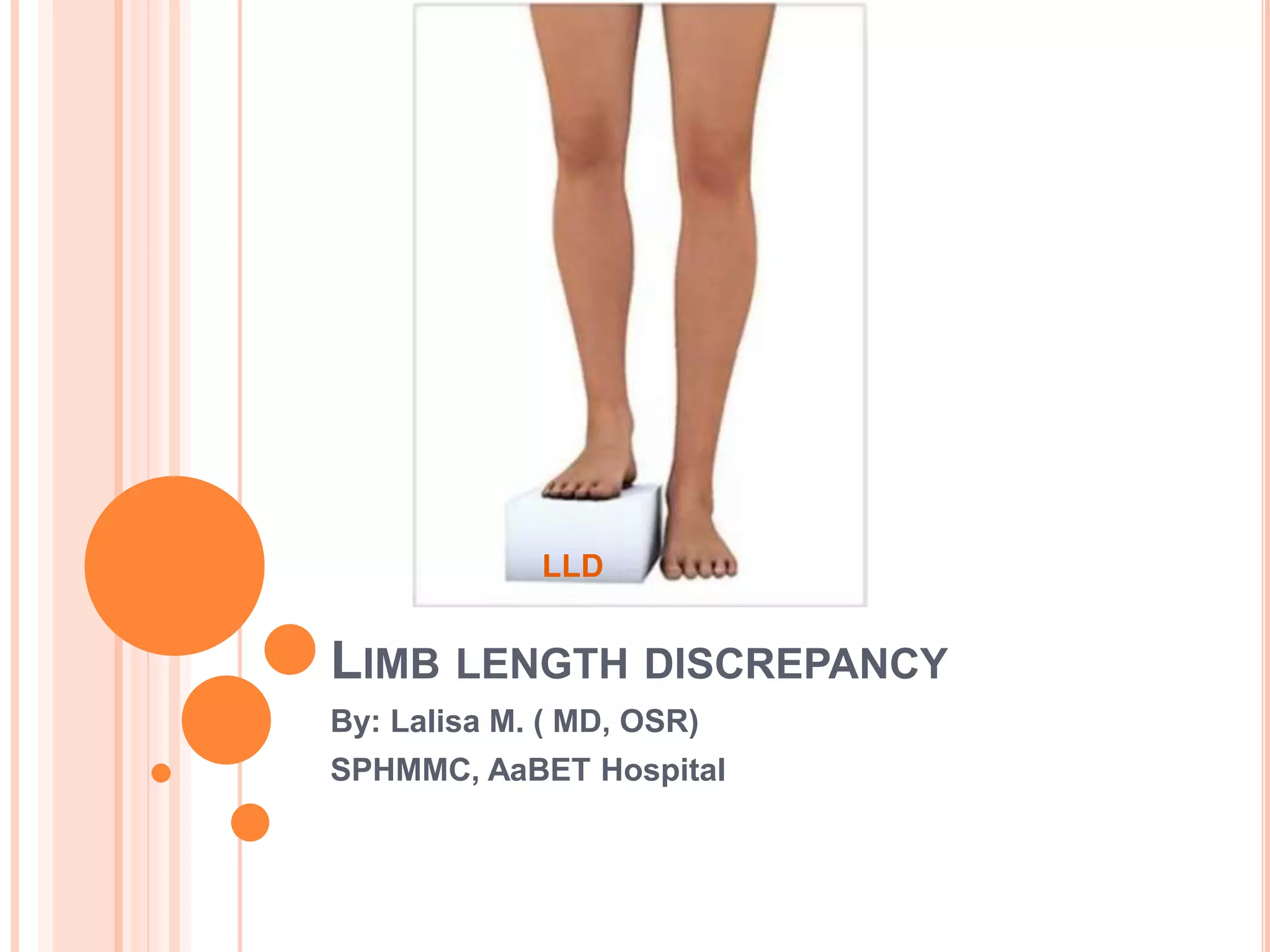
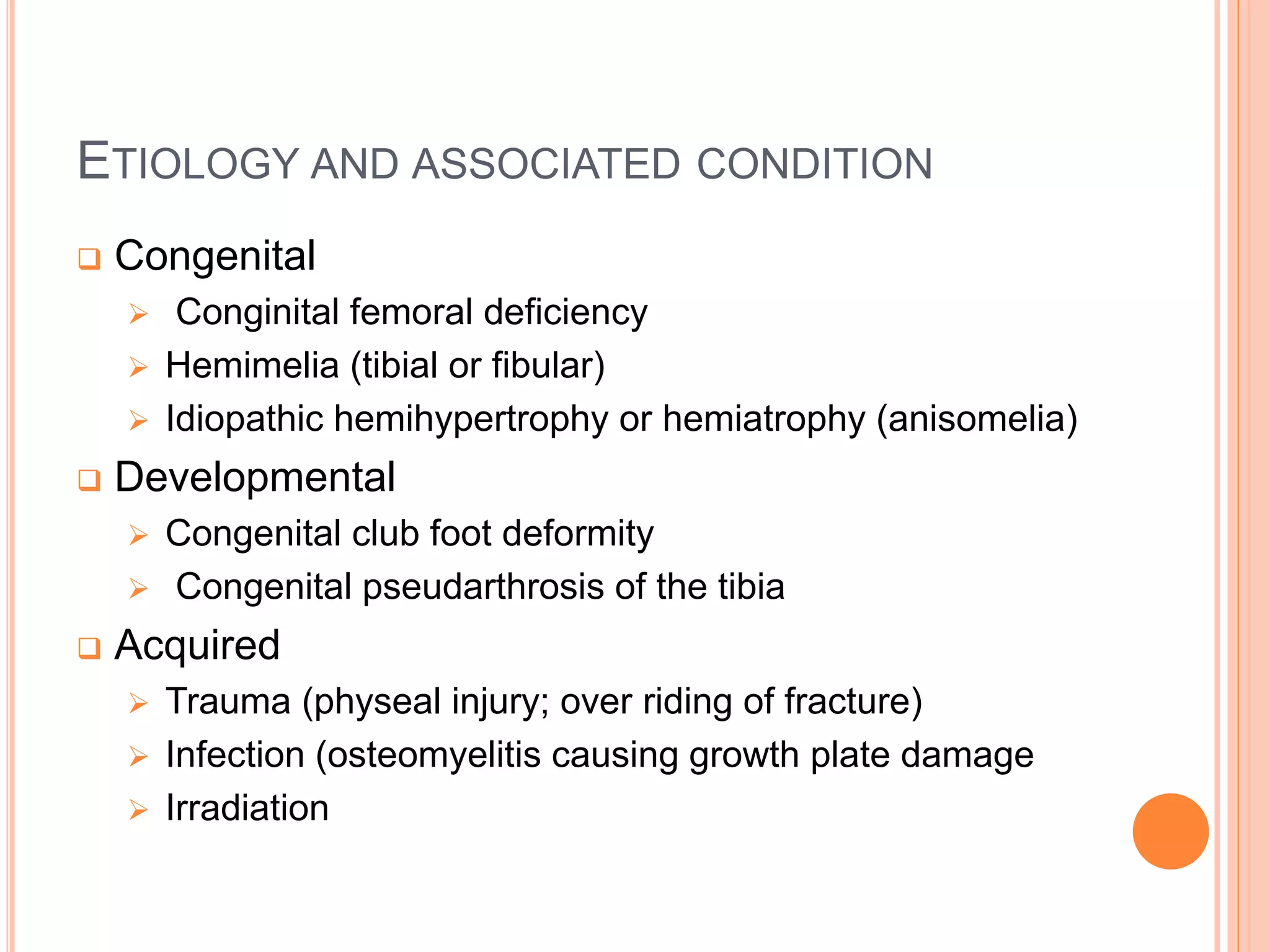
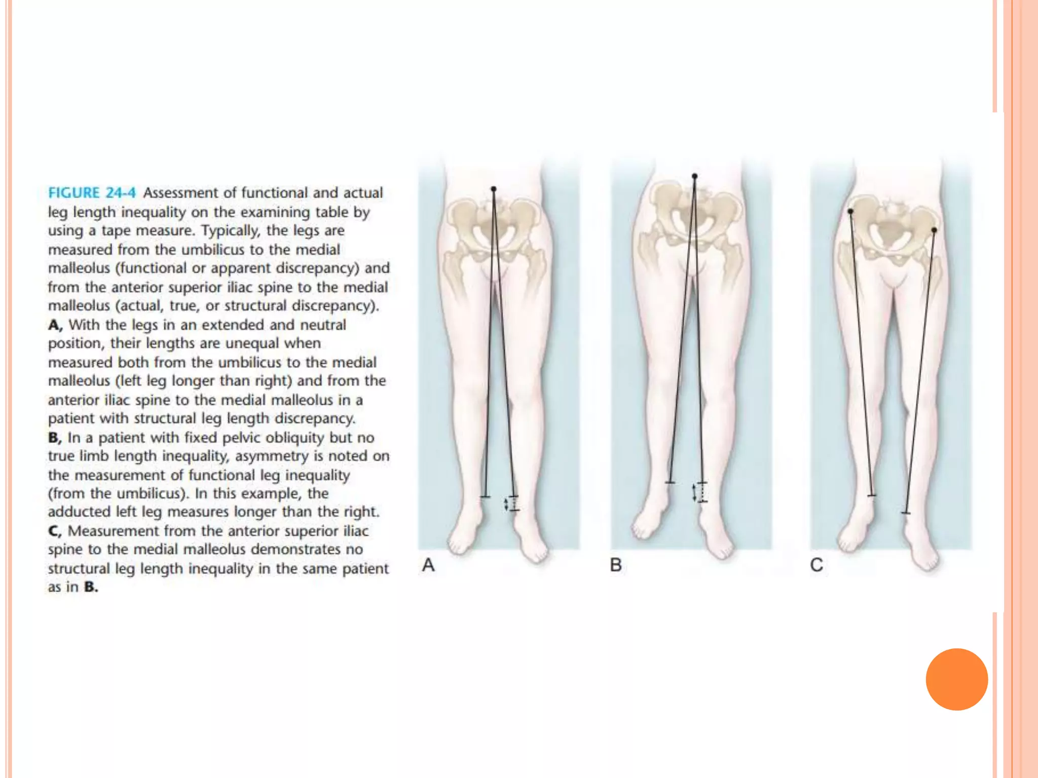
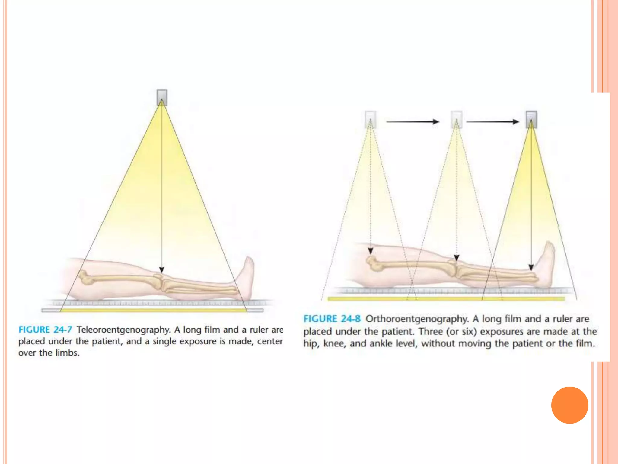
SlideShare a Scribd company logo
LIMB LENGTH DISCREPANCY
By: Lalisa M. ( MD, OSR)
SPHMMC, AaBET Hospital
LLD
OUTLINE
 Introduction
 LLI(leg length inequality)
 Etiology
 Impact of equality
 Assessment of inequality
 Prediction of leg length inequality in the skeletally immature
child
 Treatment
 Angular deformity
 Summary
 References
INTRODUCTION
 Limb length discrepancy is defined as a condition where
paired lower and upper extremity limbs have a
noticeably unequal length.
 Causes of LLD are
 Limb length inequality
 Angular deformity
 In the lower extremity, length discrepancy is not only a
cosmetic concern, but also a functional concern.
 Except in extreme cases, arm length differences cause
little or no problem in how the arms function.
 The majority of individual (up to two thirds) have some
degree of limb inequality and angular deformity.
LEG LENGTH INEQUALITY
 It is a frequent parental concern or physical finding
noted by the orthopaedic surgeon.
 Limb-length inequality of 0.5 to 2.0 cm are common
in the normal asymptomatic population.
 About one-third of population shows 0.5-1.5cm
disparities. 5% more than 1.5cm and about 1/1000
have prescribed a shoe lift.
ETIOLOGY AND ASSOCIATED CONDITION
 Congenital
 Conginital femoral deficiency
 Hemimelia (tibial or fibular)
 Idiopathic hemihypertrophy or hemiatrophy (anisomelia)
 Developmental
 Congenital club foot deformity
 Congenital pseudarthrosis of the tibia
 Acquired
 Trauma (physeal injury; over riding of fracture)
 Infection (osteomyelitis causing growth plate damage
 Irradiation
CAUSES OF LEG LENGTH INEQUALITY
IMPACT OF INEQUALITY
 Limb-length inequality of more than 2.5cm has
traditionally been considered significant. With an
increased likelihood of knee, hip, and lumbar spine
pain.
 Abnormal gait (the short leg gait is awkward)
 Increased energy expenditure because of the
excessive vertical rice and fall of the pelvis.
 Compensatory scoliosis and decreased spinal
mobility.
 Equinous contracture of ankle, callosities of foot.
CLINICAL SIGNIFICANCE OF LLD
Difference Problem Source
3mm Running injury risk Subtnick
5mm Spinal compensation Friberg
6mm Running injury risk Brody
7mm No problem <7mm Corrigan
9mm Lumbar facet angle
changes
Giles
10mm Low back pain Cyriax
15mm Compensatory
scoliosis
Gibson
20mm LE compensatory Vogel
22mm Significant scoliosis Papaloannu
40mm Requires surgical
correction
Ingram
COMPENSATORY MECHANISM OF LLI
 Circumduction
 Persistent flexion of the longer limb
 Vaulting over the longer limb
 Toe-walking on the shorter limb
 Pelvic tilt
 Shoulder tilt, unequal arm swing
TYPE OF LEG LENGTH INEQUALITY
 Structural or anatomical type
 Difference in leg length resulting from inequalities in boney
structure
 An actual shortening or lengthening of the skeletal system
occurs b/n the head of the femur and the ankle joint mortise
 Functional or apparent type
 Factor other than actual bone shortening or lengthening make
one leg shorter or longer than the other.
 Mainly due to pelvic tilt. spinal deformity
 Environmental type
 caused by the unevenness created by walking or running
tracks or along the beach.
CLASSIFICATION(MCCAW AND BATES) (1991)
 Classified according to the magnitude of the
inequality, generally expressed in cm.Described as
 Mild------less than 3cm
 Moderate------3-6 cm
 Severe------more than 6 cm
ASSEMENT OF INEQUALITY
 History
 Examination
 Imaging
• Plain radiography
 Teleoroentgenography
 Orthoroentgenography
 Scanography
• Computed radiography
 Computed tomography (CT)
• Ultrasonography
• MRI
HISTORY
 Congenital or acquired?
 Trauma/ infection?
 Progressive/ static?
 Onset & mode of deformity?
 Any syndrome associated features?
EXAMINATION
 Gait, inspection of standing child facing away from
examiner.
 Structural & functional length of lower limb.
 Determine which segment is short or long.
 Evaluate range of motion & resting position of
lumbar spine, hips, knees & ankles.
 Foot exam.
EXAMINATION CONTD'....
 Wood block test
 The client should stand facing away from the
examiner and should be undraped such that the
examiner can see the legs and waist, including the
posterior iciac spines.
 Check the knee fully extended, feet flat on the floor.
 Look at the posterior iciac spines for evidence that leg
is longer than the other.
 If descrepancy is evident, estimate discrepancy by
having the patient stand with graduated blocks under
the shorter leg until the pelvis is level.
EXAMINATION CONT'D.....
• Galeazzi (Allis) test
 Useful in young, nonambulatory or uncooperative
children.
 Alteration in the position of the joint (knee) may be
interpreted as structural discrepancy.
EXAMINATION CONT'D....
 Leg length measurements:
 Apparent length;
 From the umbilicus to the medical malleolus or
 From the xiphisternum to the medial malleolus.
 True length:
 From the ASIS to the medial malleolus after
squaring pelvis.
EXAMINATION CONT'D....
 Thigh-leg technique
 Patient placed supine with the hips and knees
flexed 90 degrees. Discrepancies are measured b/n
the table and the thigh, between the thighs at the
knees, and between the soles of the feet with the
knees even.
 Note any compensatory mechanism.
IMAGING
 Plain radiography
 Teleoroetgenography
 It is the simplest whole-leg ( entire lower extremity)
obtained with the patient supine on the radiography
table, with a long film and radiographic ruler beneath
the patient.
 Orthoroentgenography
 A technique by which three separate exposures of the
lower extremities centered over the hips, knees and
ankles are taken without moving the patient or the film
with a long film and ruler are placed under the patient.
IMAGING CONT'D....
 Scanography
 Requires movement of the x-ray tube over the
patient for three exposures, while the patient
remain motionless and the film is moved under the
patient
IMAGING CONT'D.....
 Computed Tomography
 Nowadays considered as investigation of choice
because of lower radiation exposure even when the
entire limb is exposed,
 Greater accuracy, less susceptibility to error, and
the ability to accommodate positioning difficulties
secondary to joint contractures or the presence of
external fixators.
IMAGING CONT'D....
 Ultrasonography
 Special jigs must be constructed, and the patient
must not more during the examination, main
advantage is that exposure to jonizing radiation is
not required.
PREDICTION OF LEG LENGTH INEQUALITY IN
THE SKELETALLY IMMATURE CHILD
 Longitudinal growth of long bones
 The greatest portion of the longitudinal growth of a
long bone takes place at the physes, or growth
plates.
 The epiphyses themselves grow circumferentially ,
not just at the plate.
 The major long bones; femur, tibia, fibula, humerus,
radius, and ulna have a physics at each end.
PREDICTION CONT'D....
 Longitudinal growth at physis takes place through
enchodral ossification in the horizontal zones
termed terminal, proliferative, hypertrophic &
provisional calcification.
 Horizontal growth of the physis occurs as well, in
the specialized groove of Ranvier.
 Longitudinal growth of the epiphyses themselves is
usually not taken into account when estimating the
longitudinal growth of a particular long bone.
PREDICTION CONT'D....
 Clinical and radiographic measurements of the long
bones during growth are usually made from the ends
of the physes at both ends of the long bones of the
upper and lower limb segments are known and are
limited to the physes themselves.
 In long bones with physes at each end, the
contribution of each physis to the longitudinal growth
of the bone is typically asymmetric.
 The timing of closure with cessation of longitudinal
growth in individual physes also varies by location &
individual.
PREDICTION OF GROWTH REMAINING IN THE
FEMUR AND TIBIA
 Anderson - Green - Messner Growth-Remaining
charts
 Menelaus method
 Moseley straight - line graph
 Paley multiplier method
 Automated software
PREDICTION.....
 AGM growth - Remaining chart (1963, 1864)
 Prepared by using orthoroentgenograms ( measuring
the length of the femur and tibia) hand and wrist films for
skeletal age ( using Greulich and Pyle's atlas) and other
variables were measured at each visits and recorded
against chronologic age.
 Showed that annual rate of overall growth (stature)
rapidly decreased from birth to age 6yr and was stable
from age 6 through 9years (average stature increment,
5.7 +_ 0.93cm).
 A pubertal growth spurt typically reached a maximum
b/n 10 and 12 years of age in girls and between 12 and
14 years of age in boys. The growth spurt is followed by
a final 4- year period of rapid decline in the rate of
growth until cessation of growth.
40
41
42
PREDICTION....
 Menelaus method
 Assumes growth cessation to age 16 for boys and
age 14 for girls, and bases prediction upon White
original suggestion that the distal femur grew 3/8
inch and the proximal tibia grew 1/4 inch per year.
MENELAUS METHOD…
44
PREDICTION......
 Moseley straight - line graph ( 1977)
 Constructed based on a mathematical reanalysis of
the chronologic growth data by Anderson &
colleagues.
 Intended to estimate the ultimate discrepancy in
growing children by incorporating into the calculations
skeletal maturation based on hand-wrist bone films,
growth inhibition, and relative size.
 Stated that the growth of the legs can be represented
by straight lines by a suitable manipulation of the
scale of the abscisa.
PREDICTION....
 Paley multiplier method
 Identified an arithmetic factor ( multiplier) to
calculate leg length at skeletal maturity.
 Length of each leg and the difference in leg length
at maturity can be calculated by multiplying these
measurements by the appropriate multiplier for the
subjects age and gender.
PREDICTION…
49
Automated soft-ware
TREATMENT
 Psychological and social factors
 Treating team should discuss various option, likely
problems, and outcomes with the patient and
family.
 Age- appropriate education for the child is key.
 The role of care takers, and who will be responsible
for the child's follow-up and daily care should be
established before the surgical procedure.
 Based on pretreatment evaluation, a decision not to
proceed may be made.
TREATMENT....
 Indications ( absolute)
 LLI of more than 5% ( 4cm at skeletal maturity)
 Toe-walking to compensate for LLI
TREATMENT...
 Option of treatment for LLI are;
 Orthotic management
 Shortening of the long leg
 Epiphysiodesis
 Epiphyseal stapling
 Acute shortening
 Lengthening of the short leg
 Stimulation of growth in the short leg
 Surgical lengthening of the short leg
• Acute lengthening techniques
• Gradual lengthening techniques
53
TREATMENT…
 Orthotic managent
 Shoe lift
 Considered when a child begins to toe-walk, which
is usually when LLI reaches 5% of the contralateral
side.
 Lifts of up to 1cm can be incorporated into most
shoes and larger lifts must be applied to the sole of
the shoe.
 Lifts greater than 8cm are not easy for patients to
manage and may cause them to fall over or sprain
their ankles.
Shoe lifts
TREATMENT…
 Orthotic managent…
 Ankle-foot orthosis
 Considered when a child begins to display
additional other compensatory mechanisms
(vaulting, circumducting, or increased flexion
of long leg).
 Preferred when shoe lift greater than 8cm is
required to improve gait.
TREATMENT…
 Shortening of the long leg
 Epiphysiodesis
 It is a technique to achieve permanent growth
arrest.
 Different methods exist;
 Phemister technique
 White and Stubbins technique
 Percutaneous technique
TREATMENT…
 Shortening of the long leg…
 Epiphyseal stapling or plating
 It is a technique to achieve temporary growth
arrest.
 Three evenly spaced staples placed
extraperiosteally with their tines parallel to the
physis.
 Some narrowed the indications for its use to
nearly none.
TREATMENT…
 Shortening of the long leg…
 Transphyseal screws
 It is a modification of epiphysiodesis using
percutaneously inserted transphyseal screws.
 Fully or partially threaded cancellous screws by
using crossed-scew or nonintersecting-screw
tecnique.
TREATMENT…
 Shortening of the long leg…
 Acute shortening
 A single-stage procedure to shorten longer leg
by the removal of bone and fixation.
 Relays on extent of bone to be resected and
whether a tibial or femoral segment should be
removed to gain symmetric knee height.
TREATMENT…
 Shortening of the long leg…
 Femoral shortening
 Indications;-Generally indicated only in skeletally
mature individuals with a LLI greater than 2cm.
Discrepancy localized preferably to femur to
maintain symmetric knee height after procedure.
 Shortening can be under taken in the proximal;
middle; or distal portion of the femur.
 Fixation may be achieved with a proximal
femoral blade plate; plate and screws; or IM rod.
TREATMENT…
 Shortening of the long leg…
 Tibial shortening
 Performed less frequently than femoral
shortening.
 Techniques include step-cut mid diaphyseal
shortening osteotomy with two-screw fixation;
and shortening by IM fixation.
TREATMENT…
 Lengthening of the Short Leg
 The options for lengthening the shorter limb
include;-
 Stimulation of natural growth,
 Surgical lengthening
Acute
Gradual
73
TREATMENT…
o Lengthening of the Short Leg…
 Stimulation of natural growth
 Vascularized total physeal transplantation
 Free plug of ilia crest apophyseal cartilage
 Although promising, neither technique has
broad clinical applicability at present.
TREATMENT…
 Lengthening of the Short Leg…
 Surgical Lengthening ;-
Indications:
*no absolute indication
 Expected LLD at skeletal maturity is 4cm
 Strong recommendation is when expected
shortening approaches 10% (8 cm)
75
ACUTE LENGTHENING TECHNIQUES
Transiliac acute lengthening:-
 combined with Salter innominate osteotomy for hip dysplasia
associated with shortening.
 should be limited to 2.5 to 3.0cm.
 A quadrilateral graft can be used for combined lengthening &
rotation of distal pelvic fragment to correct associated acetabular
dysplasia.
 rectangular graft can be used without associated acetabular
dysplasia.
 Iliopsoas tenotomy is mandatory.
76
77
A B
FEMORAL AND TIBIAL ACUTE
LENGTHENING:-
 Historically, the following descriptions mentioned;
 Technique of transverse osteotomy and acute
lengthening over an intramedullary rod, with locking of
the fragments by a cortical bone block. Küntscher rod,
soft tissue release, and allograft bone used.
 Method of acute lengthening of the femur with internal
fixation using a plate and screws. Here the fragments
are fixed with a long plate and screws, and the
oseotomy gap is filled with autologous bone graft from
the iliac crest.
 Complications: nerve injury, non union, mal union,
implant failure
GRADUAL LENGTHENING
TECHNIQUES
Chondrodiatasis:-
 physeal distraction without separation;
 chondrodiatasis, epiphysiolysis, epiphyseal distraction,
distraction epiphysiolysis, or epiphyseal traction.
 The challenge of ensuring the integrity of external fixation of
only the epiphysis,
 the increased risk of septic arthritis,
 The painful acute physeal disruption,
 Physeal closure frequently occurs after distraction
 Most clinical and experimental studies are not
effective
80
CALLOTASIS
 It is a more acceptable method of leg lengthening by
gradual distraction (callotasis) of a fracture callus and
involves;
 Low-energy “corticotomy” of the long bone
 Preservation of surrounding soft tissue & periosteom
 Application of external fixator
 Latent period of 3-21 days till callus formed
 Start lengthening 1mm/day
 Consolidation period = 2*distraction time
81
Consolidation
phase of
regenerate bone
82
Latent period
of the callotasis Distraction phase
EFFECTS OF GRADUAL LENGTHENING
Regenerate bone:
 Quality and quantity of newly formed bone (regenerate
bone) stimulated during limb elongation depend on;
 Rigidity of Ex-Fix
 Latency period
 Extent of damage to soft tissue
 Speed & rhythm of distraction
 Age of the patient
 Location of the osteotomy, and
 Surgical approach
83
Muscle:-Lengthening greater than 15% to 20% of
original length of the lower limb segment is associated
higher incidence of plastic deformation and
subsequent contractures especially joint spanning
muscles.
Peripheral Nerves:- nerve conduction studies and
electromyography shows deleterious effect. Excessive
gradual ([20–30 %) or acute distraction ([15 %) may
both lead to partial or complete loss of nerve
potentials 84
COMPLICATIONS OF GRADUAL LENGTHENING
 Neurovascular injury
 Incomplete Osteotomy
 Premature Consolidation
 Poor Regenerate Bone Formation
 Joint Subluxation
 Pin Site Infection
 Regenerate Bone Fracture
 Subsequent Growth Disturbance of the Lengthened
Limb 85
DEVICES FOR GRADUAL LENGTHENING
 Wagner Device
 Dynamic Axial Fixator
 Ilizarov Apparatus
 Combined Internal and External Fixation
(Lengthening Over Intramedullary Rods)
 Totally Implantable Lengthening Devices
MONOLATERAL EXTERNAL FIXATORS:
 For simple lengthening over a short to medium
distance; because they are not able to withstand
muscle forces during excessive lengthening
 The draw backs are;
 Secondary malalignment,
 premature cessation of lengthening,
 unilateral premature consolidation
 The monolateral devices used included Wagner, Orthofix
(Orthofix, Verona, Italy), and Monotube (Howmedica,
Rutherford, N.J.).
88
Wagner Device
Dynamic Axial Fixator
o The apparatus consists
of one of two types of
external connecting
bodies: a simple linear
distractor (lengthener)
with a telescoping body
(available in several
lengths).
o The fixator device is
more suitable for
fracture management.
EXTERNAL RING FIXATION:-
 Can solve any three-dimensional, multi-plane, 6-
axes deformity problem
 Hybrid wire and screw bone fixation to reduce
number of soft tissue transfixations,
 Provide greatest versatility and best mechanical
control
 Ilizarov and TSF
91
ILIZAROV APPARATUS
 Ilizarov’s circular external fixator is inherently more
complex than other devices used for either
lengthening or deformity correction.
Clinical value of the Taylor Spatial Frame: a
comparison with the Ilizarov and Orthofix fixatorsD.
Dammerer, K. Kirschbichler, L. Donnan, G. Kaufmann, M. Krismer, and R.
Biederman
Abstract
Purpose :Evaluation of the advantages and limitations of the Taylor
Spatial Frame (TSF) with regard to the healing index (HI), distraction–
consolidation time (DCT), accuracy of correction complications, and cost
of the device
Methods.Comparison of results with the traditional Ilizarov apparatus and a
unilateral Orthofix fixator in a consecutive patient series with 135 bony
deformity corrections.
Results The HI did not differ significantly between all three fixators and was
57 days/cm for all patients. The DCT was significantly shorter for the TSF
(148 days) compared to the Ilizarov fixator (204 days) and the Orthofix
device (213 days). The accuracy of deformity correction was higher for
the TSF than the other devices. The mean values of the measured angles
after correction did not differ, but the variance of the results was the
lowest. Also, the total rate of complications was considerably lower for
the TSF. The Orthofix device showed a high rate of angular deformity
during treatment, whereas both ring fixators had a relatively higher
number of pin-related problems.
ConclusionsThe findings in our patient series suggest the use of the
Orthofix apparatus for simple lengthening over short to median distances
94
LENGTHENING OVER INTRAMEDULLARY RODS
 Lengthening of the femur or tibia over
intramedullary rods, with locking of the fragments to
the rod at the completion of distraction and removal
of the external fixation device. Advantages are;
 Diminution of fixator-associated issues such as infections
and contractures,
 prevent regenerate fracture
 better tolerance and comfort.
 Maintain rotation and axial length
95
96
TOTALLY IMPLANTABLE LENGTHENING DEVICES
Three totally implantable intramedullary rods are
designed
 Albizzia nail
 intramedullary skeletal kinetic distractor (ISKD)
 Fitbone
97
SUMMARIZED GUIDANCE FOR MANAGEMENT
 <2cm…………………………………..no treatment
or shoe lift
 2-5cm………………………………….growth
modulations ( shoe lift or epiphysiodesis) limb
shortening
 > 5cm…………………………………….limb
lengthening
 >20cm……………………………….........amputati
ons and prosthetic fitting
ANGULAR DEFORMITY
 Normal Lower Extremity Alignment
 Essentially symmetric physiologic varus can be
expected between birth and 18 to 24 months of
age,
 This is followed by a valgus “deformity” that is
maximal at approximately 3 years of age and
resolves by 6 to 8 years of age.
 After “mature” lower extremity alignment has been
achieved, the legs normally look straight—that is,
the pelvis is level, and the medial femoral condyles
and the medial malleoli touch.
 Variations from this “standard” or “normal”
appearance are common.
ASSESSMENT OF DEFORMITY
 History and physical examination
 Radiographic assessment
 Careful analysis of
the mechanical axis
and joint relationships
PRINCIPLES OF DEFORMITY CORRECTION
 Angular deformity correction by osteotomy should
be performed at the level of the apex of the
deformity(metaphysis or diaphysis).
 Osteotomy is usually performed at a level remote
from the apex of angular deformity, typically in the
adjacent metaphysis(apex of angular at the level of
the physis or epiphysis).
SURGICAL OPTIONS FOR DEFORMITY
CORRECTION
 Correction by staple insertion or
hemiepiphysiodesis,
 Acute correction with internal or external fixation,
and
 Gradual correction with external fixation.
HEMIEPIPHYSEAL STAPLING
 The basic principles and techniques are the same
as for stapling to correct leg length inequality.
 The best candidates for this are patients with
angular deformities of the longer leg or very young
children who should not undergo epiphysiodesis
because of excessive growth retardation and in
whom correction by osteotomy is not desirable.
 A variation of hemiepiphyseal stapling is to use
percutaneously inserted screws across the physis
HEMIEPIPHYSIODESIS
 Can be used for any angular deformity originating in
the region of the physis if the physis on the convex
side of the deformity has adequate
growthremaining to allow angular correction.
 Useful to determine that there is not an excessive
amount of growth remaining because
hemiepiphysiodesis in such cases could lead to an
unacceptable amount of limb shortening.
 Completion of the hemiepiphysiodesis may be
required after full correction has been achieved.
ACUTE CORRECTION
 It is immediate surgical correction using opening
wedge, closing wedge, or a combination of opening
and closing osteotomies.
 The soft tissues are generally tensioned with acute
correction, even if a closing wedge osteotomy is
performed, so only a limited amount of lengthening
is achievable.
 Internal fixation after acute corrective osteotomy
may be either definitive, with plate and screws or an
intramedullary device, or partial, with percutaneous
pins supplemented with casts.
 External fixation may also be used in conjunction
with acute correction of angular deformity.
GRADUAL CORRECTION
 Angular deformities may also be corrected
gradually by osteotomy and external fixation.
 The circular ring fixator is most suitable for this
method.
 The Taylor Spatial Frame, consisting of two rings
fixed to the limb with wires or half-pins and to each
other by six adjustable struts, may also be used.
 For corrections of most lower extremity deformities,
the surgeon must rely on a computer software
program to provide the daily six-strut manipulation
to effect the desired correction.
TSF
REFERENCES
1. Tachdjchian pediatrics orthopedics 5th ed
2. Current concepts of leg lengthening ,Carol C. Hasler•
Andreas H. Krieg
3. Leg Length Discrepancy: Assessment and Secondary
Effects Ahmed A Khalifa
4. Limb Lengthening Techniques, pediatric society of
North America
5. A Straight-Line Graph for Leg-Length Discrepancies
BY C. F. MOSELEY, M.D.,C.M.,F.R.C.S.(C)*,HAMILTON,
ONTARIO, CANADA
6. Clinical value of the Taylor Spatial Frame: a
comparison with the Ilizarov and Orthofix fixators, D.
Dammerer • K. Kirschbichler • L. Donnan • G.
Kaufmann • M. Krismer • R. Biedermann
7. Comparison of three surgical epiphysiodesis
techniques for the treatment of lower limb length
discrepancy Cédric CAMPENS, Maryline MOUSNY,

More Related Content

What's hot

RSD-REFLEX SYMPATHETIC DYSTROPHY
RSD-REFLEX SYMPATHETIC DYSTROPHYRSD-REFLEX SYMPATHETIC DYSTROPHY
RSD-REFLEX SYMPATHETIC DYSTROPHY
Mohammad Akeel
 
Dupuytrens contracture
Dupuytrens contractureDupuytrens contracture
Dupuytrens contracture
orthoprince
 
hoffas fat pad syndrome.pptx
hoffas fat pad syndrome.pptxhoffas fat pad syndrome.pptx
hoffas fat pad syndrome.pptx
Collage
 

What's hot (20)

Mallet finger
Mallet fingerMallet finger
Mallet finger
 
RSD-REFLEX SYMPATHETIC DYSTROPHY
RSD-REFLEX SYMPATHETIC DYSTROPHYRSD-REFLEX SYMPATHETIC DYSTROPHY
RSD-REFLEX SYMPATHETIC DYSTROPHY
 
Shoulder impingement syndrome
Shoulder impingement syndromeShoulder impingement syndrome
Shoulder impingement syndrome
 
Limb length discrepancy
Limb length discrepancyLimb length discrepancy
Limb length discrepancy
 
Limb length discrepency
Limb length discrepencyLimb length discrepency
Limb length discrepency
 
Waddling gait- definition|role of muscle|gait analysis|kinematic and spatiote...
Waddling gait- definition|role of muscle|gait analysis|kinematic and spatiote...Waddling gait- definition|role of muscle|gait analysis|kinematic and spatiote...
Waddling gait- definition|role of muscle|gait analysis|kinematic and spatiote...
 
Pes planus / Flat Foot
Pes planus / Flat Foot Pes planus / Flat Foot
Pes planus / Flat Foot
 
Flat foot and Cavus foot
 Flat foot and Cavus foot Flat foot and Cavus foot
Flat foot and Cavus foot
 
Dupuytrens contracture
Dupuytrens contractureDupuytrens contracture
Dupuytrens contracture
 
PRINCIPLES OF TENDON TRANSFERS
PRINCIPLES OF TENDON TRANSFERSPRINCIPLES OF TENDON TRANSFERS
PRINCIPLES OF TENDON TRANSFERS
 
Achilles Tendinitis
Achilles TendinitisAchilles Tendinitis
Achilles Tendinitis
 
Pes planus
Pes planusPes planus
Pes planus
 
hoffas fat pad syndrome.pptx
hoffas fat pad syndrome.pptxhoffas fat pad syndrome.pptx
hoffas fat pad syndrome.pptx
 
post polio residual paralysis
post polio residual paralysispost polio residual paralysis
post polio residual paralysis
 
Post polio residual paralysis
Post polio residual paralysisPost polio residual paralysis
Post polio residual paralysis
 
Acl ppt
Acl pptAcl ppt
Acl ppt
 
Sprengel’s shoulder
Sprengel’s shoulderSprengel’s shoulder
Sprengel’s shoulder
 
ACL rehabilitation
ACL rehabilitationACL rehabilitation
ACL rehabilitation
 
Genu recurvatum
Genu recurvatumGenu recurvatum
Genu recurvatum
 
Scoliosis bracing
Scoliosis bracingScoliosis bracing
Scoliosis bracing
 

Similar to Limb length discrepancy

EVALUATION METHODS.presentation for evaluation
EVALUATION METHODS.presentation for evaluationEVALUATION METHODS.presentation for evaluation
EVALUATION METHODS.presentation for evaluation
PranavTrehan2
 
Presentation1.pptx, radiological imaging of anteversion angle.
Presentation1.pptx, radiological imaging of anteversion angle.Presentation1.pptx, radiological imaging of anteversion angle.
Presentation1.pptx, radiological imaging of anteversion angle.
Abdellah Nazeer
 

Similar to Limb length discrepancy (20)

lld- leg length.ppt
lld- leg length.pptlld- leg length.ppt
lld- leg length.ppt
 
LIMB-LENGTH DISCREPANCY.pptx
LIMB-LENGTH DISCREPANCY.pptxLIMB-LENGTH DISCREPANCY.pptx
LIMB-LENGTH DISCREPANCY.pptx
 
LIMB-LENGTH DISCREPANCY.pptx
LIMB-LENGTH DISCREPANCY.pptxLIMB-LENGTH DISCREPANCY.pptx
LIMB-LENGTH DISCREPANCY.pptx
 
Leg length measurements
Leg length measurementsLeg length measurements
Leg length measurements
 
Genu Valgum.pptx
Genu Valgum.pptxGenu Valgum.pptx
Genu Valgum.pptx
 
An approach to limping child
An approach to limping childAn approach to limping child
An approach to limping child
 
Skeletal dysplasia grand round
Skeletal dysplasia   grand round Skeletal dysplasia   grand round
Skeletal dysplasia grand round
 
Leg Length Discrepancy.pdf
Leg Length Discrepancy.pdfLeg Length Discrepancy.pdf
Leg Length Discrepancy.pdf
 
Adult Hip Dysplasia Presentation
Adult Hip Dysplasia PresentationAdult Hip Dysplasia Presentation
Adult Hip Dysplasia Presentation
 
Ilizarov fixator
Ilizarov fixatorIlizarov fixator
Ilizarov fixator
 
Limb length discrepancy evaluation
Limb length discrepancy evaluationLimb length discrepancy evaluation
Limb length discrepancy evaluation
 
lect 1 Anthropometry -.pdf
lect 1 Anthropometry -.pdflect 1 Anthropometry -.pdf
lect 1 Anthropometry -.pdf
 
EVALUATION METHODS.presentation for evaluation
EVALUATION METHODS.presentation for evaluationEVALUATION METHODS.presentation for evaluation
EVALUATION METHODS.presentation for evaluation
 
Congenital and developmental anomalies.2560
Congenital and developmental anomalies.2560Congenital and developmental anomalies.2560
Congenital and developmental anomalies.2560
 
Pediatric knee copy
Pediatric knee   copyPediatric knee   copy
Pediatric knee copy
 
SKELTAL DYSPLASIA.pptx
SKELTAL DYSPLASIA.pptxSKELTAL DYSPLASIA.pptx
SKELTAL DYSPLASIA.pptx
 
Angular deformities around the knee seminar
Angular deformities around the knee seminarAngular deformities around the knee seminar
Angular deformities around the knee seminar
 
Presentation1.pptx, radiological imaging of anteversion angle.
Presentation1.pptx, radiological imaging of anteversion angle.Presentation1.pptx, radiological imaging of anteversion angle.
Presentation1.pptx, radiological imaging of anteversion angle.
 
Ctev
CtevCtev
Ctev
 
Role of sonography in knee joint diseases
Role of sonography in knee joint diseasesRole of sonography in knee joint diseases
Role of sonography in knee joint diseases
 

Recently uploaded

Alcohol_Dr. Jeenal Mistry MD Pharmacology.pdf
Alcohol_Dr. Jeenal Mistry MD Pharmacology.pdfAlcohol_Dr. Jeenal Mistry MD Pharmacology.pdf
Alcohol_Dr. Jeenal Mistry MD Pharmacology.pdf
Dr Jeenal Mistry
 

Recently uploaded (20)

CURRENT HEALTH PROBLEMS AND ITS SOLUTION BY AYURVEDA.pptx
CURRENT HEALTH PROBLEMS AND ITS SOLUTION BY AYURVEDA.pptxCURRENT HEALTH PROBLEMS AND ITS SOLUTION BY AYURVEDA.pptx
CURRENT HEALTH PROBLEMS AND ITS SOLUTION BY AYURVEDA.pptx
 
"Central Hypertension"‚ in China: Towards the nation-wide use of SphygmoCor t...
"Central Hypertension"‚ in China: Towards the nation-wide use of SphygmoCor t..."Central Hypertension"‚ in China: Towards the nation-wide use of SphygmoCor t...
"Central Hypertension"‚ in China: Towards the nation-wide use of SphygmoCor t...
 
Factors Affecting child behavior in Pediatric Dentistry
Factors Affecting child behavior in Pediatric DentistryFactors Affecting child behavior in Pediatric Dentistry
Factors Affecting child behavior in Pediatric Dentistry
 
TEST BANK For Williams' Essentials of Nutrition and Diet Therapy, 13th Editio...
TEST BANK For Williams' Essentials of Nutrition and Diet Therapy, 13th Editio...TEST BANK For Williams' Essentials of Nutrition and Diet Therapy, 13th Editio...
TEST BANK For Williams' Essentials of Nutrition and Diet Therapy, 13th Editio...
 
Antiplatelets in IHD, Dose Duration, DAPT vs SAPT
Antiplatelets in IHD, Dose Duration, DAPT vs SAPTAntiplatelets in IHD, Dose Duration, DAPT vs SAPT
Antiplatelets in IHD, Dose Duration, DAPT vs SAPT
 
Fundamental of Radiobiology -SABBU.pptx
Fundamental of Radiobiology  -SABBU.pptxFundamental of Radiobiology  -SABBU.pptx
Fundamental of Radiobiology -SABBU.pptx
 
Antiulcer drugs Advance Pharmacology .pptx
Antiulcer drugs Advance Pharmacology .pptxAntiulcer drugs Advance Pharmacology .pptx
Antiulcer drugs Advance Pharmacology .pptx
 
Scientificity and feasibility study of non-invasive central arterial pressure...
Scientificity and feasibility study of non-invasive central arterial pressure...Scientificity and feasibility study of non-invasive central arterial pressure...
Scientificity and feasibility study of non-invasive central arterial pressure...
 
DECIPHERING COMMON ECG FINDINGS IN ED.pptx
DECIPHERING COMMON ECG FINDINGS IN ED.pptxDECIPHERING COMMON ECG FINDINGS IN ED.pptx
DECIPHERING COMMON ECG FINDINGS IN ED.pptx
 
Why invest into infodemic management in health emergencies
Why invest into infodemic management in health emergenciesWhy invest into infodemic management in health emergencies
Why invest into infodemic management in health emergencies
 
Evaluation of antidepressant activity of clitoris ternatea in animals
Evaluation of antidepressant activity of clitoris ternatea in animalsEvaluation of antidepressant activity of clitoris ternatea in animals
Evaluation of antidepressant activity of clitoris ternatea in animals
 
Young at heart: Cardiovascular health stations to empower healthy lifestyle b...
Young at heart: Cardiovascular health stations to empower healthy lifestyle b...Young at heart: Cardiovascular health stations to empower healthy lifestyle b...
Young at heart: Cardiovascular health stations to empower healthy lifestyle b...
 
Final CAPNOCYTOPHAGA INFECTION by Gauri Gawande.pptx
Final CAPNOCYTOPHAGA INFECTION by Gauri Gawande.pptxFinal CAPNOCYTOPHAGA INFECTION by Gauri Gawande.pptx
Final CAPNOCYTOPHAGA INFECTION by Gauri Gawande.pptx
 
Alcohol_Dr. Jeenal Mistry MD Pharmacology.pdf
Alcohol_Dr. Jeenal Mistry MD Pharmacology.pdfAlcohol_Dr. Jeenal Mistry MD Pharmacology.pdf
Alcohol_Dr. Jeenal Mistry MD Pharmacology.pdf
 
Presentació "Advancing Emergency Medicine Education through Virtual Reality"
Presentació "Advancing Emergency Medicine Education through Virtual Reality"Presentació "Advancing Emergency Medicine Education through Virtual Reality"
Presentació "Advancing Emergency Medicine Education through Virtual Reality"
 
180-hour Power Capsules For Men In Ghana
180-hour Power Capsules For Men In Ghana180-hour Power Capsules For Men In Ghana
180-hour Power Capsules For Men In Ghana
 
ANATOMY OF THE LOWER URINARY TRACT AND MALE [Autosaved] [Autosaved].pptx
ANATOMY OF THE LOWER URINARY TRACT AND MALE [Autosaved] [Autosaved].pptxANATOMY OF THE LOWER URINARY TRACT AND MALE [Autosaved] [Autosaved].pptx
ANATOMY OF THE LOWER URINARY TRACT AND MALE [Autosaved] [Autosaved].pptx
 
linearity concept of significance, standard deviation, chi square test, stude...
linearity concept of significance, standard deviation, chi square test, stude...linearity concept of significance, standard deviation, chi square test, stude...
linearity concept of significance, standard deviation, chi square test, stude...
 
Anuman- An inference for helpful in diagnosis and treatment
Anuman- An inference for helpful in diagnosis and treatmentAnuman- An inference for helpful in diagnosis and treatment
Anuman- An inference for helpful in diagnosis and treatment
 
Book Trailer: PGMEE in a Nutshell (CEE MD/MS PG Entrance Examination)
Book Trailer: PGMEE in a Nutshell (CEE MD/MS PG Entrance Examination)Book Trailer: PGMEE in a Nutshell (CEE MD/MS PG Entrance Examination)
Book Trailer: PGMEE in a Nutshell (CEE MD/MS PG Entrance Examination)
 

Limb length discrepancy

  • 1. LIMB LENGTH DISCREPANCY By: Lalisa M. ( MD, OSR) SPHMMC, AaBET Hospital LLD
  • 2. OUTLINE  Introduction  LLI(leg length inequality)  Etiology  Impact of equality  Assessment of inequality  Prediction of leg length inequality in the skeletally immature child  Treatment  Angular deformity  Summary  References
  • 3. INTRODUCTION  Limb length discrepancy is defined as a condition where paired lower and upper extremity limbs have a noticeably unequal length.  Causes of LLD are  Limb length inequality  Angular deformity  In the lower extremity, length discrepancy is not only a cosmetic concern, but also a functional concern.  Except in extreme cases, arm length differences cause little or no problem in how the arms function.  The majority of individual (up to two thirds) have some degree of limb inequality and angular deformity.
  • 4. LEG LENGTH INEQUALITY  It is a frequent parental concern or physical finding noted by the orthopaedic surgeon.  Limb-length inequality of 0.5 to 2.0 cm are common in the normal asymptomatic population.  About one-third of population shows 0.5-1.5cm disparities. 5% more than 1.5cm and about 1/1000 have prescribed a shoe lift.
  • 5. ETIOLOGY AND ASSOCIATED CONDITION  Congenital  Conginital femoral deficiency  Hemimelia (tibial or fibular)  Idiopathic hemihypertrophy or hemiatrophy (anisomelia)  Developmental  Congenital club foot deformity  Congenital pseudarthrosis of the tibia  Acquired  Trauma (physeal injury; over riding of fracture)  Infection (osteomyelitis causing growth plate damage  Irradiation
  • 6. CAUSES OF LEG LENGTH INEQUALITY
  • 7. IMPACT OF INEQUALITY  Limb-length inequality of more than 2.5cm has traditionally been considered significant. With an increased likelihood of knee, hip, and lumbar spine pain.  Abnormal gait (the short leg gait is awkward)  Increased energy expenditure because of the excessive vertical rice and fall of the pelvis.  Compensatory scoliosis and decreased spinal mobility.  Equinous contracture of ankle, callosities of foot.
  • 8. CLINICAL SIGNIFICANCE OF LLD Difference Problem Source 3mm Running injury risk Subtnick 5mm Spinal compensation Friberg 6mm Running injury risk Brody 7mm No problem <7mm Corrigan 9mm Lumbar facet angle changes Giles 10mm Low back pain Cyriax 15mm Compensatory scoliosis Gibson 20mm LE compensatory Vogel 22mm Significant scoliosis Papaloannu 40mm Requires surgical correction Ingram
  • 9.
  • 10.
  • 11. COMPENSATORY MECHANISM OF LLI  Circumduction  Persistent flexion of the longer limb  Vaulting over the longer limb  Toe-walking on the shorter limb  Pelvic tilt  Shoulder tilt, unequal arm swing
  • 12. TYPE OF LEG LENGTH INEQUALITY  Structural or anatomical type  Difference in leg length resulting from inequalities in boney structure  An actual shortening or lengthening of the skeletal system occurs b/n the head of the femur and the ankle joint mortise  Functional or apparent type  Factor other than actual bone shortening or lengthening make one leg shorter or longer than the other.  Mainly due to pelvic tilt. spinal deformity  Environmental type  caused by the unevenness created by walking or running tracks or along the beach.
  • 13. CLASSIFICATION(MCCAW AND BATES) (1991)  Classified according to the magnitude of the inequality, generally expressed in cm.Described as  Mild------less than 3cm  Moderate------3-6 cm  Severe------more than 6 cm
  • 14. ASSEMENT OF INEQUALITY  History  Examination  Imaging • Plain radiography  Teleoroentgenography  Orthoroentgenography  Scanography • Computed radiography  Computed tomography (CT) • Ultrasonography • MRI
  • 15. HISTORY  Congenital or acquired?  Trauma/ infection?  Progressive/ static?  Onset & mode of deformity?  Any syndrome associated features?
  • 16. EXAMINATION  Gait, inspection of standing child facing away from examiner.  Structural & functional length of lower limb.  Determine which segment is short or long.  Evaluate range of motion & resting position of lumbar spine, hips, knees & ankles.  Foot exam.
  • 17. EXAMINATION CONTD'....  Wood block test  The client should stand facing away from the examiner and should be undraped such that the examiner can see the legs and waist, including the posterior iciac spines.  Check the knee fully extended, feet flat on the floor.  Look at the posterior iciac spines for evidence that leg is longer than the other.  If descrepancy is evident, estimate discrepancy by having the patient stand with graduated blocks under the shorter leg until the pelvis is level.
  • 18.
  • 19. EXAMINATION CONT'D..... • Galeazzi (Allis) test  Useful in young, nonambulatory or uncooperative children.  Alteration in the position of the joint (knee) may be interpreted as structural discrepancy.
  • 20. EXAMINATION CONT'D....  Leg length measurements:  Apparent length;  From the umbilicus to the medical malleolus or  From the xiphisternum to the medial malleolus.  True length:  From the ASIS to the medial malleolus after squaring pelvis.
  • 21.
  • 22. EXAMINATION CONT'D....  Thigh-leg technique  Patient placed supine with the hips and knees flexed 90 degrees. Discrepancies are measured b/n the table and the thigh, between the thighs at the knees, and between the soles of the feet with the knees even.  Note any compensatory mechanism.
  • 23.
  • 24. IMAGING  Plain radiography  Teleoroetgenography  It is the simplest whole-leg ( entire lower extremity) obtained with the patient supine on the radiography table, with a long film and radiographic ruler beneath the patient.  Orthoroentgenography  A technique by which three separate exposures of the lower extremities centered over the hips, knees and ankles are taken without moving the patient or the film with a long film and ruler are placed under the patient.
  • 25.
  • 26. IMAGING CONT'D....  Scanography  Requires movement of the x-ray tube over the patient for three exposures, while the patient remain motionless and the film is moved under the patient
  • 27.
  • 28. IMAGING CONT'D.....  Computed Tomography  Nowadays considered as investigation of choice because of lower radiation exposure even when the entire limb is exposed,  Greater accuracy, less susceptibility to error, and the ability to accommodate positioning difficulties secondary to joint contractures or the presence of external fixators.
  • 29.
  • 30. IMAGING CONT'D....  Ultrasonography  Special jigs must be constructed, and the patient must not more during the examination, main advantage is that exposure to jonizing radiation is not required.
  • 31.
  • 32.
  • 33. PREDICTION OF LEG LENGTH INEQUALITY IN THE SKELETALLY IMMATURE CHILD  Longitudinal growth of long bones  The greatest portion of the longitudinal growth of a long bone takes place at the physes, or growth plates.  The epiphyses themselves grow circumferentially , not just at the plate.  The major long bones; femur, tibia, fibula, humerus, radius, and ulna have a physics at each end.
  • 34. PREDICTION CONT'D....  Longitudinal growth at physis takes place through enchodral ossification in the horizontal zones termed terminal, proliferative, hypertrophic & provisional calcification.  Horizontal growth of the physis occurs as well, in the specialized groove of Ranvier.  Longitudinal growth of the epiphyses themselves is usually not taken into account when estimating the longitudinal growth of a particular long bone.
  • 35. PREDICTION CONT'D....  Clinical and radiographic measurements of the long bones during growth are usually made from the ends of the physes at both ends of the long bones of the upper and lower limb segments are known and are limited to the physes themselves.  In long bones with physes at each end, the contribution of each physis to the longitudinal growth of the bone is typically asymmetric.  The timing of closure with cessation of longitudinal growth in individual physes also varies by location & individual.
  • 36. PREDICTION OF GROWTH REMAINING IN THE FEMUR AND TIBIA  Anderson - Green - Messner Growth-Remaining charts  Menelaus method  Moseley straight - line graph  Paley multiplier method  Automated software
  • 37. PREDICTION.....  AGM growth - Remaining chart (1963, 1864)  Prepared by using orthoroentgenograms ( measuring the length of the femur and tibia) hand and wrist films for skeletal age ( using Greulich and Pyle's atlas) and other variables were measured at each visits and recorded against chronologic age.  Showed that annual rate of overall growth (stature) rapidly decreased from birth to age 6yr and was stable from age 6 through 9years (average stature increment, 5.7 +_ 0.93cm).  A pubertal growth spurt typically reached a maximum b/n 10 and 12 years of age in girls and between 12 and 14 years of age in boys. The growth spurt is followed by a final 4- year period of rapid decline in the rate of growth until cessation of growth.
  • 38.
  • 39.
  • 40. 40
  • 41. 41
  • 42. 42
  • 43. PREDICTION....  Menelaus method  Assumes growth cessation to age 16 for boys and age 14 for girls, and bases prediction upon White original suggestion that the distal femur grew 3/8 inch and the proximal tibia grew 1/4 inch per year.
  • 45. PREDICTION......  Moseley straight - line graph ( 1977)  Constructed based on a mathematical reanalysis of the chronologic growth data by Anderson & colleagues.  Intended to estimate the ultimate discrepancy in growing children by incorporating into the calculations skeletal maturation based on hand-wrist bone films, growth inhibition, and relative size.  Stated that the growth of the legs can be represented by straight lines by a suitable manipulation of the scale of the abscisa.
  • 46.
  • 47. PREDICTION....  Paley multiplier method  Identified an arithmetic factor ( multiplier) to calculate leg length at skeletal maturity.  Length of each leg and the difference in leg length at maturity can be calculated by multiplying these measurements by the appropriate multiplier for the subjects age and gender.
  • 48.
  • 50. TREATMENT  Psychological and social factors  Treating team should discuss various option, likely problems, and outcomes with the patient and family.  Age- appropriate education for the child is key.  The role of care takers, and who will be responsible for the child's follow-up and daily care should be established before the surgical procedure.  Based on pretreatment evaluation, a decision not to proceed may be made.
  • 51. TREATMENT....  Indications ( absolute)  LLI of more than 5% ( 4cm at skeletal maturity)  Toe-walking to compensate for LLI
  • 52. TREATMENT...  Option of treatment for LLI are;  Orthotic management  Shortening of the long leg  Epiphysiodesis  Epiphyseal stapling  Acute shortening  Lengthening of the short leg  Stimulation of growth in the short leg  Surgical lengthening of the short leg • Acute lengthening techniques • Gradual lengthening techniques
  • 53. 53
  • 54. TREATMENT…  Orthotic managent  Shoe lift  Considered when a child begins to toe-walk, which is usually when LLI reaches 5% of the contralateral side.  Lifts of up to 1cm can be incorporated into most shoes and larger lifts must be applied to the sole of the shoe.  Lifts greater than 8cm are not easy for patients to manage and may cause them to fall over or sprain their ankles.
  • 56. TREATMENT…  Orthotic managent…  Ankle-foot orthosis  Considered when a child begins to display additional other compensatory mechanisms (vaulting, circumducting, or increased flexion of long leg).  Preferred when shoe lift greater than 8cm is required to improve gait.
  • 57.
  • 58. TREATMENT…  Shortening of the long leg  Epiphysiodesis  It is a technique to achieve permanent growth arrest.  Different methods exist;  Phemister technique  White and Stubbins technique  Percutaneous technique
  • 59.
  • 60.
  • 61. TREATMENT…  Shortening of the long leg…  Epiphyseal stapling or plating  It is a technique to achieve temporary growth arrest.  Three evenly spaced staples placed extraperiosteally with their tines parallel to the physis.  Some narrowed the indications for its use to nearly none.
  • 62.
  • 63. TREATMENT…  Shortening of the long leg…  Transphyseal screws  It is a modification of epiphysiodesis using percutaneously inserted transphyseal screws.  Fully or partially threaded cancellous screws by using crossed-scew or nonintersecting-screw tecnique.
  • 64.
  • 65.
  • 66.
  • 67. TREATMENT…  Shortening of the long leg…  Acute shortening  A single-stage procedure to shorten longer leg by the removal of bone and fixation.  Relays on extent of bone to be resected and whether a tibial or femoral segment should be removed to gain symmetric knee height.
  • 68. TREATMENT…  Shortening of the long leg…  Femoral shortening  Indications;-Generally indicated only in skeletally mature individuals with a LLI greater than 2cm. Discrepancy localized preferably to femur to maintain symmetric knee height after procedure.  Shortening can be under taken in the proximal; middle; or distal portion of the femur.  Fixation may be achieved with a proximal femoral blade plate; plate and screws; or IM rod.
  • 69.
  • 70.
  • 71. TREATMENT…  Shortening of the long leg…  Tibial shortening  Performed less frequently than femoral shortening.  Techniques include step-cut mid diaphyseal shortening osteotomy with two-screw fixation; and shortening by IM fixation.
  • 72.
  • 73. TREATMENT…  Lengthening of the Short Leg  The options for lengthening the shorter limb include;-  Stimulation of natural growth,  Surgical lengthening Acute Gradual 73
  • 74. TREATMENT… o Lengthening of the Short Leg…  Stimulation of natural growth  Vascularized total physeal transplantation  Free plug of ilia crest apophyseal cartilage  Although promising, neither technique has broad clinical applicability at present.
  • 75. TREATMENT…  Lengthening of the Short Leg…  Surgical Lengthening ;- Indications: *no absolute indication  Expected LLD at skeletal maturity is 4cm  Strong recommendation is when expected shortening approaches 10% (8 cm) 75
  • 76. ACUTE LENGTHENING TECHNIQUES Transiliac acute lengthening:-  combined with Salter innominate osteotomy for hip dysplasia associated with shortening.  should be limited to 2.5 to 3.0cm.  A quadrilateral graft can be used for combined lengthening & rotation of distal pelvic fragment to correct associated acetabular dysplasia.  rectangular graft can be used without associated acetabular dysplasia.  Iliopsoas tenotomy is mandatory. 76
  • 78. FEMORAL AND TIBIAL ACUTE LENGTHENING:-  Historically, the following descriptions mentioned;  Technique of transverse osteotomy and acute lengthening over an intramedullary rod, with locking of the fragments by a cortical bone block. Küntscher rod, soft tissue release, and allograft bone used.  Method of acute lengthening of the femur with internal fixation using a plate and screws. Here the fragments are fixed with a long plate and screws, and the oseotomy gap is filled with autologous bone graft from the iliac crest.  Complications: nerve injury, non union, mal union, implant failure
  • 79.
  • 80. GRADUAL LENGTHENING TECHNIQUES Chondrodiatasis:-  physeal distraction without separation;  chondrodiatasis, epiphysiolysis, epiphyseal distraction, distraction epiphysiolysis, or epiphyseal traction.  The challenge of ensuring the integrity of external fixation of only the epiphysis,  the increased risk of septic arthritis,  The painful acute physeal disruption,  Physeal closure frequently occurs after distraction  Most clinical and experimental studies are not effective 80
  • 81. CALLOTASIS  It is a more acceptable method of leg lengthening by gradual distraction (callotasis) of a fracture callus and involves;  Low-energy “corticotomy” of the long bone  Preservation of surrounding soft tissue & periosteom  Application of external fixator  Latent period of 3-21 days till callus formed  Start lengthening 1mm/day  Consolidation period = 2*distraction time 81
  • 82. Consolidation phase of regenerate bone 82 Latent period of the callotasis Distraction phase
  • 83. EFFECTS OF GRADUAL LENGTHENING Regenerate bone:  Quality and quantity of newly formed bone (regenerate bone) stimulated during limb elongation depend on;  Rigidity of Ex-Fix  Latency period  Extent of damage to soft tissue  Speed & rhythm of distraction  Age of the patient  Location of the osteotomy, and  Surgical approach 83
  • 84. Muscle:-Lengthening greater than 15% to 20% of original length of the lower limb segment is associated higher incidence of plastic deformation and subsequent contractures especially joint spanning muscles. Peripheral Nerves:- nerve conduction studies and electromyography shows deleterious effect. Excessive gradual ([20–30 %) or acute distraction ([15 %) may both lead to partial or complete loss of nerve potentials 84
  • 85. COMPLICATIONS OF GRADUAL LENGTHENING  Neurovascular injury  Incomplete Osteotomy  Premature Consolidation  Poor Regenerate Bone Formation  Joint Subluxation  Pin Site Infection  Regenerate Bone Fracture  Subsequent Growth Disturbance of the Lengthened Limb 85
  • 86.
  • 87. DEVICES FOR GRADUAL LENGTHENING  Wagner Device  Dynamic Axial Fixator  Ilizarov Apparatus  Combined Internal and External Fixation (Lengthening Over Intramedullary Rods)  Totally Implantable Lengthening Devices
  • 88. MONOLATERAL EXTERNAL FIXATORS:  For simple lengthening over a short to medium distance; because they are not able to withstand muscle forces during excessive lengthening  The draw backs are;  Secondary malalignment,  premature cessation of lengthening,  unilateral premature consolidation  The monolateral devices used included Wagner, Orthofix (Orthofix, Verona, Italy), and Monotube (Howmedica, Rutherford, N.J.). 88
  • 90. Dynamic Axial Fixator o The apparatus consists of one of two types of external connecting bodies: a simple linear distractor (lengthener) with a telescoping body (available in several lengths). o The fixator device is more suitable for fracture management.
  • 91. EXTERNAL RING FIXATION:-  Can solve any three-dimensional, multi-plane, 6- axes deformity problem  Hybrid wire and screw bone fixation to reduce number of soft tissue transfixations,  Provide greatest versatility and best mechanical control  Ilizarov and TSF 91
  • 92. ILIZAROV APPARATUS  Ilizarov’s circular external fixator is inherently more complex than other devices used for either lengthening or deformity correction.
  • 93.
  • 94. Clinical value of the Taylor Spatial Frame: a comparison with the Ilizarov and Orthofix fixatorsD. Dammerer, K. Kirschbichler, L. Donnan, G. Kaufmann, M. Krismer, and R. Biederman Abstract Purpose :Evaluation of the advantages and limitations of the Taylor Spatial Frame (TSF) with regard to the healing index (HI), distraction– consolidation time (DCT), accuracy of correction complications, and cost of the device Methods.Comparison of results with the traditional Ilizarov apparatus and a unilateral Orthofix fixator in a consecutive patient series with 135 bony deformity corrections. Results The HI did not differ significantly between all three fixators and was 57 days/cm for all patients. The DCT was significantly shorter for the TSF (148 days) compared to the Ilizarov fixator (204 days) and the Orthofix device (213 days). The accuracy of deformity correction was higher for the TSF than the other devices. The mean values of the measured angles after correction did not differ, but the variance of the results was the lowest. Also, the total rate of complications was considerably lower for the TSF. The Orthofix device showed a high rate of angular deformity during treatment, whereas both ring fixators had a relatively higher number of pin-related problems. ConclusionsThe findings in our patient series suggest the use of the Orthofix apparatus for simple lengthening over short to median distances 94
  • 95. LENGTHENING OVER INTRAMEDULLARY RODS  Lengthening of the femur or tibia over intramedullary rods, with locking of the fragments to the rod at the completion of distraction and removal of the external fixation device. Advantages are;  Diminution of fixator-associated issues such as infections and contractures,  prevent regenerate fracture  better tolerance and comfort.  Maintain rotation and axial length 95
  • 96. 96
  • 97. TOTALLY IMPLANTABLE LENGTHENING DEVICES Three totally implantable intramedullary rods are designed  Albizzia nail  intramedullary skeletal kinetic distractor (ISKD)  Fitbone 97
  • 98.
  • 99.
  • 100. SUMMARIZED GUIDANCE FOR MANAGEMENT  <2cm…………………………………..no treatment or shoe lift  2-5cm………………………………….growth modulations ( shoe lift or epiphysiodesis) limb shortening  > 5cm…………………………………….limb lengthening  >20cm……………………………….........amputati ons and prosthetic fitting
  • 101. ANGULAR DEFORMITY  Normal Lower Extremity Alignment  Essentially symmetric physiologic varus can be expected between birth and 18 to 24 months of age,  This is followed by a valgus “deformity” that is maximal at approximately 3 years of age and resolves by 6 to 8 years of age.  After “mature” lower extremity alignment has been achieved, the legs normally look straight—that is, the pelvis is level, and the medial femoral condyles and the medial malleoli touch.  Variations from this “standard” or “normal” appearance are common.
  • 102.
  • 103.
  • 104. ASSESSMENT OF DEFORMITY  History and physical examination  Radiographic assessment  Careful analysis of the mechanical axis and joint relationships
  • 105.
  • 106.
  • 107. PRINCIPLES OF DEFORMITY CORRECTION  Angular deformity correction by osteotomy should be performed at the level of the apex of the deformity(metaphysis or diaphysis).  Osteotomy is usually performed at a level remote from the apex of angular deformity, typically in the adjacent metaphysis(apex of angular at the level of the physis or epiphysis).
  • 108.
  • 109. SURGICAL OPTIONS FOR DEFORMITY CORRECTION  Correction by staple insertion or hemiepiphysiodesis,  Acute correction with internal or external fixation, and  Gradual correction with external fixation.
  • 110. HEMIEPIPHYSEAL STAPLING  The basic principles and techniques are the same as for stapling to correct leg length inequality.  The best candidates for this are patients with angular deformities of the longer leg or very young children who should not undergo epiphysiodesis because of excessive growth retardation and in whom correction by osteotomy is not desirable.  A variation of hemiepiphyseal stapling is to use percutaneously inserted screws across the physis
  • 111.
  • 112. HEMIEPIPHYSIODESIS  Can be used for any angular deformity originating in the region of the physis if the physis on the convex side of the deformity has adequate growthremaining to allow angular correction.  Useful to determine that there is not an excessive amount of growth remaining because hemiepiphysiodesis in such cases could lead to an unacceptable amount of limb shortening.  Completion of the hemiepiphysiodesis may be required after full correction has been achieved.
  • 113. ACUTE CORRECTION  It is immediate surgical correction using opening wedge, closing wedge, or a combination of opening and closing osteotomies.  The soft tissues are generally tensioned with acute correction, even if a closing wedge osteotomy is performed, so only a limited amount of lengthening is achievable.  Internal fixation after acute corrective osteotomy may be either definitive, with plate and screws or an intramedullary device, or partial, with percutaneous pins supplemented with casts.  External fixation may also be used in conjunction with acute correction of angular deformity.
  • 114. GRADUAL CORRECTION  Angular deformities may also be corrected gradually by osteotomy and external fixation.  The circular ring fixator is most suitable for this method.  The Taylor Spatial Frame, consisting of two rings fixed to the limb with wires or half-pins and to each other by six adjustable struts, may also be used.  For corrections of most lower extremity deformities, the surgeon must rely on a computer software program to provide the daily six-strut manipulation to effect the desired correction.
  • 115. TSF
  • 116. REFERENCES 1. Tachdjchian pediatrics orthopedics 5th ed 2. Current concepts of leg lengthening ,Carol C. Hasler• Andreas H. Krieg 3. Leg Length Discrepancy: Assessment and Secondary Effects Ahmed A Khalifa 4. Limb Lengthening Techniques, pediatric society of North America 5. A Straight-Line Graph for Leg-Length Discrepancies BY C. F. MOSELEY, M.D.,C.M.,F.R.C.S.(C)*,HAMILTON, ONTARIO, CANADA 6. Clinical value of the Taylor Spatial Frame: a comparison with the Ilizarov and Orthofix fixators, D. Dammerer • K. Kirschbichler • L. Donnan • G. Kaufmann • M. Krismer • R. Biedermann 7. Comparison of three surgical epiphysiodesis techniques for the treatment of lower limb length discrepancy Cédric CAMPENS, Maryline MOUSNY,