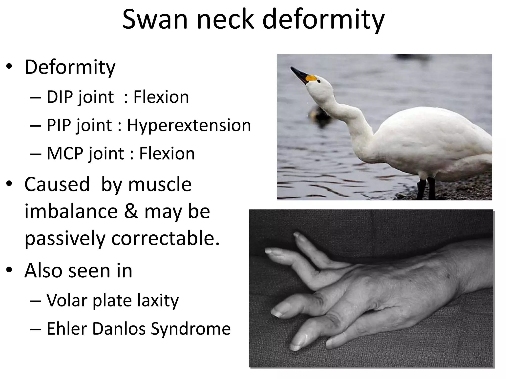
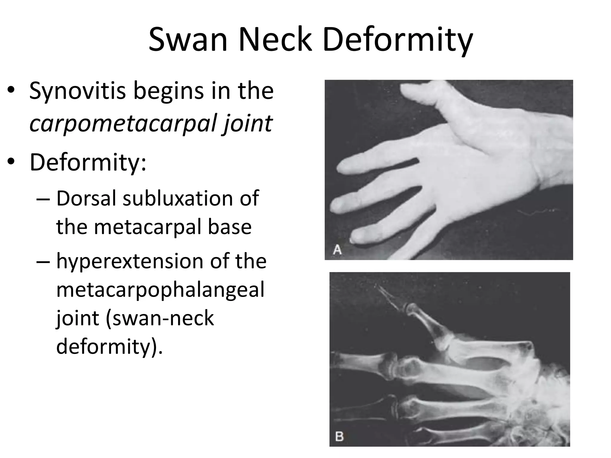
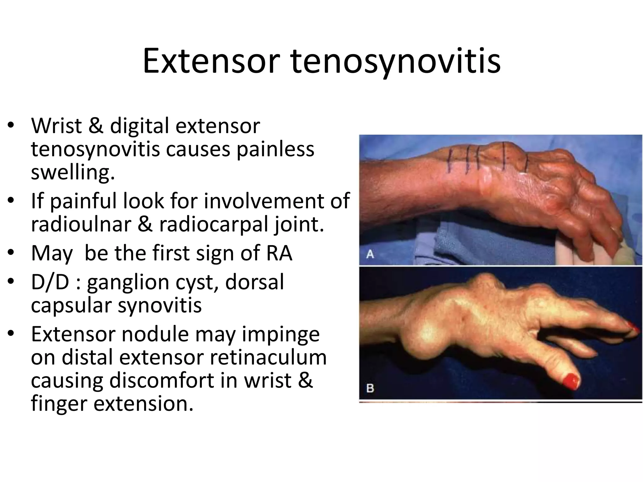
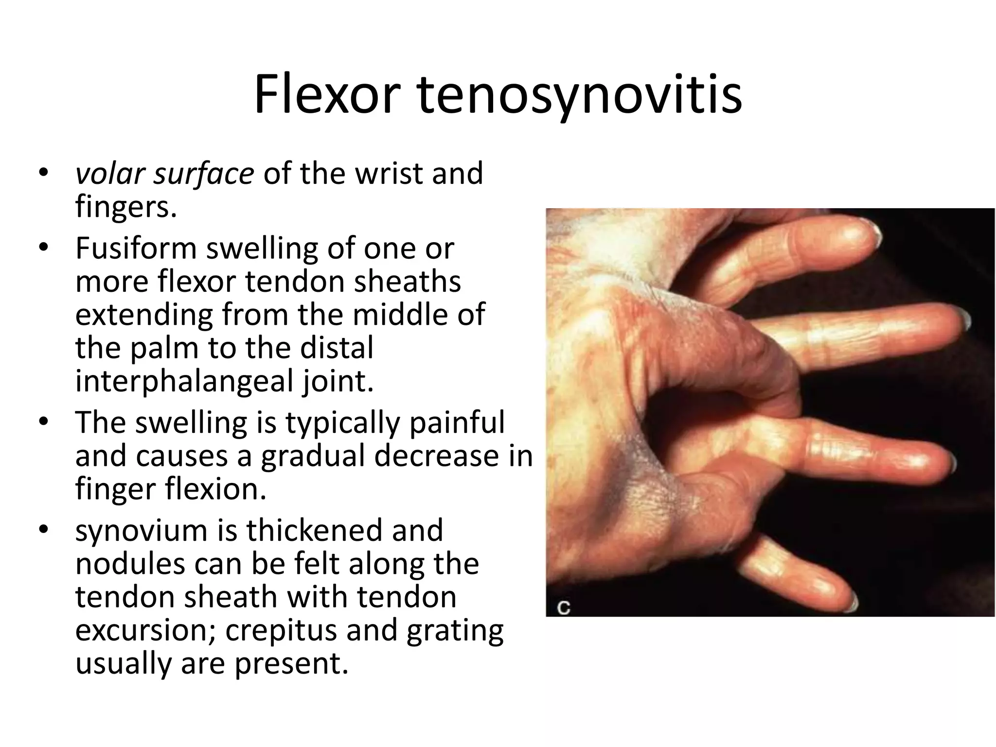
Hand Deformity in Rheumatoid 
Arthritis 
Dr Sushil Sharma 
First Year Orthopedic Resident
Introduction 
• Rheumatoid arthritis (RA) is the most common cause 
of chronic inflammatory joint disease. 
• Most typical features are a 
– symmetrical polyarthritis 
– tenosynovitis 
– morning stiffness, elevation of the erythrocyte 
sedimentation rate (ESR) 
– autoantibodies that target immunoglobulins (rheumatoid 
factors) in the serum
Stages of RA 
1. Pre Clinical 
2. Synovitis 
3. Destruction 
4. Deformity
Development of Deformity 
• As the disease progresses, the persistent 
inflammation causes joint & tendon 
destruction. 
• Erosion of the articular cartilage, tenosynovitis 
& eventually rupture of tendon occurs. 
• Combination of articular destruction, capsular 
stretching and tendon rupture leads to 
progressive instabilty & deformity of joints.
Deformities of hand 
• Def. of fingers 
• Def of thumb 
• Def. Of wrist 
• Rupture of tendons
Deformity In RA 
• MCP & Wrist affected early 
• IP jts are affected late, typically. 
• MCP- most important jt affecting function in RA. 
• Ulnar deviation & volar subluxation of fingers 
are typical deformities.
FINGER DEFORMITIES CAUSED BY 
RHEUMATOID ARTHRITIS 
• Normal forces applied to damaged joints by 
the extrinsic flexors and extensors 
• Tightness of the intrinsic muscles 
• Displacement of the lateral bands of the 
extensor hood 
• Rupture of the central slip of the hood 
• Rupture of the long extensor or long flexor 
tendons.
Deformity of Fingers 
1.INTRINSIC PLUS DEFORMITY 
2.SWAN NECK DEFORMITY 
3.BUTTON HOLE DEFORMITY 
4.ULNAR DEVIATION
Intrinsic plus deformity 
• Caused by intrinsic muscle 
tightness and contracture. 
• Deformity 
– PIP joint : Extension 
– MCP joint : Flexion 
– Thumb : Adduction 
• Volar subluxation of MCP 
joint & ulnar deviation of 
fingers 
• Bunnell test
Swan neck deformity 
• Deformity 
– DIP joint : Flexion 
– PIP joint : Hyperextension 
– MCP joint : Flexion 
• Caused by muscle 
imbalance & may be 
passively correctable. 
• Also seen in 
– Volar plate laxity 
– Ehler Danlos Syndrome
• Causes: 
– Mallet deformity associated with extensor 
tendon disruption at the DIP 
– Capsular disruption, tightening of the lateral 
bands and central tendon, and adherence of the 
lateral bands at PIP 
– Flexor tenosynovitis
BOUTONNIÈRE DEFORMITY 
(Button hole) 
• Deformity 
– PIP joint : Flexion 
– DIP joint : Hyperextension 
– MP joint : Hyperextension
Patho Anatomy 
• Synovitis of the PIP joint with a stretching out of the 
central slip, forcing the lateral bands to begin subluxate 
volarward 
• Shortening of the oblique retinacular ligaments results 
in hyperextension and limited active flexion of the DIP 
joint. 
• The flexion deformity of the PIP joint is compensated 
by extension of the MCP joint. 
• MCP joint deformity not fixed as the distal two joints.
Nalebuff and Millender Grading 
Grade Deformity PIP joint DIP Joint Radiograph 
Mild •Passively 
correctable 
(Lateral band 
subluxated volarly 
but not adherent) 
•Flexion 
deformity(15) 
•Decreased 
flexion 
Normal 
Moderate •Not correctable 
passively 
•Normal flexor 
tendon function 
•Flexor contracture 
(40) 
•Hyperextension Joint space 
preserved 
Severe •Fixed flexion 
deformity (90) 
•Ankylosis 
•Hyperextension Joint 
destruction
DIP Joint Deformity 
• mallet, hyperflexed 
distal interphalangeal 
joint 
• Due to the rupture of 
extensor slip
Ulnar drift of fingers 
• Due to 
1. metacarpophalangeal joint synovitis that 
weakens the dorsoradial capsular restraints 
2. Loosening of the metacarpophalangeal joint 
collateral ligaments results in decreased stability 
3. stretching of the flexor tunnels that permits even 
more ulnar displacement of the long flexor 
tendon
4. interosseous muscle contracture that causes 
ulnar deviation and proximal interphalangeal 
joint hyperextension as well as 
metacarpophalangeal joint flexion and 
eventually subluxation; 
5. long extensor tendon rupture at the wrist 
level that increases the possibility of 
metacarpophalangeal joint dislocations.
Ulnar Drift - Grades 
• Mild to moderate ulnar drift 
– absence of severely diseased articular surfaces or 
dislocated joints 
• Severe ulnar drift 
– one or more metacarpophalangeal joints have 
dislocated & severely diseased articular surface.
Thumb Deformity - Nalebuff Classification
Boutonniere deformity 
• Synovitis beginning in the 
metacarpophalangeal joint 
frequently leads to a 
boutonnière deformity of 
the thumb. 
• proximal phalanx : 
subluxation 
• metacarpophalangeal joint 
: flexion 
• interphalangeal joint : 
hyperextension
Swan Neck Deformity 
• Synovitis begins in the 
carpometacarpal joint 
• Deformity: 
– Dorsal subluxation of 
the metacarpal base 
– hyperextension of the 
metacarpophalangeal 
joint (swan-neck 
deformity).
Game Keeper’s Thumb 
• Synovitic destruction of the 
capsuloligamentous 
supports on the ulnar side 
of the metacarpophalangeal 
joint 
• Due to laxity of the ulnar 
collateral ligament of the 
metacarpophalangeal joint
Opera Glass Hand 
(La Main En Lorgnette) 
• Arthritis Mutilans of 
Hand 
• Shortening of fingers 
due to destruction of 
phalanges. 
• Excess skin gets folded 
transversely resembling 
‘opera glass’
Wrist Deformity 
• Rheumatoid synovitis in wrist affects 
– Ulnar styloid 
– Ulnar head 
– Mid portion of scaphoid 
• Synovitis stretches ulnar carpal ligamentous 
complex & causes ‘caput ulna syndrome’ 
– Dorsal prominence of distal ulna 
– Supination of carpus 
– Volar subluxation of ECU 
– Radial deviation of wrist
• Synovitis begins in the region of deep volar 
radiocarpal ligament & intercarpal ligament which 
results in volar subluxation of scaphoid. 
• combination of 
– rotatory subluxation of the scaphoid 
– volar subluxation of the ulnar carpus 
– dorsal subluxation of the distal ulna 
relative supination of 
the wrist
• Wrist collapse leads to 
– imbalance of the extensor 
tendons 
– radial shift of the metacarpals 
– ulnar deviation of the fingers 
• untreated, end-stage rheumatoid 
wrist is 
– Dislocated volarward 
– Complete destruction of the 
carpal bones 
– Complete dissociation of the 
radioulnar joint.
Tenosynovitis 
• Rheumatoid arthritis is a disease of the synovium. 
• Tendon sheath involvement is common and may occur 
months before the symptoms of intra-articular disease 
are noted. 
• Common sites 
– Dorsal aspect of wrist 
– Volar aspect of wrist 
– Volar aspect of digits 
• Presentation : 
– Pain 
– Tendon dysfunction 
– Tendon rupture
Extensor tenosynovitis 
• Wrist & digital extensor 
tenosynovitis causes painless 
swelling. 
• If painful look for involvement of 
radioulnar & radiocarpal joint. 
• May be the first sign of RA 
• D/D : ganglion cyst, dorsal 
capsular synovitis 
• Extensor nodule may impinge 
on distal extensor retinaculum 
causing discomfort in wrist & 
finger extension.
Extensor tendon rupture 
• Eventually tenosynovitis leads to tendon rupture 
• Major cause of deformity and disability. 
• Causes 
– Attrition rupture 
– Infiltration of synovium 
– Ischemic rupture 
• Attrition rupture occurs at 
– Distal end of the ulna 
– Lister’s tubercle (pulley for EPL gliding)
• The small finger usually is involved first and 
subsequently the ring (Vaughn-Jackson 
syndrome) and then sequentially more radial 
digital extensors. 
• The long extensor tendon of the thumb, 
because of its tortuous course, frequently 
ruptures at the Lister tubercle, where it angles 
through an enclosed tunnel or pulley.
Flexor tenosynovitis 
• volar surface of the wrist and 
fingers. 
• Fusiform swelling of one or 
more flexor tendon sheaths 
extending from the middle of 
the palm to the distal 
interphalangeal joint. 
• The swelling is typically painful 
and causes a gradual decrease in 
finger flexion. 
• synovium is thickened and 
nodules can be felt along the 
tendon sheath with tendon 
excursion; crepitus and grating 
usually are present.
Flexor tenosynovitis 
• Presentation 
– interferes with finger motion 
– Compresses the median nerve in the carpal tunnel 
– Trigger finger 
– Tendon rupture. 
• Erosion of the volar capsule and ligaments 
over radial osteophytes contribute to flexor 
pollicis longus rupture in the carpal tunnel 
(Mannerfelt lesion).
Flexor tendon rupture 
• Not as common as extensor tendon rupture 
but is much more difficult to treat surgically. 
• Sites: 
– Digit (infiltrative tenosynovitis) 
– Wrist (FPL tendon : Most common tendon to 
rupture) 
• Infiltration, weakening, and eventual rupture 
of the profundus tendons may likewise occur 
and are more obvious and disabling clinically.
Thank You

Hand deformity in rheumatoid arthritis

  • 1.
    Hand Deformity inRheumatoid Arthritis Dr Sushil Sharma First Year Orthopedic Resident
  • 2.
    Introduction • Rheumatoidarthritis (RA) is the most common cause of chronic inflammatory joint disease. • Most typical features are a – symmetrical polyarthritis – tenosynovitis – morning stiffness, elevation of the erythrocyte sedimentation rate (ESR) – autoantibodies that target immunoglobulins (rheumatoid factors) in the serum
  • 3.
    Stages of RA 1. Pre Clinical 2. Synovitis 3. Destruction 4. Deformity
  • 4.
    Development of Deformity • As the disease progresses, the persistent inflammation causes joint & tendon destruction. • Erosion of the articular cartilage, tenosynovitis & eventually rupture of tendon occurs. • Combination of articular destruction, capsular stretching and tendon rupture leads to progressive instabilty & deformity of joints.
  • 5.
    Deformities of hand • Def. of fingers • Def of thumb • Def. Of wrist • Rupture of tendons
  • 6.
    Deformity In RA • MCP & Wrist affected early • IP jts are affected late, typically. • MCP- most important jt affecting function in RA. • Ulnar deviation & volar subluxation of fingers are typical deformities.
  • 7.
    FINGER DEFORMITIES CAUSEDBY RHEUMATOID ARTHRITIS • Normal forces applied to damaged joints by the extrinsic flexors and extensors • Tightness of the intrinsic muscles • Displacement of the lateral bands of the extensor hood • Rupture of the central slip of the hood • Rupture of the long extensor or long flexor tendons.
  • 8.
    Deformity of Fingers 1.INTRINSIC PLUS DEFORMITY 2.SWAN NECK DEFORMITY 3.BUTTON HOLE DEFORMITY 4.ULNAR DEVIATION
  • 9.
    Intrinsic plus deformity • Caused by intrinsic muscle tightness and contracture. • Deformity – PIP joint : Extension – MCP joint : Flexion – Thumb : Adduction • Volar subluxation of MCP joint & ulnar deviation of fingers • Bunnell test
  • 10.
    Swan neck deformity • Deformity – DIP joint : Flexion – PIP joint : Hyperextension – MCP joint : Flexion • Caused by muscle imbalance & may be passively correctable. • Also seen in – Volar plate laxity – Ehler Danlos Syndrome
  • 11.
    • Causes: –Mallet deformity associated with extensor tendon disruption at the DIP – Capsular disruption, tightening of the lateral bands and central tendon, and adherence of the lateral bands at PIP – Flexor tenosynovitis
  • 13.
    BOUTONNIÈRE DEFORMITY (Buttonhole) • Deformity – PIP joint : Flexion – DIP joint : Hyperextension – MP joint : Hyperextension
  • 14.
    Patho Anatomy •Synovitis of the PIP joint with a stretching out of the central slip, forcing the lateral bands to begin subluxate volarward • Shortening of the oblique retinacular ligaments results in hyperextension and limited active flexion of the DIP joint. • The flexion deformity of the PIP joint is compensated by extension of the MCP joint. • MCP joint deformity not fixed as the distal two joints.
  • 16.
    Nalebuff and MillenderGrading Grade Deformity PIP joint DIP Joint Radiograph Mild •Passively correctable (Lateral band subluxated volarly but not adherent) •Flexion deformity(15) •Decreased flexion Normal Moderate •Not correctable passively •Normal flexor tendon function •Flexor contracture (40) •Hyperextension Joint space preserved Severe •Fixed flexion deformity (90) •Ankylosis •Hyperextension Joint destruction
  • 17.
    DIP Joint Deformity • mallet, hyperflexed distal interphalangeal joint • Due to the rupture of extensor slip
  • 18.
    Ulnar drift offingers • Due to 1. metacarpophalangeal joint synovitis that weakens the dorsoradial capsular restraints 2. Loosening of the metacarpophalangeal joint collateral ligaments results in decreased stability 3. stretching of the flexor tunnels that permits even more ulnar displacement of the long flexor tendon
  • 19.
    4. interosseous musclecontracture that causes ulnar deviation and proximal interphalangeal joint hyperextension as well as metacarpophalangeal joint flexion and eventually subluxation; 5. long extensor tendon rupture at the wrist level that increases the possibility of metacarpophalangeal joint dislocations.
  • 21.
    Ulnar Drift -Grades • Mild to moderate ulnar drift – absence of severely diseased articular surfaces or dislocated joints • Severe ulnar drift – one or more metacarpophalangeal joints have dislocated & severely diseased articular surface.
  • 22.
    Thumb Deformity -Nalebuff Classification
  • 23.
    Boutonniere deformity •Synovitis beginning in the metacarpophalangeal joint frequently leads to a boutonnière deformity of the thumb. • proximal phalanx : subluxation • metacarpophalangeal joint : flexion • interphalangeal joint : hyperextension
  • 24.
    Swan Neck Deformity • Synovitis begins in the carpometacarpal joint • Deformity: – Dorsal subluxation of the metacarpal base – hyperextension of the metacarpophalangeal joint (swan-neck deformity).
  • 25.
    Game Keeper’s Thumb • Synovitic destruction of the capsuloligamentous supports on the ulnar side of the metacarpophalangeal joint • Due to laxity of the ulnar collateral ligament of the metacarpophalangeal joint
  • 26.
    Opera Glass Hand (La Main En Lorgnette) • Arthritis Mutilans of Hand • Shortening of fingers due to destruction of phalanges. • Excess skin gets folded transversely resembling ‘opera glass’
  • 27.
    Wrist Deformity •Rheumatoid synovitis in wrist affects – Ulnar styloid – Ulnar head – Mid portion of scaphoid • Synovitis stretches ulnar carpal ligamentous complex & causes ‘caput ulna syndrome’ – Dorsal prominence of distal ulna – Supination of carpus – Volar subluxation of ECU – Radial deviation of wrist
  • 28.
    • Synovitis beginsin the region of deep volar radiocarpal ligament & intercarpal ligament which results in volar subluxation of scaphoid. • combination of – rotatory subluxation of the scaphoid – volar subluxation of the ulnar carpus – dorsal subluxation of the distal ulna relative supination of the wrist
  • 29.
    • Wrist collapseleads to – imbalance of the extensor tendons – radial shift of the metacarpals – ulnar deviation of the fingers • untreated, end-stage rheumatoid wrist is – Dislocated volarward – Complete destruction of the carpal bones – Complete dissociation of the radioulnar joint.
  • 30.
    Tenosynovitis • Rheumatoidarthritis is a disease of the synovium. • Tendon sheath involvement is common and may occur months before the symptoms of intra-articular disease are noted. • Common sites – Dorsal aspect of wrist – Volar aspect of wrist – Volar aspect of digits • Presentation : – Pain – Tendon dysfunction – Tendon rupture
  • 31.
    Extensor tenosynovitis •Wrist & digital extensor tenosynovitis causes painless swelling. • If painful look for involvement of radioulnar & radiocarpal joint. • May be the first sign of RA • D/D : ganglion cyst, dorsal capsular synovitis • Extensor nodule may impinge on distal extensor retinaculum causing discomfort in wrist & finger extension.
  • 32.
    Extensor tendon rupture • Eventually tenosynovitis leads to tendon rupture • Major cause of deformity and disability. • Causes – Attrition rupture – Infiltration of synovium – Ischemic rupture • Attrition rupture occurs at – Distal end of the ulna – Lister’s tubercle (pulley for EPL gliding)
  • 33.
    • The smallfinger usually is involved first and subsequently the ring (Vaughn-Jackson syndrome) and then sequentially more radial digital extensors. • The long extensor tendon of the thumb, because of its tortuous course, frequently ruptures at the Lister tubercle, where it angles through an enclosed tunnel or pulley.
  • 35.
    Flexor tenosynovitis •volar surface of the wrist and fingers. • Fusiform swelling of one or more flexor tendon sheaths extending from the middle of the palm to the distal interphalangeal joint. • The swelling is typically painful and causes a gradual decrease in finger flexion. • synovium is thickened and nodules can be felt along the tendon sheath with tendon excursion; crepitus and grating usually are present.
  • 36.
    Flexor tenosynovitis •Presentation – interferes with finger motion – Compresses the median nerve in the carpal tunnel – Trigger finger – Tendon rupture. • Erosion of the volar capsule and ligaments over radial osteophytes contribute to flexor pollicis longus rupture in the carpal tunnel (Mannerfelt lesion).
  • 37.
    Flexor tendon rupture • Not as common as extensor tendon rupture but is much more difficult to treat surgically. • Sites: – Digit (infiltrative tenosynovitis) – Wrist (FPL tendon : Most common tendon to rupture) • Infiltration, weakening, and eventual rupture of the profundus tendons may likewise occur and are more obvious and disabling clinically.
  • 38.