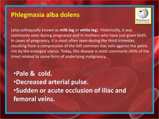
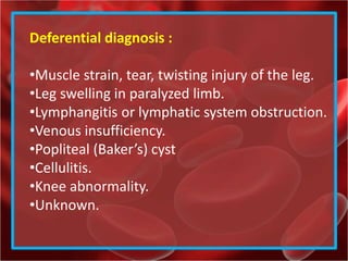
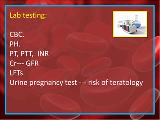
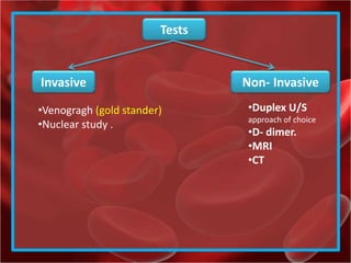
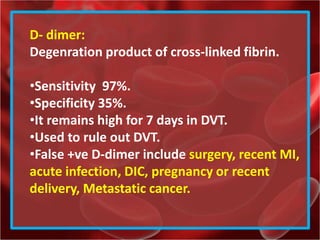
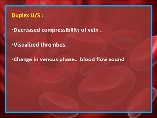
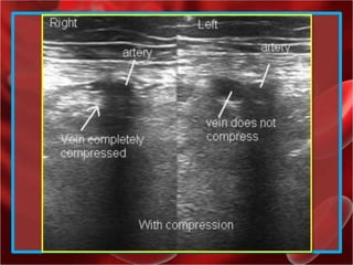
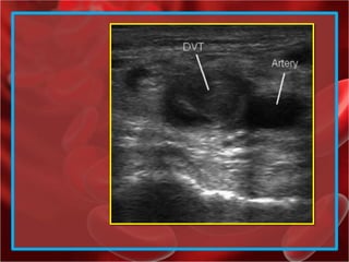
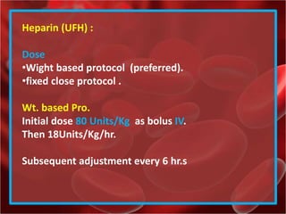
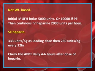
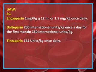
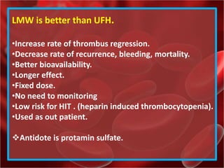
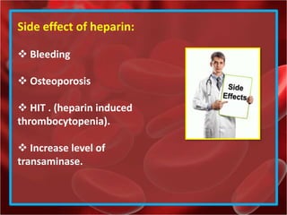
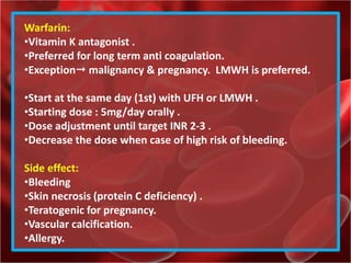
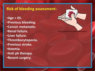
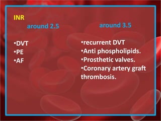
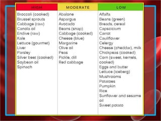
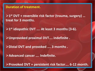
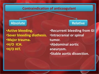
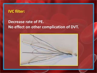
Deep vein thrombosis
Done by:
Mohammed A Qazzaz
Definition :
Formation of a blood clot in one of the deep
veins of the body.
Deep veins of the body:
•Leg
•Arm
•Pelvis
VTE
DVT and pulmonary embolism
DVT of the lower limb.
•Distal / calf
•Proximal / popliteal, femoral, iliac veins
That was an old classification. But now its no
longer used…
The used to treat just the proximal DVT.. But
now we treat both.
PE:
•< 2% orginated from the upper arm DVT
•90% is from the leg DVT
Incidence:
2-3 / 1000 .
Men > women
>45 year old.
Risk Factors :
hypercoaguilable
Venous stasis :
Advanced age.
obesity.
Immobilization
paralysis.
Pregnancy & post partum.
Endothelial Injury:
Surgery .
Catherter
Trauma.
vasculits
Virchow's triad
Hypercoaguliablity:
Cancer- chemotherapy.
Estrogen / OCP
Nephrotic syndrome.
Sepsis
HRT
Antiphospholibid
Hyperhomocystinuria.
Thrombophilia
•Ant thrombin deficiency.
•Protein C deficiency.
•Protein S Deficiency.
•Factor V Leiden.
Signs:
Calf tenderness.
Pitting Edema.
Circumferences increased > 3cm.
Temperature.
Superficial venous dilatation.
Homan’s sign .
Pratt’s sign.
Search for stigmata of PE
examin for signs of underlying
factors.
Symptoms:
Swelling .
Pain .
Redness/ erythema.
Phlegmasia alba dolens
(also colloquially known as milk leg or white leg). Historically, it was
commonly seen during pregnancy and in mothers who have just given birth.
In cases of pregnancy, it is most often seen during the third trimester,
resulting from a compression of the left common iliac vein against the pelvic
rim by the enlarged uterus. Today, this disease is most commonly (40% of the
time) related to some form of underlying malignancy.
•Pale & cold.
•Decreased arterial pulse.
•Sudden or acute occlusion of iliac and
femoral veins.
Phlegmasia cerulea dolens
(literally: painful blue edema) is an uncommon severe form of deep venous
thrombosis which results from extensive thrombotic occlusion (blockage by
a thrombus) of the major and the collateral veins of an extremity. it is
characterized by sudden severe pain, swelling, cyanosis and edema of the
affected limb. There is a high risk of massive pulmonary embolism, even
under anticoagulation. Foot gangrene may also occur. An underlying
malignancy is found in 50% of cases. Usually, it occurs in those afflicted by a
life-threatening illness.
•Sever leg pain, swelling, cyanosis, edema.
•Venous gangrene
•Compartment syndrome.
•circulation collapse and shock .
•PE.
Deferential diagnosis :
•Muscle strain, tear, twisting injury of the leg.
•Leg swelling in paralyzed limb.
•Lymphangitis or lymphatic system obstruction.
•Venous insufficiency.
•Popliteal (Baker’s) cyst
•Cellulitis.
•Knee abnormality.
•Unknown.
Lab testing:
CBC.
PH.
PT, PTT, INR
Cr--- GFR
LFTs
Urine pregnancy test --- risk of teratology
Tests
Invasive Non- Invasive
•Duplex U/S
approach of choice
•D- dimer.
•MRI
•CT
•Venogragh (gold stander)
•Nuclear study .
D- dimer:
Degenration product of cross-linked fibrin.
•Sensitivity 97%.
•Specificity 35%.
•It remains high for 7 days in DVT.
•Used to rule out DVT.
•False +ve D-dimer include surgery, recent MI,
acute infection, DIC, pregnancy or recent
delivery, Metastatic cancer.
Duplex U/S :
•Decreased compressibility of vein .
•Visualized thrombus.
•Change in venous phase… blood flow sound
Venogragh :
•Gold stander.
•When U/S –ve + high probability . Use it.
•Side effect.
Phlebitis.
Anaphylaxis.
MRI :
•Detect legs, pelvis, pulmonary thrombi.
•Sensitivity and spasticity high
Venogragh
Treatment :
Selection of agents
•LMW heparin
•UF heparin.
•Fondaprinux.
•Oral Factor X inhibitors.
•Oral direct thrombin inhibitor.
Heparin (UFH) :
Dose
•Wight based protocol (preferred).
•fixed close protocol .
Wt. based Pro.
Initial dose 80 Units/Kg as bolus IV.
Then 18Units/Kg/hr.
Subsequent adjustment every 6 hr.s
Not Wt. based.
Initial IV UFH bolus 5000 units. Or 10000 if PE
Then continous IV heparine 2000 units per hour.
SC heparin.
333 units/Kg as loading dose then 250 units/Kg
every 12hr
Check the APPT daily 4-6 hours after dose of
heparin.
LMW:
SC.
Enoxaparin 1mg/Kg q 12 hr. or 1.5 mg/Kg once daily.
Dalteparin 200 international units/kg once a day for
the first month; 150 international units/kg.
Tinzaparin 175 Units/kg once daily.
LMW is better than UFH.
•Increase rate of thrombus regression.
•Decrease rate of recurrence, bleeding, mortality.
•Better bioavailability.
•Longer effect.
•Fixed dose.
•No need to monitoring
•Low risk for HIT . (heparin induced thrombocytopenia).
•Used as out patient.
Antidote is protamin sulfate.
Side effect of heparin:
 Bleeding
 Osteoporosis
 HIT . (heparin induced
thrombocytopenia).
 Increase level of
transaminase.
Warfarin:
•Vitamin K antagonist .
•Preferred for long term anti coagulation.
•Exception malignancy & pregnancy. LMWH is preferred.
•Start at the same day (1st) with UFH or LMWH .
•Starting dose : 5mg/day orally .
•Dose adjustment until target INR 2-3 .
•Decrease the dose when case of high risk of bleeding.
Side effect:
•Bleeding
•Skin necrosis (protein C deficiency) .
•Teratogenic for pregnancy.
•Vascular calcification.
•Allergy.
Risk of bleeding assessment:
•Age > 65.
•Previous bleeding.
•Cancer metastatic.
•Renal failure.
•Liver failure.
•Thrombocytopenia.
•Previous stroke.
•Anemia.
•Anti ph therapy.
•Recent surgery.
INR
around 2.5
•DVT
•PE
•AF
around 3.5
•recurrent DVT
•Anti phospholipids.
•Prosthetic valves.
•Coronary artery graft
thrombosis.
Warfarin:
•Vitamin K antagonist . Avoid rich food of Vit K.
•Avoid drugs that interact with warfarin.
•Avoid the IM injection.
•Tell your surgeon about your warfarin thereby.
Duration of treatment.
1st DVT + reversible risk factor (trauma, surgery) …
treat for 3 months.
1st idiopathic DVT …. At least 3 months (3-6).
Unprovoked proximal DVT…. indefinite .
Distal DVT and provoked …. 3 months .
Advanced cancer …. Indefinite.
Provoked DVT + persistent risk factor…. 6-12 month.
Contraindication of anticoagulant
Absolute Relative
•Recurrent bleeding from GI
•Intracranial or spinal
tumor.
•Abdominal aortic
aneurysm.
•Stable aortic dissection.
•Active bleeding.
•Sever bleeding diathesis.
•Major trauma.
•H/O ICH.
•H/O HIT.
IVC filter:
Decrease rate of PE.
No effect on other complication of DVT.
DVT.. Deep vein thrombosis.

DVT.. Deep vein thrombosis.

  • 1.
    Deep vein thrombosis Doneby: Mohammed A Qazzaz
  • 2.
    Definition : Formation ofa blood clot in one of the deep veins of the body. Deep veins of the body: •Leg •Arm •Pelvis VTE DVT and pulmonary embolism
  • 3.
    DVT of thelower limb. •Distal / calf •Proximal / popliteal, femoral, iliac veins That was an old classification. But now its no longer used… The used to treat just the proximal DVT.. But now we treat both.
  • 4.
    PE: •< 2% orginatedfrom the upper arm DVT •90% is from the leg DVT Incidence: 2-3 / 1000 . Men > women >45 year old.
  • 5.
    Risk Factors : hypercoaguilable Venousstasis : Advanced age. obesity. Immobilization paralysis. Pregnancy & post partum. Endothelial Injury: Surgery . Catherter Trauma. vasculits Virchow's triad
  • 6.
    Hypercoaguliablity: Cancer- chemotherapy. Estrogen /OCP Nephrotic syndrome. Sepsis HRT Antiphospholibid Hyperhomocystinuria. Thrombophilia •Ant thrombin deficiency. •Protein C deficiency. •Protein S Deficiency. •Factor V Leiden.
  • 7.
    Signs: Calf tenderness. Pitting Edema. Circumferencesincreased > 3cm. Temperature. Superficial venous dilatation. Homan’s sign . Pratt’s sign. Search for stigmata of PE examin for signs of underlying factors.
  • 8.
  • 10.
    Phlegmasia alba dolens (alsocolloquially known as milk leg or white leg). Historically, it was commonly seen during pregnancy and in mothers who have just given birth. In cases of pregnancy, it is most often seen during the third trimester, resulting from a compression of the left common iliac vein against the pelvic rim by the enlarged uterus. Today, this disease is most commonly (40% of the time) related to some form of underlying malignancy. •Pale & cold. •Decreased arterial pulse. •Sudden or acute occlusion of iliac and femoral veins.
  • 11.
    Phlegmasia cerulea dolens (literally:painful blue edema) is an uncommon severe form of deep venous thrombosis which results from extensive thrombotic occlusion (blockage by a thrombus) of the major and the collateral veins of an extremity. it is characterized by sudden severe pain, swelling, cyanosis and edema of the affected limb. There is a high risk of massive pulmonary embolism, even under anticoagulation. Foot gangrene may also occur. An underlying malignancy is found in 50% of cases. Usually, it occurs in those afflicted by a life-threatening illness. •Sever leg pain, swelling, cyanosis, edema. •Venous gangrene •Compartment syndrome. •circulation collapse and shock . •PE.
  • 12.
    Deferential diagnosis : •Musclestrain, tear, twisting injury of the leg. •Leg swelling in paralyzed limb. •Lymphangitis or lymphatic system obstruction. •Venous insufficiency. •Popliteal (Baker’s) cyst •Cellulitis. •Knee abnormality. •Unknown.
  • 13.
    Lab testing: CBC. PH. PT, PTT,INR Cr--- GFR LFTs Urine pregnancy test --- risk of teratology
  • 14.
    Tests Invasive Non- Invasive •DuplexU/S approach of choice •D- dimer. •MRI •CT •Venogragh (gold stander) •Nuclear study .
  • 15.
    D- dimer: Degenration productof cross-linked fibrin. •Sensitivity 97%. •Specificity 35%. •It remains high for 7 days in DVT. •Used to rule out DVT. •False +ve D-dimer include surgery, recent MI, acute infection, DIC, pregnancy or recent delivery, Metastatic cancer.
  • 16.
    Duplex U/S : •Decreasedcompressibility of vein . •Visualized thrombus. •Change in venous phase… blood flow sound
  • 19.
    Venogragh : •Gold stander. •WhenU/S –ve + high probability . Use it. •Side effect. Phlebitis. Anaphylaxis. MRI : •Detect legs, pelvis, pulmonary thrombi. •Sensitivity and spasticity high
  • 20.
  • 21.
    Treatment : Selection ofagents •LMW heparin •UF heparin. •Fondaprinux. •Oral Factor X inhibitors. •Oral direct thrombin inhibitor.
  • 22.
    Heparin (UFH) : Dose •Wightbased protocol (preferred). •fixed close protocol . Wt. based Pro. Initial dose 80 Units/Kg as bolus IV. Then 18Units/Kg/hr. Subsequent adjustment every 6 hr.s
  • 23.
    Not Wt. based. InitialIV UFH bolus 5000 units. Or 10000 if PE Then continous IV heparine 2000 units per hour. SC heparin. 333 units/Kg as loading dose then 250 units/Kg every 12hr Check the APPT daily 4-6 hours after dose of heparin.
  • 24.
    LMW: SC. Enoxaparin 1mg/Kg q12 hr. or 1.5 mg/Kg once daily. Dalteparin 200 international units/kg once a day for the first month; 150 international units/kg. Tinzaparin 175 Units/kg once daily.
  • 25.
    LMW is betterthan UFH. •Increase rate of thrombus regression. •Decrease rate of recurrence, bleeding, mortality. •Better bioavailability. •Longer effect. •Fixed dose. •No need to monitoring •Low risk for HIT . (heparin induced thrombocytopenia). •Used as out patient. Antidote is protamin sulfate.
  • 26.
    Side effect ofheparin:  Bleeding  Osteoporosis  HIT . (heparin induced thrombocytopenia).  Increase level of transaminase.
  • 27.
    Warfarin: •Vitamin K antagonist. •Preferred for long term anti coagulation. •Exception malignancy & pregnancy. LMWH is preferred. •Start at the same day (1st) with UFH or LMWH . •Starting dose : 5mg/day orally . •Dose adjustment until target INR 2-3 . •Decrease the dose when case of high risk of bleeding. Side effect: •Bleeding •Skin necrosis (protein C deficiency) . •Teratogenic for pregnancy. •Vascular calcification. •Allergy.
  • 28.
    Risk of bleedingassessment: •Age > 65. •Previous bleeding. •Cancer metastatic. •Renal failure. •Liver failure. •Thrombocytopenia. •Previous stroke. •Anemia. •Anti ph therapy. •Recent surgery.
  • 29.
    INR around 2.5 •DVT •PE •AF around 3.5 •recurrentDVT •Anti phospholipids. •Prosthetic valves. •Coronary artery graft thrombosis.
  • 30.
    Warfarin: •Vitamin K antagonist. Avoid rich food of Vit K. •Avoid drugs that interact with warfarin. •Avoid the IM injection. •Tell your surgeon about your warfarin thereby.
  • 33.
    Duration of treatment. 1stDVT + reversible risk factor (trauma, surgery) … treat for 3 months. 1st idiopathic DVT …. At least 3 months (3-6). Unprovoked proximal DVT…. indefinite . Distal DVT and provoked …. 3 months . Advanced cancer …. Indefinite. Provoked DVT + persistent risk factor…. 6-12 month.
  • 34.
    Contraindication of anticoagulant AbsoluteRelative •Recurrent bleeding from GI •Intracranial or spinal tumor. •Abdominal aortic aneurysm. •Stable aortic dissection. •Active bleeding. •Sever bleeding diathesis. •Major trauma. •H/O ICH. •H/O HIT.
  • 35.
    IVC filter: Decrease rateof PE. No effect on other complication of DVT.