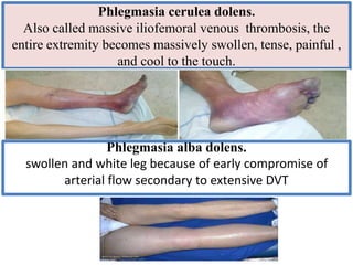
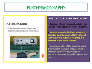
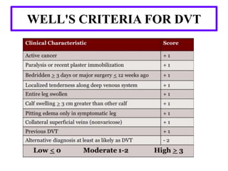
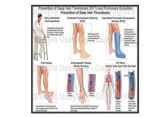
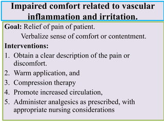
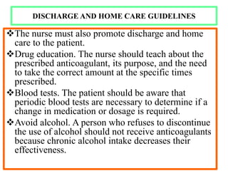
MANAGEMENT OF PATIENTS
WITH
DEEP- VEIN -THROMBOSIS &
ANEURYSMS
INTRODUCTION
DEEP VEINTHROMBOSIS
• Formation of a blood clot in one of the deep veins
of the body, usually in the leg.
EPIDEMIOLOGY
 7% diagnosed and treated.
 34% sudden pulmonary embolism.
 59% as undetected Venous thromboembolism related
deaths 3,00,000/annum.
INCIDENCE:-
 An annual incidence of symptomatic Venous
thromboembolism as 17.46 per 100,00 admissions
Without prophylaxis the incidence of deep vein thrombosis is
about
14% in gynecological surgery,
22% in neurosurgery.
26% in abdominal surgery.
45%-60% in patients undergoing hip and knee surgeries,
15% to 40% Urologic surgery.
RISK FACTORS
VIRCHOW TRIAD
HYPERCOAGULABLE STATE OF
MALIGNANCY
• Up to 15% of cancer patients presents with VTE.
• VTE is not equally common in all types of cancer.
• Highest incidence is found in mucin-producing
adenocarcinomas, pancreas and gastrointestinal
tract, lung cancer, and ovarian cancer.
When legs are
inactive or calf
muscle pump is
ineffective blood
pools by gravity
Platelet
aggregation
including
thrombin,fibrin,
catecholamines
PATHOPHYSIOLOGY
Platelets
+collagen=ADP
ADP helps
formation of
platelet plug
called thrombi
Size vary from 1
mm to long
tubular masses
filling main veins
If large thrombi
obstructs major vein
,venous pressure and
volume increases
Thrombi can
travel and can
lead to
pulmonary
embolism
If thrombi in the
small veins
,collateral venous
channels relieve
increased pressure
and volume
Inflammator
y process
destroy
valves of the
veins-venous
insufficiency
PATHOPHYSIOLOGY
CLINICAL MANIFESTATIONS
• Swelling with pitting
edema
• Calf pain & Tenderness
• Foot gangrene
• Superficial venous
dilatation
Phlegmasia cerulea dolens.
Also called massive iliofemoral venous thrombosis, the
entire extremity becomes massively swollen, tense, painful ,
and cool to the touch.
Phlegmasia alba dolens.
swollen and white leg because of early compromise of
arterial flow secondary to extensive DVT
Venous gangrene
• Hydrostatic pressure in arterial and venous capillaries exceeds
• fluid sequestration in the interstitium.
• Circulatory shock, and arterial insufficiency which causes gangrene.
40-60% also have capillary involvement irreversible
venous gangrene.
PHYSICAL EXAMINATION
• Palpate distal pulses and evaluate
capillary refill to assess limb perfusion.
• Move and palpate all joints to detect
acute arthritis or other joint
pathology.
• Neurologic evaluation may detect
 Nerve root irritation
Sensory, motor, and reflex deficits
should be noted.
Homan’s sign: Pain in the posterior calf or
knee with forced dorsiflexion of the foot.
MOSES’S SIGN
• Neuhof’s sign -Thickening and deep
tenderness elicited while palpating deep in
calf muscles
• Lintons sign: After applying torniquet at
saphenofemoral junction patient made to
walk. then limb is elevated in supine
position prominent superficial veins will be
observed Search for signs of PE such as
tachycardia, tachypnoea and chest findings
(rare).
DIAGNOSTIC STUDIES
– Clinical examination alone is able to confirm only
20-30% of cases of DVT.
– Blood Tests:
D-dimer: It is specific degradation product of
cross-linked fibrin. Because concurrent
production and breakdown of clot characterize
thrombosis, patients with thromboembolism
disease have elevated levels of D-dimer.
– Imaging Studies
IMAGING STUDIES
INVASIVE
Venography
Radio-labeled fibrinogen.
NONINVASIVE
Ultrasound
Plethysmography
VENOGRAPHY
ADVANTAGES:
• It is useful if the patient has a high clinical
probability of thrombosis and a negative
ultrasound
• It is also valuable in symptomatic patients
with a history of prior thrombosis in whom
the ultrasound is non-diagnostic
DISADVANTAGE:
• It can primary cause of DVT in 3% of
patients who undergo this diagnostic
procedure.
• An invasive and expensive.
• Although Venography was once considered
the gold standard for diagnosis of DVT,
today it is more commonly used in research
environments and less frequently utilized in
clinical practice.
NUCLEAR MEDICINE STUDIES
• Because the radioactive
isotope incorporates into a
growing thrombus, this test
can distinguish new clot
from an old clot.
• Nuclear medicine studies
done with iodine 125-
labeled fibrinogen.
• More commonly used in
research
PLETHYSMOGRAPHY
IMPEDANCE PLETHYSMOGRAPHY:
 Principle- Blood volume changes in the
leg lead to changes in electrical resistance.
 Venous return in the lower extremity is
occluded by inflation of a thigh cuff, and
then the cuff is released, resulting in a
decrease in calf blood volume.
 Any obstruction of the proximal veins
diminishes the volume change, which is
detected by measuring changes in
electrical resistance (impedance) over the
calf.
ULTRASONOGRAPHY
• Colour- flow Duplex
scanning is the imaging
test of choice for
patients with suspected
DVT.
• Ultrasound can also
distinguish other causes
of leg swelling, such as
tumor, popliteal cyst,
abscess, aneurysm, or
hematoma.
MAGNETIC RESONANCE IMAGING
• It detects leg. pelvis, and pulmonary
thrombi and is 97% sensitive and
95% specific for DVT.
• It distinguishes a mature from an
immature clot.
• MRI is safe in all stages of
pregnancy.
• Test may not be appropriate for
patients with pacemakers or other
metallic implants, it can be an
effective diagnostic option for some
patients
WELL'S CRITERIA FOR DVT
MANAGEMENT
EMERGENCY DEPARTMENT CARE
The primary objectives of the treatment of DVT are to
• Prevent pulmonary embolism
• Reduce morbidity, and
• Prevent or minimize the risk of developing the post phlebitis
syndrome
GENERAL THERAPEUTIC MEASURES:
 Bed rest
 Encourage the patient to perform gentle foot exercises every hour,
 Increase fluid intake up to 2L/day unless contraindicated
 Avoid deep palpation
SPECIFIC MANAGEMENT:
 It further includes
 Pharmacological Therapy
 Thrombolytic therapy
 Endovascular Surgery for DVT.
PHARMACOLOGICALTHERAPY
UNFRACTIONED HEPARIN:
Heparin prevents extension of the
thrombus
MECHANISM OF ACTION:
Heparins anticoagulant effect is
related directly to its activation of
antithrombin III. Antithrombin III,
the body's primary anticoagulant,
inactivates thrombin and inhibits the
activity of activated factor X. factor
IX in the coagulation process
SIDE EFFECTS:.
•Bleeding.
•Osteoporosis.
•Thrombocytopenia
•Skin lesions-
urticaria, papules,
necrosis.
•Hypoaldosteronism,
hyperkalemia
CONTRAINDICATIONS
Bleeding disorders, Severe
hypertension , Threatened abortion
DOSE:- IV bolus dose of 5.000 to
10,000 units followed by an
infusion of 1000 units per hour
other method of initiating therapy
is to begin with, loading dose of
50-100 units of heparin followed
by a constant infusion of 1525
units/hr.
LOW MOLECULAR WEIGHT HEPARIN
Selectively inhibit factor X
Superior bioavailability
Superior or equivalent safety and efficacy
Subcutaneous once or twice daily dosing
No laboratory monitoring
Les phlebotomy (no monitoring/no intravenous
line).
WARFARIN
This vitamin K antagonist prevents carboxylation activation of coagulation factors II, VII, IX,
and Х
The full effect of warfarin requires at least 5 days, even if the prothrombin time used for
monitoring, becomes elevated more rapidly.
 Warfarin Dosing in an average-size adult, warfarin is often initiated in a dose of 5 mg.
The prothrombin time is standardized by calculating the international normalized ratio (INR)
which assesses the anticoagulant effect of warfarin.The target INR is usually 2.5, with a range of 2.0-
3.0. The warfarin dose is usually titrated empirically to achieve the target INR.
ANTIFACTOR Xa
 Fondaparinux, an anti-Xa Penta saccharide, is administered as a weight-based once-
daily subcutaneous injection in a prefilled syringe.
 No laboratory monitoring is required.
 Fondaparinux is synthesized in a laboratory and, unlike LMWH or UFH, is not derived
from animal products,
 It does not cause heparin-induced thrombocytopenia.
 The dose must be adjusted downward for patients with renal dysfunction.
INDICATIONS:-
• When anticoagulant therapy is ineffective, unsafe, or
contraindicated.
• The major surgical procedures for DVT are clot removal and
partial interruption of the inferior vena cava to prevent
pulmonary embolism
ENDOVASCULAR SURGERY FOR DVT
CATHETER DIRECTED THROMBOLYSIS
 In this procedure, surgeon injects clot
dissolving drops through a catheter directly
into the clot.
 Successful clot lysis in 85%
 Better 1-yr patency,
 Long-term symptom resolution.
OPEN THROMBECTOMY:
 It is occasionally recommended, also called as
venous thrombectomy
FILTERS FOR DVT
• INDICATIONS:
 Contraindication to anticoagulation
 Significant bleeding complication of
anticoagulation therapy.
 Pulmonary embolism with contraindication to
anticoagulation
 Recurrent thrombo-embolic complication
despite adequate anticoagulation therapy.
 Inferior vena cava filters reduce the rate of
pulmonary embolism but have no effect on the
other complications of deep vein thrombosis.
Thrombolysis should be considered in patients
with major proximal vein thrombosis and
threatened venous infarction.
PREVENTION OF DVT
PROPHYLAXIS:
Indicated in who underwent major abdominal trauma or
orthopedic surgery or patient having prolonged
immobilization (> 3 days).
BENEFITS OF VTE PROPHYLAXIS.
 Improved patient outcomes.
 Reduced costs.
METHODS OF DVT PREVENTION:
– Ambulation early and often (simplest and most cost effective
means to reduce risk of DVT)
– Mechanical methods like Intermittent External Compression
Devices also called sequential compression devices or SCDs.
They increase rate/velocity of venous blood and reduce pooling in
the peripheral veins. Compression should begin pre-operatively
and be continued until the patient is fully ambulatory
– Pharmacological therapy.
• Deep vein thrombosis can be prevented, especially if
patients who are considered high risk are identified
and preventive measures are instituted without
delay.
• Graduated compression stockings. Compression
stockings prevent dislodgement of the thrombus.
• Pneumatic compression device. Intermittent
pneumatic compression devices increase blood
velocity beyond that produced by the stockings.
• Leg exercises. Encourage early mobilization and
leg exercises to keep the blood circulating
adequately
COMPLICATION
• Bleeding.
• Thrombocytopenia.
• Drug interactions.
NURSING DIAGNOSIS AND INTERVENTION
NURSING ASSESSMENT:
• Assessment of a patient with deep vein thrombosis
include:
– Presenting signs and symptoms. If a patient present with
signs and symptoms of DVT, carry out an assessment of
general medical history and a physical examination to
exclude other causes.
– Wells diagnostic algorithm. Because of the unreliability of
clinical features, Well's diagnostic algorithm has been
validated whereby patients are classified as having a high,
intermediate, or low probability of developing DVT.
ASSESSING AND MONITORING ANTICOAGULANT THERAPY
 Haemorrhage, continuous intravenous infusion by electronic infusion device
is the preferred method of administering un fractionated heparin.
 Dosage calculations are based on the patient's weight, and any possible
bleeding tendencies are detected by a pre treatment clotting profile
 Periodic coagulation tests and haematocrit levels are obtained. Heparin is in
the effective, or therapeutic ,range when the partial thromboplastin time is
1.5 times the control.
 Intermittent intravenous injection is another means of administering heparin;
a dilute solution of heparin is administered every 4 hours. Administration
may be facilitated by using a heparin lock, an intravenous catheter or a
small, butterfly-type scalp vein needle with an injection site at the end of the
tubing.
 Oral anticoagulants, such as warfarin, are monitored by the prothrombin time or inr.
Because their effect is delayed for 3 to 5 days, they are usually administered with
 Heparin until desired anticoagulation has been achieved (ie, when the prothrombin
time is 1.5 to 2 times normal or the INR is 2.0 to 3.0)
Ineffective tissue perfusion related to
interruption of venous blood flow
Goal: Demonstrate increased perfusion as individually
appropriate.
• Interventions:
– Provide comfort.
– Elevation of the affected extremity.
– Graduated compression stockings.
– Discourage standing still or sitting for prolonged periods.
– Maintain warm temperature and avoid chilling.
– Counsel in ways to avoid emotional upsets; stress
management,
– Encourage avoidance of leg crossing.
Impaired comfort related to vascular
inflammation and irritation.
Goal: Relief of pain of patient.
Verbalize sense of comfort or contentment.
Interventions:
1. Obtain a clear description of the pain or
discomfort.
2. Warm application, and
3. Compression therapy
4. Promote increased circulation,
5. Administer analgesics as prescribed, with
appropriate nursing considerations
• Risk for impaired skin integrity related to compromised
circulation.
Goal: Attainment/maintenance of tissue integrity. Maintain position of
function and skin integrity as evidenced by absence of contractures,
footdrop, decubitus, and so forth.
Interventions:Instruct in ways to avoid trauma to extremities.
• Encourage meticulous hygiene; bathing with neutral soaps, applying
lotions, carefully trimming nails.
Deficient knowledge regarding pathophysiology of condition related
to lack of information and misinterpretation
Goal: Verbalize understanding of condition, therapy, regimen, side
effects of medications, and when to contact the healthcare provider.
Interventions: Include family/significant others in teaching program.
– Provide written instructions about foot care, leg care, and exercise
program.
– Assist to obtain properly fitting clothing, shoes, stockings.
MONITORING AND MANAGING POTENTIAL COMPLICATIONS OF
HEPARIN THERAPY :
BLEEDING:
Bleeding from the
kidneys is detected by
microscopic
examination of the
urine
 Bruises, nosebleeds,
and bleeding gums are
also early signs.
To reverse the effects
of heparin promptly,
intravenous injections
of protamine sulfate
may be administered.
THROMBOCYTOPENI
A
•It may develop in
patients who receive
heparin for more than
5 days or on re
administration after a
brief interruption of
heparin therapy.
•Beginning warfarin
concomitantly with
heparin can provide a
stable INR or
prothrombin time by
day 5 of heparin
treatment.
DRUG INTERACTIONS:
•Herbal and nutritional
supplements
• salicylates, anabolic steroids,
chloral hydrate, glucagon,
chloramphenicol, neomycin,
quinidine, phenylbutazone
(Butazolidin), coenzyme ,
garlic, and vitamin E; those that
decrease the anticoagulant
effect include phenytoin,
barbiturates, diuretics, estrogen,
and vitamin C.
DISCHARGE AND HOME CARE GUIDELINES
The nurse must also promote discharge and home
care to the patient.
Drug education. The nurse should teach about the
prescribed anticoagulant, its purpose, and the need
to take the correct amount at the specific times
prescribed.
Blood tests. The patient should be aware that
periodic blood tests are necessary to determine if a
change in medication or dosage is required.
Avoid alcohol. A person who refuses to discontinue
the use of alcohol should not receive anticoagulants
because chronic alcohol intake decreases their
effectiveness.
ANEURYSM
• INTRODUCTION:
– An aneurysm is a localized sac or
dilation formed at a weak point in
the wall of the artery.
– Because of the high pressure in
the arterial system, aneurysms
can enlarge, producing
complications by compressing
surrounding structures.
• Aneurysm is the second most
frequent disease of the aorta
after atherosclerosis
DEFINITION:
• According to American heart association an
aneurysm occurs when part of an artery wall
weakens, allowing it to widen abnormally or balloon
out.
CLASSIFICATION:
It can be based on:
• Based upon etiological factors.
• Based upon the layers of vessel wall involved.
• Based upon morphology.
• Based upon location.
• Based upon pathological mechanism.
BASED UPON ETIOLOGICAL FACTORS
1. CONGENITAL: Primary connective tissue disorders (Marfan's
syndrome , tuberous sclerosis, Turner's syndrome, Menkes'
syndrome)
2. MECHANICAL (HEMODYNAMIC): Post stenotic and arterio
venous fistula and amputation- related
3. TRAUMATIC (PSEUDOANEURYSMS): Penetrating arterial
injuries, blunt arterial injuries, pseudo aneurysms.
4. INFLAMMATORY (NONINFECTIOUS): Associated with
arteritis ( giant cell arteritis, systemic lupus erythematosus,
Kawasaki's disease) and periarterial inflammation (ie, pancreatitis)
5. INFECTIOUS (MYCOTIC): Bacterial, fungal, spirochete
infections.
6. PREGNANCY-RELATED DEGENERATIVE: Nonspecific,
inflammatory variant
7. ANASTOMOTIC (POSTARTERIOTOMY) AND GRAFT
ANEURYSMS: Infection, arterial wall failure, suture failure, graft
failure.
BASED UPON THE LAYERS OF VESSEL WALL INVOLVED
TRUE ANEURYSM:
A true aneurysm is that which involve
all three layers of the artery that is
intima, media and adventitia,
FALSE ANEURYSM:
It is also called as pseudo aneurysm. It
is collection of blood leaking
completely out of vessel, but confined
next to the vessel by surrounding
tissue. It occurs like when a blood
vessel wall is injured like during
coronary angiography, arterial grafting
or because of direct trauma from knife
or bullet
BASED UPON MORPHOLOGY (SHAPE AND SIZE OF
ANEURYSM)
• SACCULAR ANEURYSM: They are
spherical in shape and involve only a
portion of the vessel wall, they vary in
size and are often filled either partially
or fully by a thrombus
• FUSIFORM ANEURYSM: A localized
dilation of an artery in which the entire
circumference of the vessel is distended.
The result is an elongated, tubular, or
spindle like swelling
• BERRY ANEURYSM: A berry
aneurysm, which looks like a berry on a
narrow stem is the most common type of
brain aneurysm. When a berry aneurysm
ruptures, blood from the artery moves
into the brain causing subarachnoid
haemorrhage.
BASED UPON LOCATION
INTRACRANIAL ANEURYSM:
• It is defined as weakness in the cerebral artery or vein causes a
localized dilatation or ballooning of the blood vessel
CLASSIFICATION:
– SMALLANEURYSM: These have diameter of less than 15mm.
– LARGE ANEURYSM: These have diameter of 15-25mm.
– GAINT ANEURYSM: These have diameter of 25-50mm.
– SUPER GAINT ANEURYSM: These have diameter of more than
50mm.
SIGNS AND SYMPTOMS BEFORE LARGE ANEURYSM
RUPTURES
 Severe headache.
 Nausea and vomiting.
 Vision and speech impairment.
COMPLICATIONS OF INTRACRANIAL ANEURYSM
• The aneurysm ruptures it can lead to hemorrhage
stroke.
• A ruptured aneurysm quickly become life
threatening and requires prompt medical
treatment
• aneurysm size is an important risk factor for
aneurysmal bleeding.
AORTIC ANEURYSM
• It is defined as out
pouching or dilatation of
aortic wall. The common
types of aortic aneurysm
are as:
• Thoracic aortic
aneurysm.
• Abdominal aortic
aneurysm.
A. THORACIC AORTIC ANEURYSM
 It is primarily in the thorax.
 It is the ballooning of the upper aspect of aorta, above the diaphragm.
 The principal causes of death due to thoracic aneurysmal disease are dissection and
rupture.
 Once rupture occurs the mortality rate is 50 to 80%.
SIGNS AND SYMPTOMS OF THORACIC AORTIC ANEURYS:
 Compression of Superior vena cava may produce congestion of head, neck and upper
extremities.
 Pulse and BP differences.
 Chest pain.
 Abnormal pulsation apparent on chest.
 Shortness of breath,Cough.
 Hoarseness.
 Dysphagia
 Weakness or complete loss of the voice (aphonia), resulting from pressure against the left
recurrent laryngeal nerve.
COMPLICATION OF THORACIC ANEURYSM:
 Untreated or unrecognized aneurysm can be fatal due to dissection or popping of the aneurysm leading
to nearly instant death.
A. ABDOMINALAORTIC ANEURYSM
 It is enlargement in the area in lower part of aorta, the major
vessel that supply blood to major part of body It is defined
by increase in size 3 cm.
 Most common form of aortic aneurysm.
SIGNS AND SYMITOMS:
 Pulsating feel near the navel
 Back pain
 Deep constant pain in abdomen
 Vast majority of aneurysm are symptomatic unless raptured,
Rupture may result in pain in the abdomen or back, low
blood pressure and often result in death
3. OTHER ANEURYSM:
 Aneurysms may also arise in the peripheral vessels, most
often as a result of atherosclerosis. These may involve such
vessels as the subclavian artery, renal artery, femoral artery,
or (most frequently) popliteal artery
 Between 50% and 60% of popliteal aneurysms are bilateral
and may be associated with abdominal aortic aneurysms.
 The aneurysm produces a pulsating muss and disturbs
peripheral circulation distal to it. Pain and swelling develop
because of pressure on adjacent nerves and veins
BASED UPON PATHOLOGICAL MECHANISM
• ATHEROSCLEROTIC ANEURYSM:
– Atherosclerotic Aneurysms are aneurysms caused by
atherosclerosis and typically occur in the abdominal aorta.
– Inflammation associated with atherosclerosis leads to
destruction, thinning and thus weakening of vascular wall,
specifically the tunica media, which leads to aneurysmal
dilation of the vessel
• SYPHILITIC ANEURYSM:
 Syphilitic Aneurysms arise during tertiary syphilis due
to chronic inflammation in the tunica adventitia of large
elastic arteries, particularly the aorta, Ischemia of the
tunica media, combined with further syphilitic invasion
into the tunica media itself, results in medial
destruction and weakening, ultimately causing dilation
and aneurysm formation
ETIOLOGY/RISK FACTORS
Atherosclerosis may contribute to weakening of
blood vessel
Infection which affects the arterial wall.
Syphilis.
Marfan syndrome (An inherited disorder that affects
connective tissue).
Penetrating or blunt trauma,
High blood pressure it may enlarge or weaken the
blood vessel
Older age (greater than 60).
Male sex
Smoking
PATHOPHYSIOLOGY:
Alteration
in tunica
media(smo
oth muscle
cell and
extra
cellular
matrix)
Weakening
and
degeneratio
n of a blood
vessel wall
Hypertensive
injury may
compound
there
degeneration
and accelerate
and expansion
of aneurysm.
As the
aneurysm
expands the
wall tension
increases.
Further
weakening
of vessel
walls
If not treated :
it rupture.
DIAGNOSIS
PHYSICAL EXAMINATION:
 In abdominal aortic aneurysm with palpation, pulsating mass in middle of a
patients abdomen.
 Bruits may be auscultated with a stethoscope placed over the aneurysm.
 Blue toe syndrome.
 Patients may be hypotensive,.
X-RAY:
 CXR Shows any widening of the thoracic aorta, widening of
mediastinum.
 Abdominal X-ray may show calcification within the wall of abdominal
aortic aneurysm.
ULTRASONOGRAPHY
CT ANGIOGRAPHY: It is gold standard of diagnosis. It can measure the
anterior to posterior length and the cross sectional diameter.
MRI: It is used to assess the location and severity of aneurysm.
ECG
Echo-cardiogram
MANAGEMENT OF ANEURYSM
The goal of management is to prevent the aneurysm from rupturing. The
management includes
1. Conservative management.
2. Surgical management.
CONSERVATIVE MANAGEMENT:
It is for the patients with small aneurysm less than 4cm. It includes
– Risk factor modification.
– Decreasing blood pressure.
– Monitoring aneurysm size every six months using USG, MRI and CT scan.
SURGICAL MANAGEMENT:
INDICATIONS:
 Persistent Pain
 Aortic valve involvement.
 Coronary artery involvement.
 Diameter greater than 5.5 cm or rapidly expanding greater than .5 cm per
year for patients with no co-morbidities.
 Symptomatic patients should undergo aneurysm resection, regardless of
aneurysm size.
 In case of acute rupture, emergency surgery is indicated.
TYPES
• OPEN SURGERY
• ENDOVASCULAR REPAIR.
OPEN SURGERY:
• Abdominal Aortic Aneurysm (AAA) Open surgical Repair It
further includes:
– Clipping the aneurysm.
– Wrapping the aneurysm.
– Coiling the aneurysm.
CLIPPING THE ANEURYSM
• Most common procedure used for
intracranial aneurysm.
• the goal of clipping is to place a small
metallic clip or clips along the neck
of the aneurysm.
• This prevents blood from entering
into the aneurysmal sac so that there
is no longer pose a risk for bleeding.
• Once an aneurysm is clipped the clips
remains in place for life.
• The aneurysm will shrink and the
scar down permanently after clipping.
WRAPPING THE ANEURYSM
 It is mainly used for fusiform
aneurysm as they are challenging to
treat with direct clipping, Wrapping
includes wrapping of the aneurysm
with biological or synthetic wrap and
prevent rupture.
COILING OF ANEURYSM:
 A coil is used to occlude the
aneurysm, coils are made of coils,
spring like platinum.
 The softness of the coil allows it to
assume the irregular shape of the
aneurysm.
 A catheter is introduced in to the
femoral artery and threaded up to the
cerebral blood vessel Platinum coils
attached to a thin wire are inserted
into the catheter and then placed in
the aneurysm until the aneurysm is
filled with the coil.
 So it packing prevents the blood
from circulating through the
aneurysm and reduce the risk of
rupture.
ENDO VASCULAR GRAFT PROCEDURE:
– involves the placement of a suture less aortic graft into
the abdominal aorta inside the aneurysm via femoral
artery cut down
– After the graft is delivered to the predetermined point,
the graft is pressed or implanted against the vessel wall
by balloon inflation.
– The blood then flows through the vascular graft, thus
preventing the expansion of the aneurysm due to
pressure, and the aneurysmal wall begin to shrink over
time because the blood is now being diverted through
the endograft.
NURSING MANAGEMENT
Anxiety related to Close monitoring by medical or nursing
staff ,Fear of death ,Impending surgery ,Multiple tests and
procedures, Sudden onset of illness as evidenced by
Constant demands, Increased alertness,
Nursing Interventions
• Assess the client’s anxiety level (mild, severe). Note
signs and symptoms, especially nonverbal
communication.
• Acknowledge awareness of the client’s anxiety.
• Provide a quiet, private place for significant others to
wait.
• Reduce unnecessary external stimuli.
• Explain all procedures as appropriate, using simple,
concrete words.
Knowledge deficit related to Unfamiliarity with surgical procedure and
hospital care as evidenced by verbalization of need for information
• Assess the client’s knowledge of the disease and treatment
options.
• Instruct medically treated clients about the following:
 Goals of therapy (avoidance of excess BP and strain on the disease arterial wall)
 Importance of follow-up computed tomography scanning
 Signs and symptoms to report
 Side effects of the drug
 Use of antihypertensive medications as prescribed; importance of compliance
• Instruct surgical clients about the following:
 Activity restrictions
 Avoidance activities that are isometric or abruptly can raise BP (e.g., lifting and
carrying of heavy objects, straining for bowel movements)
 Signs and symptoms to report
 Wound care
Risk for Decreased Cardiac Output May be related to
Progressive dissection, Rupture of the aorta ,Side effects
of medications
Nursing Interventions
• Asess for signs of myocardial ischemia: chest pain, tachycardia,
or ST-segment and T-wave changes on electrocardiogram (ECG).
• Assess the client’s hemodynamic status. Monitor for signs of
decreasing cardiac output, such as tachycardia, decreased urine
output, and restlessness.
• Administer medications, intravenous fluids, and blood as ordered.
• Send a blood specimen for type and crossmatch and other routine
preoperative blood work.
• Prepare the client for surgical repair
Research Input
.Khalid AnwerAl-Mugheed, Nurhan Bayraktar conducted a study to determine the knowledge
and practices of deep vein thrombosis risks and prophylaxis among nurses and to investigate
the relation between descriptive characteristics and knowledge and practices of nurses. This
descriptive study was conducted with the registered nurses who currently work in a university
hospital of Northern Cyprus. Total 165 voluntary nurses composed the sample of the study. A
questionnaire prepared by the researchers on the basis of the literature was used as the data
collection tool in this study. After having obtained the ethical approval, data were collected
through the self-completion method in July 2017. Statistical Package of Social Sciences (SPSS)
software, version 20.0, was used to analyze the collected data. Descriptive statistics and
Pearson chi-square tests were used in analysis of the data. The results of the study showed that
nurses had a high level of general knowledge on deep vein thrombosis. However, they had
inadequate knowledge on deep vein thrombosis risks, preventive measures, and poor practices
with respect to the prevention of deep vein thrombosis. The study found statistically significant
differences in terms of educational levels and experiences of the nurses with different items at
risk factor, prevention, and practices on deep vein thrombosis. The study also demonstrated
that the nurses with a bachelor's degree had more correct knowledge than the ones graduated
from the health-care vocational high school; the nurses with 6–10 years of experience had, on
the other hand, higher correct knowledge rates than other groups, in terms of some items.
Based on the results of the study, implementation of comprehensive, systematic, and
continuous educational programs to enhance the knowledge and practices of the nurses on
deep vein thrombosis was recommended
Pettersson, Monica PhD, MScN, RN; Bergbom, Ingegerd conducted a s
study to assess patients’ experiences of living with the knowledge that
they have an aneurysm for which they are receiving conservative
treatment.A qualitative, phenomenological-hermeneutic approach was
used. Interviews were conducted between April 2007 and December
2008 with 10 patients diagnosed with abdominal aortic aneurysm less
than 55 mm. The interpretation and analysis process involved 3 steps:
(1) naive reading and understanding, (2) structural analysis, and (3)
comprehensive understanding.Five themes based on subthemes were
identified: (a) sudden knowledge of a hitherto undetected condition, (b)
putting your life in someone else’s hands, (c) waiting in limbo—feeling
secure despite concerns, (d) life is at stake, and (e) feeling obliged not
to cause worry.Living with a diagnosis of abdominal aortic aneurysm
implies awareness of having an invisible, life-threatening disease and a
sense of being subjected to suffering. They found that patients searched
for answers about how to influence the growth of the aneurysm in their
everyday life. They avoided thoughts about the aneurysm and struggled
to live life as usual.
.
EVALUATION
• Below is a patient who is at risk for aortic
aneurysm. Identify the disease
Enlist the main areas of discharge teaching to a patient with DVT
o Drug education. The nurse should teach about the prescribed anticoagulant, its
purpose, and the need to take the correct amount at the specific times
prescribed.
o Blood tests.
o Avoid alcohol..
DVT AND ANEURYSMS.pptx

DVT AND ANEURYSMS.pptx

  • 1.
    MANAGEMENT OF PATIENTS WITH DEEP-VEIN -THROMBOSIS & ANEURYSMS
  • 2.
  • 3.
    DEEP VEINTHROMBOSIS • Formationof a blood clot in one of the deep veins of the body, usually in the leg.
  • 4.
    EPIDEMIOLOGY  7% diagnosedand treated.  34% sudden pulmonary embolism.  59% as undetected Venous thromboembolism related deaths 3,00,000/annum. INCIDENCE:-  An annual incidence of symptomatic Venous thromboembolism as 17.46 per 100,00 admissions Without prophylaxis the incidence of deep vein thrombosis is about 14% in gynecological surgery, 22% in neurosurgery. 26% in abdominal surgery. 45%-60% in patients undergoing hip and knee surgeries, 15% to 40% Urologic surgery.
  • 5.
  • 6.
  • 7.
    HYPERCOAGULABLE STATE OF MALIGNANCY •Up to 15% of cancer patients presents with VTE. • VTE is not equally common in all types of cancer. • Highest incidence is found in mucin-producing adenocarcinomas, pancreas and gastrointestinal tract, lung cancer, and ovarian cancer.
  • 8.
    When legs are inactiveor calf muscle pump is ineffective blood pools by gravity Platelet aggregation including thrombin,fibrin, catecholamines PATHOPHYSIOLOGY Platelets +collagen=ADP ADP helps formation of platelet plug called thrombi
  • 9.
    Size vary from1 mm to long tubular masses filling main veins If large thrombi obstructs major vein ,venous pressure and volume increases Thrombi can travel and can lead to pulmonary embolism If thrombi in the small veins ,collateral venous channels relieve increased pressure and volume Inflammator y process destroy valves of the veins-venous insufficiency
  • 10.
  • 11.
    CLINICAL MANIFESTATIONS • Swellingwith pitting edema • Calf pain & Tenderness • Foot gangrene • Superficial venous dilatation
  • 13.
    Phlegmasia cerulea dolens. Alsocalled massive iliofemoral venous thrombosis, the entire extremity becomes massively swollen, tense, painful , and cool to the touch. Phlegmasia alba dolens. swollen and white leg because of early compromise of arterial flow secondary to extensive DVT
  • 14.
    Venous gangrene • Hydrostaticpressure in arterial and venous capillaries exceeds • fluid sequestration in the interstitium. • Circulatory shock, and arterial insufficiency which causes gangrene. 40-60% also have capillary involvement irreversible venous gangrene.
  • 15.
    PHYSICAL EXAMINATION • Palpatedistal pulses and evaluate capillary refill to assess limb perfusion. • Move and palpate all joints to detect acute arthritis or other joint pathology. • Neurologic evaluation may detect  Nerve root irritation Sensory, motor, and reflex deficits should be noted.
  • 16.
    Homan’s sign: Painin the posterior calf or knee with forced dorsiflexion of the foot.
  • 17.
  • 18.
    • Neuhof’s sign-Thickening and deep tenderness elicited while palpating deep in calf muscles • Lintons sign: After applying torniquet at saphenofemoral junction patient made to walk. then limb is elevated in supine position prominent superficial veins will be observed Search for signs of PE such as tachycardia, tachypnoea and chest findings (rare).
  • 19.
    DIAGNOSTIC STUDIES – Clinicalexamination alone is able to confirm only 20-30% of cases of DVT. – Blood Tests: D-dimer: It is specific degradation product of cross-linked fibrin. Because concurrent production and breakdown of clot characterize thrombosis, patients with thromboembolism disease have elevated levels of D-dimer. – Imaging Studies
  • 20.
  • 21.
    VENOGRAPHY ADVANTAGES: • It isuseful if the patient has a high clinical probability of thrombosis and a negative ultrasound • It is also valuable in symptomatic patients with a history of prior thrombosis in whom the ultrasound is non-diagnostic DISADVANTAGE: • It can primary cause of DVT in 3% of patients who undergo this diagnostic procedure. • An invasive and expensive. • Although Venography was once considered the gold standard for diagnosis of DVT, today it is more commonly used in research environments and less frequently utilized in clinical practice.
  • 22.
    NUCLEAR MEDICINE STUDIES •Because the radioactive isotope incorporates into a growing thrombus, this test can distinguish new clot from an old clot. • Nuclear medicine studies done with iodine 125- labeled fibrinogen. • More commonly used in research
  • 23.
    PLETHYSMOGRAPHY IMPEDANCE PLETHYSMOGRAPHY:  Principle-Blood volume changes in the leg lead to changes in electrical resistance.  Venous return in the lower extremity is occluded by inflation of a thigh cuff, and then the cuff is released, resulting in a decrease in calf blood volume.  Any obstruction of the proximal veins diminishes the volume change, which is detected by measuring changes in electrical resistance (impedance) over the calf.
  • 24.
    ULTRASONOGRAPHY • Colour- flowDuplex scanning is the imaging test of choice for patients with suspected DVT. • Ultrasound can also distinguish other causes of leg swelling, such as tumor, popliteal cyst, abscess, aneurysm, or hematoma.
  • 25.
    MAGNETIC RESONANCE IMAGING •It detects leg. pelvis, and pulmonary thrombi and is 97% sensitive and 95% specific for DVT. • It distinguishes a mature from an immature clot. • MRI is safe in all stages of pregnancy. • Test may not be appropriate for patients with pacemakers or other metallic implants, it can be an effective diagnostic option for some patients
  • 26.
  • 27.
    MANAGEMENT EMERGENCY DEPARTMENT CARE Theprimary objectives of the treatment of DVT are to • Prevent pulmonary embolism • Reduce morbidity, and • Prevent or minimize the risk of developing the post phlebitis syndrome GENERAL THERAPEUTIC MEASURES:  Bed rest  Encourage the patient to perform gentle foot exercises every hour,  Increase fluid intake up to 2L/day unless contraindicated  Avoid deep palpation SPECIFIC MANAGEMENT:  It further includes  Pharmacological Therapy  Thrombolytic therapy  Endovascular Surgery for DVT.
  • 28.
    PHARMACOLOGICALTHERAPY UNFRACTIONED HEPARIN: Heparin preventsextension of the thrombus MECHANISM OF ACTION: Heparins anticoagulant effect is related directly to its activation of antithrombin III. Antithrombin III, the body's primary anticoagulant, inactivates thrombin and inhibits the activity of activated factor X. factor IX in the coagulation process SIDE EFFECTS:. •Bleeding. •Osteoporosis. •Thrombocytopenia •Skin lesions- urticaria, papules, necrosis. •Hypoaldosteronism, hyperkalemia CONTRAINDICATIONS Bleeding disorders, Severe hypertension , Threatened abortion DOSE:- IV bolus dose of 5.000 to 10,000 units followed by an infusion of 1000 units per hour other method of initiating therapy is to begin with, loading dose of 50-100 units of heparin followed by a constant infusion of 1525 units/hr. LOW MOLECULAR WEIGHT HEPARIN Selectively inhibit factor X Superior bioavailability Superior or equivalent safety and efficacy Subcutaneous once or twice daily dosing No laboratory monitoring Les phlebotomy (no monitoring/no intravenous line).
  • 29.
    WARFARIN This vitamin Kantagonist prevents carboxylation activation of coagulation factors II, VII, IX, and Х The full effect of warfarin requires at least 5 days, even if the prothrombin time used for monitoring, becomes elevated more rapidly.  Warfarin Dosing in an average-size adult, warfarin is often initiated in a dose of 5 mg. The prothrombin time is standardized by calculating the international normalized ratio (INR) which assesses the anticoagulant effect of warfarin.The target INR is usually 2.5, with a range of 2.0- 3.0. The warfarin dose is usually titrated empirically to achieve the target INR. ANTIFACTOR Xa  Fondaparinux, an anti-Xa Penta saccharide, is administered as a weight-based once- daily subcutaneous injection in a prefilled syringe.  No laboratory monitoring is required.  Fondaparinux is synthesized in a laboratory and, unlike LMWH or UFH, is not derived from animal products,  It does not cause heparin-induced thrombocytopenia.  The dose must be adjusted downward for patients with renal dysfunction.
  • 30.
    INDICATIONS:- • When anticoagulanttherapy is ineffective, unsafe, or contraindicated. • The major surgical procedures for DVT are clot removal and partial interruption of the inferior vena cava to prevent pulmonary embolism ENDOVASCULAR SURGERY FOR DVT
  • 31.
    CATHETER DIRECTED THROMBOLYSIS In this procedure, surgeon injects clot dissolving drops through a catheter directly into the clot.  Successful clot lysis in 85%  Better 1-yr patency,  Long-term symptom resolution. OPEN THROMBECTOMY:  It is occasionally recommended, also called as venous thrombectomy
  • 32.
    FILTERS FOR DVT •INDICATIONS:  Contraindication to anticoagulation  Significant bleeding complication of anticoagulation therapy.  Pulmonary embolism with contraindication to anticoagulation  Recurrent thrombo-embolic complication despite adequate anticoagulation therapy.  Inferior vena cava filters reduce the rate of pulmonary embolism but have no effect on the other complications of deep vein thrombosis. Thrombolysis should be considered in patients with major proximal vein thrombosis and threatened venous infarction.
  • 33.
    PREVENTION OF DVT PROPHYLAXIS: Indicatedin who underwent major abdominal trauma or orthopedic surgery or patient having prolonged immobilization (> 3 days). BENEFITS OF VTE PROPHYLAXIS.  Improved patient outcomes.  Reduced costs. METHODS OF DVT PREVENTION: – Ambulation early and often (simplest and most cost effective means to reduce risk of DVT) – Mechanical methods like Intermittent External Compression Devices also called sequential compression devices or SCDs. They increase rate/velocity of venous blood and reduce pooling in the peripheral veins. Compression should begin pre-operatively and be continued until the patient is fully ambulatory – Pharmacological therapy.
  • 35.
    • Deep veinthrombosis can be prevented, especially if patients who are considered high risk are identified and preventive measures are instituted without delay. • Graduated compression stockings. Compression stockings prevent dislodgement of the thrombus. • Pneumatic compression device. Intermittent pneumatic compression devices increase blood velocity beyond that produced by the stockings. • Leg exercises. Encourage early mobilization and leg exercises to keep the blood circulating adequately
  • 36.
  • 37.
    NURSING DIAGNOSIS ANDINTERVENTION NURSING ASSESSMENT: • Assessment of a patient with deep vein thrombosis include: – Presenting signs and symptoms. If a patient present with signs and symptoms of DVT, carry out an assessment of general medical history and a physical examination to exclude other causes. – Wells diagnostic algorithm. Because of the unreliability of clinical features, Well's diagnostic algorithm has been validated whereby patients are classified as having a high, intermediate, or low probability of developing DVT.
  • 38.
    ASSESSING AND MONITORINGANTICOAGULANT THERAPY  Haemorrhage, continuous intravenous infusion by electronic infusion device is the preferred method of administering un fractionated heparin.  Dosage calculations are based on the patient's weight, and any possible bleeding tendencies are detected by a pre treatment clotting profile  Periodic coagulation tests and haematocrit levels are obtained. Heparin is in the effective, or therapeutic ,range when the partial thromboplastin time is 1.5 times the control.  Intermittent intravenous injection is another means of administering heparin; a dilute solution of heparin is administered every 4 hours. Administration may be facilitated by using a heparin lock, an intravenous catheter or a small, butterfly-type scalp vein needle with an injection site at the end of the tubing.  Oral anticoagulants, such as warfarin, are monitored by the prothrombin time or inr. Because their effect is delayed for 3 to 5 days, they are usually administered with  Heparin until desired anticoagulation has been achieved (ie, when the prothrombin time is 1.5 to 2 times normal or the INR is 2.0 to 3.0)
  • 39.
    Ineffective tissue perfusionrelated to interruption of venous blood flow Goal: Demonstrate increased perfusion as individually appropriate. • Interventions: – Provide comfort. – Elevation of the affected extremity. – Graduated compression stockings. – Discourage standing still or sitting for prolonged periods. – Maintain warm temperature and avoid chilling. – Counsel in ways to avoid emotional upsets; stress management, – Encourage avoidance of leg crossing.
  • 40.
    Impaired comfort relatedto vascular inflammation and irritation. Goal: Relief of pain of patient. Verbalize sense of comfort or contentment. Interventions: 1. Obtain a clear description of the pain or discomfort. 2. Warm application, and 3. Compression therapy 4. Promote increased circulation, 5. Administer analgesics as prescribed, with appropriate nursing considerations
  • 41.
    • Risk forimpaired skin integrity related to compromised circulation. Goal: Attainment/maintenance of tissue integrity. Maintain position of function and skin integrity as evidenced by absence of contractures, footdrop, decubitus, and so forth. Interventions:Instruct in ways to avoid trauma to extremities. • Encourage meticulous hygiene; bathing with neutral soaps, applying lotions, carefully trimming nails. Deficient knowledge regarding pathophysiology of condition related to lack of information and misinterpretation Goal: Verbalize understanding of condition, therapy, regimen, side effects of medications, and when to contact the healthcare provider. Interventions: Include family/significant others in teaching program. – Provide written instructions about foot care, leg care, and exercise program. – Assist to obtain properly fitting clothing, shoes, stockings.
  • 42.
    MONITORING AND MANAGINGPOTENTIAL COMPLICATIONS OF HEPARIN THERAPY : BLEEDING: Bleeding from the kidneys is detected by microscopic examination of the urine  Bruises, nosebleeds, and bleeding gums are also early signs. To reverse the effects of heparin promptly, intravenous injections of protamine sulfate may be administered. THROMBOCYTOPENI A •It may develop in patients who receive heparin for more than 5 days or on re administration after a brief interruption of heparin therapy. •Beginning warfarin concomitantly with heparin can provide a stable INR or prothrombin time by day 5 of heparin treatment. DRUG INTERACTIONS: •Herbal and nutritional supplements • salicylates, anabolic steroids, chloral hydrate, glucagon, chloramphenicol, neomycin, quinidine, phenylbutazone (Butazolidin), coenzyme , garlic, and vitamin E; those that decrease the anticoagulant effect include phenytoin, barbiturates, diuretics, estrogen, and vitamin C.
  • 43.
    DISCHARGE AND HOMECARE GUIDELINES The nurse must also promote discharge and home care to the patient. Drug education. The nurse should teach about the prescribed anticoagulant, its purpose, and the need to take the correct amount at the specific times prescribed. Blood tests. The patient should be aware that periodic blood tests are necessary to determine if a change in medication or dosage is required. Avoid alcohol. A person who refuses to discontinue the use of alcohol should not receive anticoagulants because chronic alcohol intake decreases their effectiveness.
  • 44.
    ANEURYSM • INTRODUCTION: – Ananeurysm is a localized sac or dilation formed at a weak point in the wall of the artery. – Because of the high pressure in the arterial system, aneurysms can enlarge, producing complications by compressing surrounding structures. • Aneurysm is the second most frequent disease of the aorta after atherosclerosis
  • 45.
    DEFINITION: • According toAmerican heart association an aneurysm occurs when part of an artery wall weakens, allowing it to widen abnormally or balloon out. CLASSIFICATION: It can be based on: • Based upon etiological factors. • Based upon the layers of vessel wall involved. • Based upon morphology. • Based upon location. • Based upon pathological mechanism.
  • 46.
    BASED UPON ETIOLOGICALFACTORS 1. CONGENITAL: Primary connective tissue disorders (Marfan's syndrome , tuberous sclerosis, Turner's syndrome, Menkes' syndrome) 2. MECHANICAL (HEMODYNAMIC): Post stenotic and arterio venous fistula and amputation- related 3. TRAUMATIC (PSEUDOANEURYSMS): Penetrating arterial injuries, blunt arterial injuries, pseudo aneurysms. 4. INFLAMMATORY (NONINFECTIOUS): Associated with arteritis ( giant cell arteritis, systemic lupus erythematosus, Kawasaki's disease) and periarterial inflammation (ie, pancreatitis) 5. INFECTIOUS (MYCOTIC): Bacterial, fungal, spirochete infections. 6. PREGNANCY-RELATED DEGENERATIVE: Nonspecific, inflammatory variant 7. ANASTOMOTIC (POSTARTERIOTOMY) AND GRAFT ANEURYSMS: Infection, arterial wall failure, suture failure, graft failure.
  • 47.
    BASED UPON THELAYERS OF VESSEL WALL INVOLVED TRUE ANEURYSM: A true aneurysm is that which involve all three layers of the artery that is intima, media and adventitia, FALSE ANEURYSM: It is also called as pseudo aneurysm. It is collection of blood leaking completely out of vessel, but confined next to the vessel by surrounding tissue. It occurs like when a blood vessel wall is injured like during coronary angiography, arterial grafting or because of direct trauma from knife or bullet
  • 48.
    BASED UPON MORPHOLOGY(SHAPE AND SIZE OF ANEURYSM) • SACCULAR ANEURYSM: They are spherical in shape and involve only a portion of the vessel wall, they vary in size and are often filled either partially or fully by a thrombus • FUSIFORM ANEURYSM: A localized dilation of an artery in which the entire circumference of the vessel is distended. The result is an elongated, tubular, or spindle like swelling • BERRY ANEURYSM: A berry aneurysm, which looks like a berry on a narrow stem is the most common type of brain aneurysm. When a berry aneurysm ruptures, blood from the artery moves into the brain causing subarachnoid haemorrhage.
  • 49.
    BASED UPON LOCATION INTRACRANIALANEURYSM: • It is defined as weakness in the cerebral artery or vein causes a localized dilatation or ballooning of the blood vessel CLASSIFICATION: – SMALLANEURYSM: These have diameter of less than 15mm. – LARGE ANEURYSM: These have diameter of 15-25mm. – GAINT ANEURYSM: These have diameter of 25-50mm. – SUPER GAINT ANEURYSM: These have diameter of more than 50mm. SIGNS AND SYMPTOMS BEFORE LARGE ANEURYSM RUPTURES  Severe headache.  Nausea and vomiting.  Vision and speech impairment.
  • 50.
    COMPLICATIONS OF INTRACRANIALANEURYSM • The aneurysm ruptures it can lead to hemorrhage stroke. • A ruptured aneurysm quickly become life threatening and requires prompt medical treatment • aneurysm size is an important risk factor for aneurysmal bleeding.
  • 51.
    AORTIC ANEURYSM • Itis defined as out pouching or dilatation of aortic wall. The common types of aortic aneurysm are as: • Thoracic aortic aneurysm. • Abdominal aortic aneurysm.
  • 52.
    A. THORACIC AORTICANEURYSM  It is primarily in the thorax.  It is the ballooning of the upper aspect of aorta, above the diaphragm.  The principal causes of death due to thoracic aneurysmal disease are dissection and rupture.  Once rupture occurs the mortality rate is 50 to 80%. SIGNS AND SYMPTOMS OF THORACIC AORTIC ANEURYS:  Compression of Superior vena cava may produce congestion of head, neck and upper extremities.  Pulse and BP differences.  Chest pain.  Abnormal pulsation apparent on chest.  Shortness of breath,Cough.  Hoarseness.  Dysphagia  Weakness or complete loss of the voice (aphonia), resulting from pressure against the left recurrent laryngeal nerve. COMPLICATION OF THORACIC ANEURYSM:  Untreated or unrecognized aneurysm can be fatal due to dissection or popping of the aneurysm leading to nearly instant death.
  • 54.
    A. ABDOMINALAORTIC ANEURYSM It is enlargement in the area in lower part of aorta, the major vessel that supply blood to major part of body It is defined by increase in size 3 cm.  Most common form of aortic aneurysm. SIGNS AND SYMITOMS:  Pulsating feel near the navel  Back pain  Deep constant pain in abdomen  Vast majority of aneurysm are symptomatic unless raptured, Rupture may result in pain in the abdomen or back, low blood pressure and often result in death 3. OTHER ANEURYSM:  Aneurysms may also arise in the peripheral vessels, most often as a result of atherosclerosis. These may involve such vessels as the subclavian artery, renal artery, femoral artery, or (most frequently) popliteal artery  Between 50% and 60% of popliteal aneurysms are bilateral and may be associated with abdominal aortic aneurysms.  The aneurysm produces a pulsating muss and disturbs peripheral circulation distal to it. Pain and swelling develop because of pressure on adjacent nerves and veins
  • 55.
    BASED UPON PATHOLOGICALMECHANISM • ATHEROSCLEROTIC ANEURYSM: – Atherosclerotic Aneurysms are aneurysms caused by atherosclerosis and typically occur in the abdominal aorta. – Inflammation associated with atherosclerosis leads to destruction, thinning and thus weakening of vascular wall, specifically the tunica media, which leads to aneurysmal dilation of the vessel • SYPHILITIC ANEURYSM:  Syphilitic Aneurysms arise during tertiary syphilis due to chronic inflammation in the tunica adventitia of large elastic arteries, particularly the aorta, Ischemia of the tunica media, combined with further syphilitic invasion into the tunica media itself, results in medial destruction and weakening, ultimately causing dilation and aneurysm formation
  • 56.
    ETIOLOGY/RISK FACTORS Atherosclerosis maycontribute to weakening of blood vessel Infection which affects the arterial wall. Syphilis. Marfan syndrome (An inherited disorder that affects connective tissue). Penetrating or blunt trauma, High blood pressure it may enlarge or weaken the blood vessel Older age (greater than 60). Male sex Smoking
  • 57.
    PATHOPHYSIOLOGY: Alteration in tunica media(smo oth muscle celland extra cellular matrix) Weakening and degeneratio n of a blood vessel wall Hypertensive injury may compound there degeneration and accelerate and expansion of aneurysm. As the aneurysm expands the wall tension increases. Further weakening of vessel walls If not treated : it rupture.
  • 58.
    DIAGNOSIS PHYSICAL EXAMINATION:  Inabdominal aortic aneurysm with palpation, pulsating mass in middle of a patients abdomen.  Bruits may be auscultated with a stethoscope placed over the aneurysm.  Blue toe syndrome.  Patients may be hypotensive,. X-RAY:  CXR Shows any widening of the thoracic aorta, widening of mediastinum.  Abdominal X-ray may show calcification within the wall of abdominal aortic aneurysm. ULTRASONOGRAPHY CT ANGIOGRAPHY: It is gold standard of diagnosis. It can measure the anterior to posterior length and the cross sectional diameter. MRI: It is used to assess the location and severity of aneurysm. ECG Echo-cardiogram
  • 59.
    MANAGEMENT OF ANEURYSM Thegoal of management is to prevent the aneurysm from rupturing. The management includes 1. Conservative management. 2. Surgical management. CONSERVATIVE MANAGEMENT: It is for the patients with small aneurysm less than 4cm. It includes – Risk factor modification. – Decreasing blood pressure. – Monitoring aneurysm size every six months using USG, MRI and CT scan. SURGICAL MANAGEMENT: INDICATIONS:  Persistent Pain  Aortic valve involvement.  Coronary artery involvement.  Diameter greater than 5.5 cm or rapidly expanding greater than .5 cm per year for patients with no co-morbidities.  Symptomatic patients should undergo aneurysm resection, regardless of aneurysm size.  In case of acute rupture, emergency surgery is indicated.
  • 60.
    TYPES • OPEN SURGERY •ENDOVASCULAR REPAIR. OPEN SURGERY: • Abdominal Aortic Aneurysm (AAA) Open surgical Repair It further includes: – Clipping the aneurysm. – Wrapping the aneurysm. – Coiling the aneurysm.
  • 61.
    CLIPPING THE ANEURYSM •Most common procedure used for intracranial aneurysm. • the goal of clipping is to place a small metallic clip or clips along the neck of the aneurysm. • This prevents blood from entering into the aneurysmal sac so that there is no longer pose a risk for bleeding. • Once an aneurysm is clipped the clips remains in place for life. • The aneurysm will shrink and the scar down permanently after clipping.
  • 62.
    WRAPPING THE ANEURYSM It is mainly used for fusiform aneurysm as they are challenging to treat with direct clipping, Wrapping includes wrapping of the aneurysm with biological or synthetic wrap and prevent rupture. COILING OF ANEURYSM:  A coil is used to occlude the aneurysm, coils are made of coils, spring like platinum.  The softness of the coil allows it to assume the irregular shape of the aneurysm.  A catheter is introduced in to the femoral artery and threaded up to the cerebral blood vessel Platinum coils attached to a thin wire are inserted into the catheter and then placed in the aneurysm until the aneurysm is filled with the coil.  So it packing prevents the blood from circulating through the aneurysm and reduce the risk of rupture.
  • 63.
    ENDO VASCULAR GRAFTPROCEDURE: – involves the placement of a suture less aortic graft into the abdominal aorta inside the aneurysm via femoral artery cut down – After the graft is delivered to the predetermined point, the graft is pressed or implanted against the vessel wall by balloon inflation. – The blood then flows through the vascular graft, thus preventing the expansion of the aneurysm due to pressure, and the aneurysmal wall begin to shrink over time because the blood is now being diverted through the endograft.
  • 64.
  • 65.
    Anxiety related toClose monitoring by medical or nursing staff ,Fear of death ,Impending surgery ,Multiple tests and procedures, Sudden onset of illness as evidenced by Constant demands, Increased alertness, Nursing Interventions • Assess the client’s anxiety level (mild, severe). Note signs and symptoms, especially nonverbal communication. • Acknowledge awareness of the client’s anxiety. • Provide a quiet, private place for significant others to wait. • Reduce unnecessary external stimuli. • Explain all procedures as appropriate, using simple, concrete words.
  • 66.
    Knowledge deficit relatedto Unfamiliarity with surgical procedure and hospital care as evidenced by verbalization of need for information • Assess the client’s knowledge of the disease and treatment options. • Instruct medically treated clients about the following:  Goals of therapy (avoidance of excess BP and strain on the disease arterial wall)  Importance of follow-up computed tomography scanning  Signs and symptoms to report  Side effects of the drug  Use of antihypertensive medications as prescribed; importance of compliance • Instruct surgical clients about the following:  Activity restrictions  Avoidance activities that are isometric or abruptly can raise BP (e.g., lifting and carrying of heavy objects, straining for bowel movements)  Signs and symptoms to report  Wound care
  • 67.
    Risk for DecreasedCardiac Output May be related to Progressive dissection, Rupture of the aorta ,Side effects of medications Nursing Interventions • Asess for signs of myocardial ischemia: chest pain, tachycardia, or ST-segment and T-wave changes on electrocardiogram (ECG). • Assess the client’s hemodynamic status. Monitor for signs of decreasing cardiac output, such as tachycardia, decreased urine output, and restlessness. • Administer medications, intravenous fluids, and blood as ordered. • Send a blood specimen for type and crossmatch and other routine preoperative blood work. • Prepare the client for surgical repair
  • 68.
    Research Input .Khalid AnwerAl-Mugheed,Nurhan Bayraktar conducted a study to determine the knowledge and practices of deep vein thrombosis risks and prophylaxis among nurses and to investigate the relation between descriptive characteristics and knowledge and practices of nurses. This descriptive study was conducted with the registered nurses who currently work in a university hospital of Northern Cyprus. Total 165 voluntary nurses composed the sample of the study. A questionnaire prepared by the researchers on the basis of the literature was used as the data collection tool in this study. After having obtained the ethical approval, data were collected through the self-completion method in July 2017. Statistical Package of Social Sciences (SPSS) software, version 20.0, was used to analyze the collected data. Descriptive statistics and Pearson chi-square tests were used in analysis of the data. The results of the study showed that nurses had a high level of general knowledge on deep vein thrombosis. However, they had inadequate knowledge on deep vein thrombosis risks, preventive measures, and poor practices with respect to the prevention of deep vein thrombosis. The study found statistically significant differences in terms of educational levels and experiences of the nurses with different items at risk factor, prevention, and practices on deep vein thrombosis. The study also demonstrated that the nurses with a bachelor's degree had more correct knowledge than the ones graduated from the health-care vocational high school; the nurses with 6–10 years of experience had, on the other hand, higher correct knowledge rates than other groups, in terms of some items. Based on the results of the study, implementation of comprehensive, systematic, and continuous educational programs to enhance the knowledge and practices of the nurses on deep vein thrombosis was recommended
  • 69.
    Pettersson, Monica PhD,MScN, RN; Bergbom, Ingegerd conducted a s study to assess patients’ experiences of living with the knowledge that they have an aneurysm for which they are receiving conservative treatment.A qualitative, phenomenological-hermeneutic approach was used. Interviews were conducted between April 2007 and December 2008 with 10 patients diagnosed with abdominal aortic aneurysm less than 55 mm. The interpretation and analysis process involved 3 steps: (1) naive reading and understanding, (2) structural analysis, and (3) comprehensive understanding.Five themes based on subthemes were identified: (a) sudden knowledge of a hitherto undetected condition, (b) putting your life in someone else’s hands, (c) waiting in limbo—feeling secure despite concerns, (d) life is at stake, and (e) feeling obliged not to cause worry.Living with a diagnosis of abdominal aortic aneurysm implies awareness of having an invisible, life-threatening disease and a sense of being subjected to suffering. They found that patients searched for answers about how to influence the growth of the aneurysm in their everyday life. They avoided thoughts about the aneurysm and struggled to live life as usual. .
  • 70.
    EVALUATION • Below isa patient who is at risk for aortic aneurysm. Identify the disease Enlist the main areas of discharge teaching to a patient with DVT o Drug education. The nurse should teach about the prescribed anticoagulant, its purpose, and the need to take the correct amount at the specific times prescribed. o Blood tests. o Avoid alcohol..