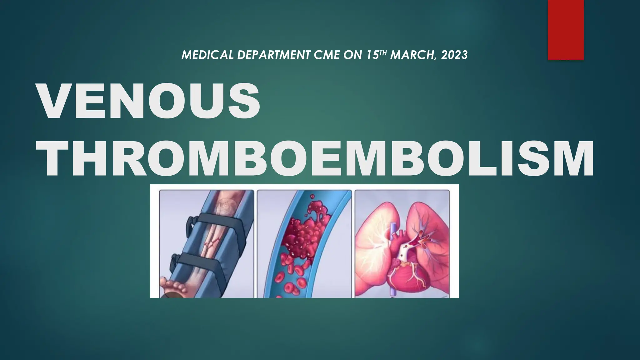
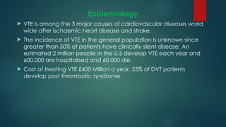
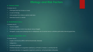
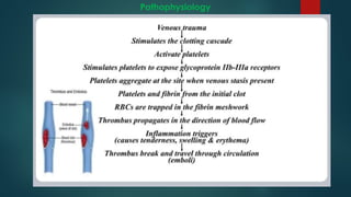
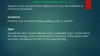
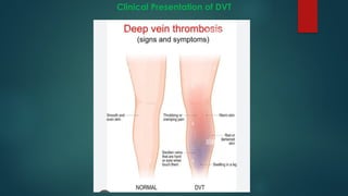
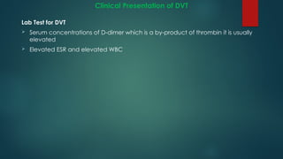
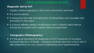
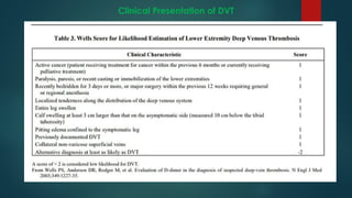
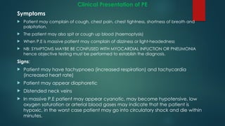
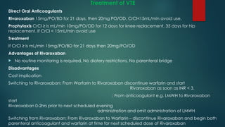
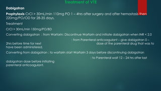
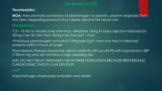
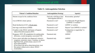
VENOUS
THROMBOEMBOLISM
MEDICAL DEPARTMENT CME ON 15TH
MARCH, 2023
Definition
 Venus Thromboembolism (VTE) is a condition in which a
blood clot (Thrombus) forms in the vein which in some
cases breaks free and enters the circulation as an
embolus, finally lodging in and completely obstructing a
blood vessel e.g in lungs causing pulmonary embolism
 VTE includes:
Deep vein thrombosis (DVT)
Pulmonary embolism (PE)
Deep Vein Thrombosis (DVT)
 It’s the most common type of VTE
 It is the formation of a blood clot in one of the deep veins in the
body usually in the leg.
 DVT
 Below knee (Distal)
 Above knee (Proximal)
 Atypical (e.g Arm)
Deep Vein Thrombosis (DVT)
Pulmonary Embolism (PE)
 It is a serious and life threatening condition, it happens
when the underlying blood clot in the leg(DVT) dislodges
as an embolus and blocks blood vessels in the lung.
 A massive Pulmonary Embolism can cause collapse and
death.
Pulmonary Embolism (PE)
Epidemiology
 VTE is among the 3 major causes of cardiovascular diseases world
wide after ischaemic heart disease and stroke.
 The incidence of VTE in the general population is unknown since
greater than 50% of patients have clinically silent disease. An
estimated 2 million people in the U.S develop VTE each year and
600,000 are hospitalised and 60,000 die.
 Cost of treating VTE £400 Million a year. 25% of DVT patients
develop post thrombotic syndrome.
Etiology and Risk Factors
 More than 100 years ago Rudolf Virchow described a triad of
factors that are thought to contribute to thrombosis
a) Venous stasis
b) Endothelial injury
c) Hypercoagulable
Virchow triad
Etiology and Risk Factors
a. Venous stasis
It maybe due to:
 Prolonged bed rest (4 days or more)
 A cast on the leg
 Limb paralysis from stroke or spinal code injury
 Extended travel in a vehicle
b. Endotheleal injury
It maybe due to:
 Trauma, Surgery
 Invasive procedure which may disrupt venous integrity
 Iatrogenic causes are increasing due to widespread use of central venous catheters particularly internal jugular lines.
c. Hypercoagulable state
It maybe due to:
 Surgery and trauma (40% of all thromboembolic diseases)
 Malignancy
 Increased oestrogen
 Inherited disorders of coagulation (deficiency of Protein S, Protein C, anti-thrombin III)
 Acquired disorders of coagulation (nephrotic syndrome, anti-phospholipid antibodies)
Pathophysiology
Clinical Presentation of DVT
Majority have asymptomatic disease and may die suddenly of
Pulmonary Embolism
Symptoms
Patients may complain of leg swelling, pain or warmth
Signs:
Superficial veins maybe dilated and a ‘palpable cord’ maybe felt in
the affected leg, their could be pain in the back of the knee when
examiner dorsiflexes the foot of the affected leg.
Clinical Presentation of DVT
Lab Test for DVT
 Serum concentrations of D-dimer which is a by-product of thrombin it is usually
elevated
 Elevated ESR and elevated WBC
Clinical Presentation of DVT
Diagnostic test for DVT
 Duplex ultrasonography is the most commonly used test
 It is non-inversive
 It measures the rate and direction of blood flow and visualize clot
formation in the veins
 It cannot reliably detect small blood clot in diastal veins hence
should be coupled with careful clinical assessment
 Venography (Phlebography)
 It’s the goal standard for diagnosis of DVT however it’s invasive,
involves injection of Radio – opaque contrast dye into a foot vein it
also expensive an can cause anaphylaxis and nephrotoxicity
Clinical Presentation of DVT
Clinical Presentation of PE
Symptoms
 Patient may complain of cough, chest pain, chest tightness, shortness of breath and
palpitation.
 The patient may also spit or cough up blood (haemoptysis)
 When P.E is massive patient may complain of dizziness or light-headedness
 NB: SYMPTOMS MAYBE BE CONFUSED WITH MYOCARDIAL INFUCTION OR PNEUMONIA
hence objective testing must be performed to establish the diagnosis.
Signs:
 Patient may have tachypnoea (increased respiration) and tachycardia
(increased heart rate)
 Patient may appear diaphoretic
 Distended neck veins
 In massive P.E patient may appear cyanotic, may become hypotensive, low
oxygen saturation or arterial blood gases may indicate that the patient is
hypoxic, in the worst case patient may go into circulatory shock and die within
minutes.
Clinical Presentation of PE
Lab tests for P.E
Elevated serum concentrations of D-dimer
Raised ESR
Raised WBCs
Diagnostic Tests for P.E
 Ventilation perfusion (V/Q) and computed tomography CT scan are the most
commonly used test to diagnose P.E.
 A V/Q scan measures the distribution of blood and air flow in the lungs.
 A large mismatch between blood and air flow in one area of the lung shows
there is a high probability that the patient has P.E.
 Spiral CT scans can detect emboli in the Pulmonary Arteries.
 Pulmonary Angiography
 Is the goal standard for diagnosis of P.E but it is invasive and costly
Clinical Presentation of PE
Treatment of VTE
Goals of Treatment
1. To allow normal circulation in the limbs
2. To prevent damage to the valves of the veins thus reducing the
risk of swollen post phlebitic limb
3. To prevent associated PE and recurrence of either PE or DVT
4. To provide medication without adverse effects
5. To improve the quality of life
Treatment of VTE
Prophylaxis of VTE
Mechanical
1. Leg elevation
2. Graded compression stocking
3. Early ambulation
4. Pneumatic compression boots
Treatment of VTE
Classification of drugs
LMWH Heparin(enoxparin,
Dalteparin)
Sythetic (Fondaparinux)
Heparin(ufh)
Lepirudin, Bivalirudin,
Argatroban
Oral
Anticoagulants
Direct thrombin
inhibitors
Indirect thrombin
inhibitors
Thrombolytics
Direct oral anticoagulant (DOAC)
Streptokinase, Urokinase, alteplase, reteplase
Parenteral
coagulant
Indirect oral anticoagulant
Dabigatran, Rivaroxaban,
Apixaban
Warfarin
Treatment of VTE
Heparin (UFH)
 Prophylaxis of DVT and PE
5000 units SC 8 to 12 hourly or 7500 units SC 12 hourly
Treatment of DVT and PE
Dose
1. 80 units/kg IV bolus, then continuous infusion of 18units/kg/hr or
2. 5000 units IV bolus, then continuous infusion of 1300 units/hr or
3. 250 unit/kg (17,500 units) SQ then 250 units/kg 12 hourly.
ADRs
 Bleeding, Heparin induced thrombocytopenia (HIT),
 Hypersensitivity reactions, osteoporosis, reversible alopecia
ANTIDOTE
Protamine sulphate (1-1.5mg per 100 USP units of heparin not to exceed 50mg) Monitor
APTT 5 – 15 mins after dose, then 2-8hrs
Treatment of VTE
Low molecular weight heparin
Enoxaparin
Prophylaxis 40mg/sc/OD
Treatment 1mg/kg/sc BD or 1.5mg/kg/sc (administered at same time each day.
Dalteparin 2500 IU/SC/OD phoprophilaxis 100 units/kg 12 hourly for treatment
ADRs
Bleeding (but less than with unfractionated heparin)
HIT, Several neurological injury for patients undergoing spinal puncture.
Antidotes:
Protamine sulphate 1 mg per mg of enoxaparin (if enoxaparin overdose given
within 8 hours > 8hrs you give 0.5mg per mg of enoxaparin .
Treatment of VTE
5. Indirect Oral Anticoagulants
They interfere with synthesis of Vitamin K dependant factors in the liver
Warfarin
Prophylaxis 2- 5mg PO for 2 days.
Treatment
2 – 5mg PO, maintenance dose is 2 & 10mg per day.
Initiate Warfarin on day 1 or 2 of parenteral anticoagulation therapy(e.g LMWH or UFH), overlap
Warfarin and parenteral anticoagulant for at least five days until desired INR (>2.0) maintained for 24
hours then discontinue the parenteral therapy.
Target INR 2-3, dose adjustment is 10-15% of the total weekly dose
ADRs
Bleeding, skin necrosis, purple toe syndrome, teratogenisity, osteoporosis, leucopenia
Antidote is Vitamin K – if INR is 4.5 – 10, no bleeding, 1-2.5mg/PO/OD. If INR is > 10 and no bleeding 2.5
– 5mg PO/OD. Major bleeding + any elevated INR give prothrombin complex concentrate (PCC) with
vitamin K 5-10mg/IV diluted in 50mls IV fluid and infused over 20mins.
Treatment of VTE
Direct Oral Anticoagulants
Rivaroxaban 15mg/PO/BD for 21 days, then 20mg PO/OD, CrCl<15mL/min avoid use.
Prophylaxis CrCl ≥ is mL/min 10mg/PO/OD for 12 days for knee replacement, 35 days for hip
replacement. If CrCl < 15mL/min avoid use
Treatment
If CrCl ≥ is mL/min 15mg/PO/BD for 21 days then 20mg/PO/OD
Advantages of Rivaroxaban
 No routine monitoring is required, No diatery restrictions, No parenteral bridge
Disadvantages
Cost implication
Switching to Rivaroxaban: From Warfarin to Rivaroxaban discontinue warfarin and start
Rivaroxaban as soon as INR < 3.
: From anticoagulant e.g. LMWH to Rivaroxaban
start
Rivaroxaban 0-2hrs prior to next scheduled evening
administration and omit administration of LMWH
Switching from Rivaroxaban: From Rivaroxaban to Warfarin – discontinue Rivaroxaban and begin both
parenteral anticoagulant and warfarin at time for next scheduled dose of Rivaroxaban
Treatment of VTE
Apixaban
Prophylaxis 2.5mg PO/BD for 35 days
Treatment:
10mg/PO/BD for 7 days then 5mg/BD
Switching from Apixaban to Warfarin, discontinue Apixaban and begin both parenteral anticoagulant
and warfarin at the time the next dose of apixaban would have been taken.
Discontinue parenteral anticoagulant when INR reaches unacceptable level.
Antidote for Rivaroxaban and Apixaban is Andexanet (Andexa)
IV bolus 400mg IV targeting infusion rate of 30mg/min
Treatment of VTE
Dabigatran
Prophylaxis CrCl > 30mL/min 110mg PO 1 – 4hrs after surgery and after hemostasis then
220mg/PO/OD for 28-35 days.
Treatment
CrCl > 30mL/min 150mg/PO/BD
Converting dabigatran : from Warfarin: Discontinue Warfarin and initiate dabigatran when INR < 2.0
: from Parenteral anticoagulant – give dabigatran 0 –
2hrs before time for next dose of the parenteral drug that was to
have been administered.
Converting from dabigatran : to warfarin start Warfarin 3 days before discontinuing dabigatran
: to Parenteral wait 12 – 24 hrs after last
dabigatran dose before initiating
parenteral anticoagulant.
Treatment of VTE
Thrombolytics
MOA: they promote conversion of plasminogen to plasmin, plasmin degrades fibrin
into fibrin degrading products thus rapidly dissolve the blood clot.
Streptokinase
7.5 – 15 lac IU infused over one hour, alteplase 15mg IV bolus injection followed by
50mg over 30 mins then 35mg over the next 1 hour.
t-PA(tissue plasminogen activator) 0.9mg per kg/IV over one hour in selected
patients within 3 hours of onset.
Thrombolytic therapy should be used in patients with acute PE with hypotension SBP
< 90mm Hg who do not have a high bleeding risk.
N/B: DO NOT DELAY THROMBOLYSIS IN THESE POPULATION BECAUSE IRREVERSABLE
CARDIOGENIC SHOCK CAN DEVELOP.
ADR
Haemorrhage anaphylaxis embolism and stroke
Treatment of VTE
Treatment of VTE
Thrombectomy, Embolectomy, IVC filter and Ligation
These are used in cases where anticoagulation is contraindicated or
there has been a complication of anticoagulation or where
anticoagulation has failed.
Treatment of VTE
Patient Care
 Patient should be monitored for resolution of symptoms
 Development of recurrent thrombosis and symptoms of post thrombotic
syndrome
 Haemoglobin, Haematocrit and BP should be monitored carefully to detect
bleeding from anticoagulant therapy.
 Coagulation tests (APTT, PT, INR) should be performed prior to initiating therapy to
establish the patient baseline values and to guide on dose adjustments.
 Patients taking Warfarin should be question about medication adherence and
symptoms related to bleeding and thromboembolic complications.
Treatment of VTE
Duration of Treatment
 All VTE patients anticoagulant is recommended for a minimum of 3 complete
months (active treatment).
 Surgery provoked DVT or PE 3 months.
 1st
unprovoked DVT or PE with high bleeding risk, 3 months.
 1st
unprovoked distal DVT regardless of bleeding risk, 3 months
 2nd
unprovoked DVT or PE with low or moderate bleeding risk – extended
treatments
 2nd
unprovoked DVT or PE with high bleeding risk, 3 months.
 DVT or PE and active cancer – extended treatment with periodic risk benefit
analysis.
 Indication for indefinite treatment duration – persistent or paroxysmal nonvalvular
AF in patients with high risk of stroke i.e. ischemic stroke, TIA or systemic embolism
or patient with 2 of the following age >75, impaired left ventricular systolic
function, history of hypertension or DM.
Complications of VTE
 Recurrent VTE
 Varicose veins
 Chronic venous insufficiency (CVI).
 Post phlebitis syndrome (Pain edema, ulceration and pigmentation).
 Chronic pulmonary hypertension and vascular corpalmonale.
 Death due to massive or sub massive PE.
 Risk must be regularly reassessed
 - A bleed will physiologically trigger clot formation
 References
- European Society for Vascular Surgery 2021 Guidelines for VTE.
- Textbook of Pharmaco Therapy.
- American College of Chest physician guidelines for VTE prevention.
THANK YOU!
Questions?

Venous Thromboembolism Presentation.pptx

  • 1.
  • 2.
    Definition  Venus Thromboembolism(VTE) is a condition in which a blood clot (Thrombus) forms in the vein which in some cases breaks free and enters the circulation as an embolus, finally lodging in and completely obstructing a blood vessel e.g in lungs causing pulmonary embolism  VTE includes: Deep vein thrombosis (DVT) Pulmonary embolism (PE)
  • 3.
    Deep Vein Thrombosis(DVT)  It’s the most common type of VTE  It is the formation of a blood clot in one of the deep veins in the body usually in the leg.  DVT  Below knee (Distal)  Above knee (Proximal)  Atypical (e.g Arm)
  • 4.
  • 5.
    Pulmonary Embolism (PE) It is a serious and life threatening condition, it happens when the underlying blood clot in the leg(DVT) dislodges as an embolus and blocks blood vessels in the lung.  A massive Pulmonary Embolism can cause collapse and death.
  • 6.
  • 7.
    Epidemiology  VTE isamong the 3 major causes of cardiovascular diseases world wide after ischaemic heart disease and stroke.  The incidence of VTE in the general population is unknown since greater than 50% of patients have clinically silent disease. An estimated 2 million people in the U.S develop VTE each year and 600,000 are hospitalised and 60,000 die.  Cost of treating VTE £400 Million a year. 25% of DVT patients develop post thrombotic syndrome.
  • 8.
    Etiology and RiskFactors  More than 100 years ago Rudolf Virchow described a triad of factors that are thought to contribute to thrombosis a) Venous stasis b) Endothelial injury c) Hypercoagulable Virchow triad
  • 9.
    Etiology and RiskFactors a. Venous stasis It maybe due to:  Prolonged bed rest (4 days or more)  A cast on the leg  Limb paralysis from stroke or spinal code injury  Extended travel in a vehicle b. Endotheleal injury It maybe due to:  Trauma, Surgery  Invasive procedure which may disrupt venous integrity  Iatrogenic causes are increasing due to widespread use of central venous catheters particularly internal jugular lines. c. Hypercoagulable state It maybe due to:  Surgery and trauma (40% of all thromboembolic diseases)  Malignancy  Increased oestrogen  Inherited disorders of coagulation (deficiency of Protein S, Protein C, anti-thrombin III)  Acquired disorders of coagulation (nephrotic syndrome, anti-phospholipid antibodies)
  • 10.
  • 11.
    Clinical Presentation ofDVT Majority have asymptomatic disease and may die suddenly of Pulmonary Embolism Symptoms Patients may complain of leg swelling, pain or warmth Signs: Superficial veins maybe dilated and a ‘palpable cord’ maybe felt in the affected leg, their could be pain in the back of the knee when examiner dorsiflexes the foot of the affected leg.
  • 12.
  • 13.
    Clinical Presentation ofDVT Lab Test for DVT  Serum concentrations of D-dimer which is a by-product of thrombin it is usually elevated  Elevated ESR and elevated WBC
  • 14.
    Clinical Presentation ofDVT Diagnostic test for DVT  Duplex ultrasonography is the most commonly used test  It is non-inversive  It measures the rate and direction of blood flow and visualize clot formation in the veins  It cannot reliably detect small blood clot in diastal veins hence should be coupled with careful clinical assessment  Venography (Phlebography)  It’s the goal standard for diagnosis of DVT however it’s invasive, involves injection of Radio – opaque contrast dye into a foot vein it also expensive an can cause anaphylaxis and nephrotoxicity
  • 15.
  • 16.
    Clinical Presentation ofPE Symptoms  Patient may complain of cough, chest pain, chest tightness, shortness of breath and palpitation.  The patient may also spit or cough up blood (haemoptysis)  When P.E is massive patient may complain of dizziness or light-headedness  NB: SYMPTOMS MAYBE BE CONFUSED WITH MYOCARDIAL INFUCTION OR PNEUMONIA hence objective testing must be performed to establish the diagnosis. Signs:  Patient may have tachypnoea (increased respiration) and tachycardia (increased heart rate)  Patient may appear diaphoretic  Distended neck veins  In massive P.E patient may appear cyanotic, may become hypotensive, low oxygen saturation or arterial blood gases may indicate that the patient is hypoxic, in the worst case patient may go into circulatory shock and die within minutes.
  • 17.
    Clinical Presentation ofPE Lab tests for P.E Elevated serum concentrations of D-dimer Raised ESR Raised WBCs Diagnostic Tests for P.E  Ventilation perfusion (V/Q) and computed tomography CT scan are the most commonly used test to diagnose P.E.  A V/Q scan measures the distribution of blood and air flow in the lungs.  A large mismatch between blood and air flow in one area of the lung shows there is a high probability that the patient has P.E.  Spiral CT scans can detect emboli in the Pulmonary Arteries.  Pulmonary Angiography  Is the goal standard for diagnosis of P.E but it is invasive and costly
  • 18.
  • 19.
    Treatment of VTE Goalsof Treatment 1. To allow normal circulation in the limbs 2. To prevent damage to the valves of the veins thus reducing the risk of swollen post phlebitic limb 3. To prevent associated PE and recurrence of either PE or DVT 4. To provide medication without adverse effects 5. To improve the quality of life
  • 20.
    Treatment of VTE Prophylaxisof VTE Mechanical 1. Leg elevation 2. Graded compression stocking 3. Early ambulation 4. Pneumatic compression boots
  • 21.
    Treatment of VTE Classificationof drugs LMWH Heparin(enoxparin, Dalteparin) Sythetic (Fondaparinux) Heparin(ufh) Lepirudin, Bivalirudin, Argatroban Oral Anticoagulants Direct thrombin inhibitors Indirect thrombin inhibitors Thrombolytics Direct oral anticoagulant (DOAC) Streptokinase, Urokinase, alteplase, reteplase Parenteral coagulant Indirect oral anticoagulant Dabigatran, Rivaroxaban, Apixaban Warfarin
  • 22.
    Treatment of VTE Heparin(UFH)  Prophylaxis of DVT and PE 5000 units SC 8 to 12 hourly or 7500 units SC 12 hourly Treatment of DVT and PE Dose 1. 80 units/kg IV bolus, then continuous infusion of 18units/kg/hr or 2. 5000 units IV bolus, then continuous infusion of 1300 units/hr or 3. 250 unit/kg (17,500 units) SQ then 250 units/kg 12 hourly. ADRs  Bleeding, Heparin induced thrombocytopenia (HIT),  Hypersensitivity reactions, osteoporosis, reversible alopecia ANTIDOTE Protamine sulphate (1-1.5mg per 100 USP units of heparin not to exceed 50mg) Monitor APTT 5 – 15 mins after dose, then 2-8hrs
  • 23.
    Treatment of VTE Lowmolecular weight heparin Enoxaparin Prophylaxis 40mg/sc/OD Treatment 1mg/kg/sc BD or 1.5mg/kg/sc (administered at same time each day. Dalteparin 2500 IU/SC/OD phoprophilaxis 100 units/kg 12 hourly for treatment ADRs Bleeding (but less than with unfractionated heparin) HIT, Several neurological injury for patients undergoing spinal puncture. Antidotes: Protamine sulphate 1 mg per mg of enoxaparin (if enoxaparin overdose given within 8 hours > 8hrs you give 0.5mg per mg of enoxaparin .
  • 24.
    Treatment of VTE 5.Indirect Oral Anticoagulants They interfere with synthesis of Vitamin K dependant factors in the liver Warfarin Prophylaxis 2- 5mg PO for 2 days. Treatment 2 – 5mg PO, maintenance dose is 2 & 10mg per day. Initiate Warfarin on day 1 or 2 of parenteral anticoagulation therapy(e.g LMWH or UFH), overlap Warfarin and parenteral anticoagulant for at least five days until desired INR (>2.0) maintained for 24 hours then discontinue the parenteral therapy. Target INR 2-3, dose adjustment is 10-15% of the total weekly dose ADRs Bleeding, skin necrosis, purple toe syndrome, teratogenisity, osteoporosis, leucopenia Antidote is Vitamin K – if INR is 4.5 – 10, no bleeding, 1-2.5mg/PO/OD. If INR is > 10 and no bleeding 2.5 – 5mg PO/OD. Major bleeding + any elevated INR give prothrombin complex concentrate (PCC) with vitamin K 5-10mg/IV diluted in 50mls IV fluid and infused over 20mins.
  • 25.
    Treatment of VTE DirectOral Anticoagulants Rivaroxaban 15mg/PO/BD for 21 days, then 20mg PO/OD, CrCl<15mL/min avoid use. Prophylaxis CrCl ≥ is mL/min 10mg/PO/OD for 12 days for knee replacement, 35 days for hip replacement. If CrCl < 15mL/min avoid use Treatment If CrCl ≥ is mL/min 15mg/PO/BD for 21 days then 20mg/PO/OD Advantages of Rivaroxaban  No routine monitoring is required, No diatery restrictions, No parenteral bridge Disadvantages Cost implication Switching to Rivaroxaban: From Warfarin to Rivaroxaban discontinue warfarin and start Rivaroxaban as soon as INR < 3. : From anticoagulant e.g. LMWH to Rivaroxaban start Rivaroxaban 0-2hrs prior to next scheduled evening administration and omit administration of LMWH Switching from Rivaroxaban: From Rivaroxaban to Warfarin – discontinue Rivaroxaban and begin both parenteral anticoagulant and warfarin at time for next scheduled dose of Rivaroxaban
  • 26.
    Treatment of VTE Apixaban Prophylaxis2.5mg PO/BD for 35 days Treatment: 10mg/PO/BD for 7 days then 5mg/BD Switching from Apixaban to Warfarin, discontinue Apixaban and begin both parenteral anticoagulant and warfarin at the time the next dose of apixaban would have been taken. Discontinue parenteral anticoagulant when INR reaches unacceptable level. Antidote for Rivaroxaban and Apixaban is Andexanet (Andexa) IV bolus 400mg IV targeting infusion rate of 30mg/min
  • 27.
    Treatment of VTE Dabigatran ProphylaxisCrCl > 30mL/min 110mg PO 1 – 4hrs after surgery and after hemostasis then 220mg/PO/OD for 28-35 days. Treatment CrCl > 30mL/min 150mg/PO/BD Converting dabigatran : from Warfarin: Discontinue Warfarin and initiate dabigatran when INR < 2.0 : from Parenteral anticoagulant – give dabigatran 0 – 2hrs before time for next dose of the parenteral drug that was to have been administered. Converting from dabigatran : to warfarin start Warfarin 3 days before discontinuing dabigatran : to Parenteral wait 12 – 24 hrs after last dabigatran dose before initiating parenteral anticoagulant.
  • 28.
    Treatment of VTE Thrombolytics MOA:they promote conversion of plasminogen to plasmin, plasmin degrades fibrin into fibrin degrading products thus rapidly dissolve the blood clot. Streptokinase 7.5 – 15 lac IU infused over one hour, alteplase 15mg IV bolus injection followed by 50mg over 30 mins then 35mg over the next 1 hour. t-PA(tissue plasminogen activator) 0.9mg per kg/IV over one hour in selected patients within 3 hours of onset. Thrombolytic therapy should be used in patients with acute PE with hypotension SBP < 90mm Hg who do not have a high bleeding risk. N/B: DO NOT DELAY THROMBOLYSIS IN THESE POPULATION BECAUSE IRREVERSABLE CARDIOGENIC SHOCK CAN DEVELOP. ADR Haemorrhage anaphylaxis embolism and stroke
  • 29.
  • 30.
    Treatment of VTE Thrombectomy,Embolectomy, IVC filter and Ligation These are used in cases where anticoagulation is contraindicated or there has been a complication of anticoagulation or where anticoagulation has failed.
  • 31.
    Treatment of VTE PatientCare  Patient should be monitored for resolution of symptoms  Development of recurrent thrombosis and symptoms of post thrombotic syndrome  Haemoglobin, Haematocrit and BP should be monitored carefully to detect bleeding from anticoagulant therapy.  Coagulation tests (APTT, PT, INR) should be performed prior to initiating therapy to establish the patient baseline values and to guide on dose adjustments.  Patients taking Warfarin should be question about medication adherence and symptoms related to bleeding and thromboembolic complications.
  • 32.
    Treatment of VTE Durationof Treatment  All VTE patients anticoagulant is recommended for a minimum of 3 complete months (active treatment).  Surgery provoked DVT or PE 3 months.  1st unprovoked DVT or PE with high bleeding risk, 3 months.  1st unprovoked distal DVT regardless of bleeding risk, 3 months  2nd unprovoked DVT or PE with low or moderate bleeding risk – extended treatments  2nd unprovoked DVT or PE with high bleeding risk, 3 months.  DVT or PE and active cancer – extended treatment with periodic risk benefit analysis.  Indication for indefinite treatment duration – persistent or paroxysmal nonvalvular AF in patients with high risk of stroke i.e. ischemic stroke, TIA or systemic embolism or patient with 2 of the following age >75, impaired left ventricular systolic function, history of hypertension or DM.
  • 33.
    Complications of VTE Recurrent VTE  Varicose veins  Chronic venous insufficiency (CVI).  Post phlebitis syndrome (Pain edema, ulceration and pigmentation).  Chronic pulmonary hypertension and vascular corpalmonale.  Death due to massive or sub massive PE.
  • 34.
     Risk mustbe regularly reassessed  - A bleed will physiologically trigger clot formation
  • 35.
     References - EuropeanSociety for Vascular Surgery 2021 Guidelines for VTE. - Textbook of Pharmaco Therapy. - American College of Chest physician guidelines for VTE prevention.
  • 36.