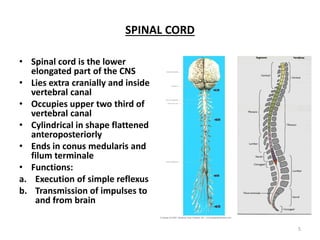
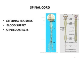
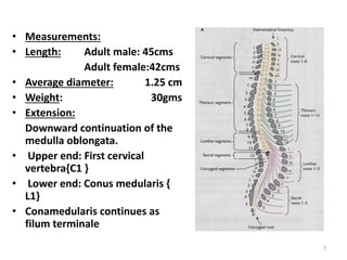
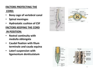
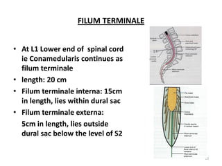
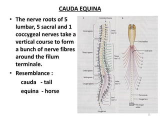
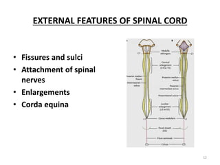
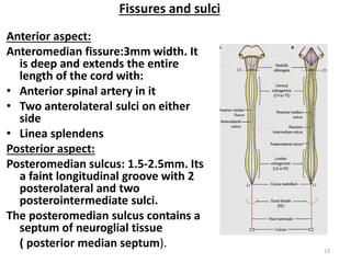
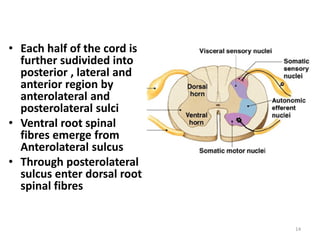
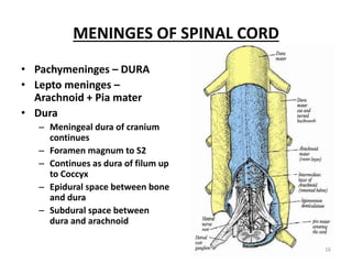
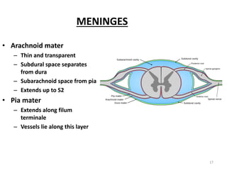
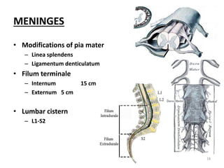
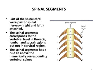
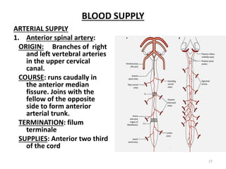
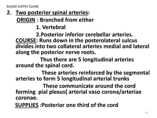
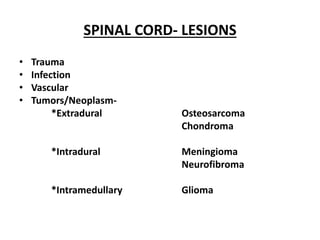
Spinal cord- gross anatomy,
internal features, blood supply
Dr.G.Arthi
1
NERVOUS SYSTEM
• Neurons
• Neuroglial cells
• Meninges
• Neurons+neuroglial cells
– Inside cns=grey mater/nuclei
– Outside cns=ganglia
• Nerve fibers
– Inside cns – tracts/
– Outside cns - nerves
2
INTRODUCTION
NERVOUS SYSTEM
CENTRAL
BRAIN SPINAL CORD
PERIPHERAL
CRANIAL-12
SPINAL-31
AUTONOMIC
SYMPATHETIC PARASYMPATHETIC
3
GROSS FEATURES
• Elongated cylindrical portion of
CNS
– Outside the cranium
– Inside the vertebral column
• In fetus occupies whole Vertebral
column till 3rd month IUL
• Adult upper 2/3 of vertebral
column
4
SPINAL CORD
• Spinal cord is the lower
elongated part of the CNS
• Lies extra cranially and inside
vertebral canal
• Occupies upper two third of
vertebral canal
• Cylindrical in shape flattened
anteroposteriorly
• Ends in conus medularis and
filum terminale
• Functions:
a. Execution of simple reflexus
b. Transmission of impulses to
and from brain
5
SPINAL CORD
• EXTERNAL FEATURES
• BLOOD SUPPLY
• APPLIED ASPECTS
6
• Measurements:
• Length: Adult male: 45cms
Adult female:42cms
• Average diameter: 1.25 cm
• Weight: 30gms
• Extension:
Downward continuation of the
medulla oblongata.
• Upper end: First cervical
vertebra{C1 }
• Lower end: Conus medularis {
L1}
• Conamedularis continues as
filum terminale
7
FACTORS PROTECTING THE
CORD:
• Bony cage of vertebral canal
• Spinal meninges
• Hydrostatic cushion of CSF
FACTORS KEEPING THE CORD
IN POSITION:
• Rostral continuity with
medulla oblongata
• Caudal fixation with filum
terminale and cauda equina
• Laterl suspension with
ligamentum denticulatum
8
ENLARGEMENTS
• ENLARGEMENTS
contains more motor neurons to
supply the limbs
• Cervical:
Extends from C5 to T1 segments
to form brachial plexus
widest circumferance-38mm
at C6
• Lumbosacral:
Extends from L2toS3 to form
lumbosacral plexus.
Widest circumferance -35mm at
S1
9
FILUM TERMINALE
• At L1 Lower end of spinal cord
ie Conamedularis continues as
filum terminale
• length: 20 cm
• Filum terminale interna: 15cm
in length, lies within dural sac
• Filum terminale externa:
5cm in length, lies outside
dural sac below the level of S2
10
CAUDA EQUINA
• The nerve roots of 5
lumbar, 5 sacral and 1
coccygeal nerves take a
vertical course to form
a bunch of nerve fibres
around the filum
terminale.
• Resemblance :
cauda - tail
equina - horse
11
EXTERNAL FEATURES OF SPINAL CORD
• Fissures and sulci
• Attachment of spinal
nerves
• Enlargements
• Corda equina
12
Fissures and sulci
Anterior aspect:
Anteromedian fissure:3mm width. It
is deep and extends the entire
length of the cord with:
• Anterior spinal artery in it
• Two anterolateral sulci on either
side
• Linea splendens
Posterior aspect:
Posteromedian sulcus: 1.5-2.5mm. Its
a faint longitudinal groove with 2
posterolateral and two
posterointermediate sulci.
The posteromedian sulcus contains a
septum of neuroglial tissue
( posterior median septum). 13
• Each half of the cord is
further sudivided into
posterior , lateral and
anterior region by
anterolateral and
posterolateral sulci
• Ventral root spinal
fibres emerge from
Anterolateral sulcus
• Through posterolateral
sulcus enter dorsal root
spinal fibres
14
ATTACHMENT OF SPINAL NERVES
• Emerges 31 pairs of
spinal nerves from the
side.
• Cervical -8, thoracic-
12,lumbar-5,sacral-
5,coccygeal-1.
Spinal nerve
attachments
anterior posterior
motor sensory
15
MENINGES OF SPINAL CORD
• Pachymeninges – DURA
• Lepto meninges –
Arachnoid + Pia mater
• Dura
– Meningeal dura of cranium
continues
– Foramen magnum to S2
– Continues as dura of filum up
to Coccyx
– Epidural space between bone
and dura
– Subdural space between
dura and arachnoid
16
MENINGES
• Arachnoid mater
– Thin and transparent
– Subdural space separates
from dura
– Subarachnoid space from pia
– Extends up to S2
• Pia mater
– Extends along filum
terminale
– Vessels lie along this layer
17
MENINGES
• Modifications of pia mater
– Linea splendens
– Ligamentum denticulatum
• Filum terminale
– Internum 15 cm
– Externum 5 cm
• Lumbar cistern
– L1-S2
18
SPINAL SEGMENTS
• Part of the spinal cord
were pair of spinal
nerves– { right and left }
attached.
• The spinal segments
corresponds to the
vertebral level in thoracic,
lumbar and sacral regions
but not in cervical region.
• The spinal segments has a
rule lie above the
numerically corresponding
vertebral spines
19
SPINAL SEGMENT
• Segment of cord with a
pair of spinal nerves
• 31 segments
• Cervical 8
• Thoracic 12
• Lumbar 5
• Sacral 5
• Coccygeal 1
20
LOCALIZATION OF SPINAL SEGMENTS
• Localization of spinal segment is important to locate
cord lesion or the site of surgical approach
Region Spine Cord segment
Cervical C6 +1 C7
Upper thoracic T3 +2 T5
Lower thoracic T7 +3 T10
Sacral T12-L1 S1-S5
21
REGIONS
SPINAL
SEGMENTS
VERTEBRAL
LEVEL
GENERAL
RULE
Upper
cervical
C2 C2 Same level
Lower
cervical
C6 C5 One
vertebra
above
Upper
thoracic
T5 T3 Two above
Lower
thoracic
T10 T7 Three above
lumbar L1 TO L5 T10 TO T11 Three to five
above
Sacral and
coccygeal
S1 TO S5
C X 1
T12 TO L1 Six to ten
above
22
SPINAL CORD
EXTERNAL AND INTERNAL FEATURES
• White mater
• Anterior, lateral and
posterior funiculus
• Grey mater
• Anterior, posterior and
lateral horns
23
MACROSCOPIC APPEARANCE
cervical
Thoracic
Lumbar sacral
24
CHARACTERSTIC FEATURES OF SPINAL SEGMENTS AT
VARIOUS T.S LEVELS
25
CHARACTERSTIC FEATURES OF SPINAL SEGMENTS AT
VARIOUS T.S LEVELS
Features
Levels
Cervical Thoracic Lumbar Sacral
Outline of section Oval Oval to circular Nearly circular Circular to
quadrilateral
Gray matter
•Posterior horn
•Lateral horn
•Anterior horn
Large
Slender, extends
far posteriorly
Massive
Absent
Small
Slender
Slender
Present
Large
Bulbous
Bulbous
Present only in
L1,L2
Relatively large
Massive
Massive
Present only in
S2,3,4
Reticular formation W ell developed Poorly
developed
Absent Absent
Amount of white
matter
Massive ++++ Large+++ Less+++ Very less+
Postero intermediate
sulcus
Present through
out
Present in
upper 7 T seg
Absent Absent
26
BLOOD SUPPLY
ARTERIAL SUPPLY
1. Anterior spinal artery:
ORIGIN: Branches of right
and left vertebral arteries
in the upper cervical
canal.
COURSE: runs caudally in
the anterior median
fissure. Joins with the
fellow of the opposite
side to form anterior
arterial trunk.
TERMINATION: filum
terminale
SUPPLIES: Anterior two third
of the cord
27
BLOOD SUPPLY Contd
2. Two posterior spinal arteries:
ORIGIN : Branched from either
1. Vertebral
2.Posterior inferior cerebellar arteries.
COURSE: Runs down in the posterolateral sulcus
divides into two collateral arteries medial and lateral
along the posterior nerve roots.
Thus there are 5 longitudinal arteries
around the spinal cord.
These arteries reinforced by the segmental
arteries to form 5 longitudinal arterial trunks
These communicate around the cord
forming pial plexus{ arterial vaso corona/arteriae
coronae.
SUPPLIES :Posterior one third of the cord
28
BLOOD SUPPLY Contd
3. Segmental arteries:
Branches of
Deep cervical,
Ascending cervical,
Intercostal and
Lumbar
Segmental arterial feeders reach the cord as anterior and posterior
radicular arteries.{8 to 12}
ANTERIOR RADICULAR ARTERIES: Larger and less in number.
T1 and T11 are very large – Arteria radicularis magna
{Artery of adamkiewicz}
POSTERIOR RADICULAR ARTERIES: Smaller and more in number.
FEEDER ARTERIES: Great anterior medullary artery of
Adamkiewicz-arises from aorta at T12 or L1 vertebral level
unilateral left side
Importance: major source of blood to the lower 2/3 of the
spinal cord
29
BLOOD SUPPLY Contd
• Anterior arterial trunk-
formed by union of two
anterior spinal arteries
arising from vertebral
arteries and a branch of
Arteria magna
• Posterior spinal trunk-
formed by two posterior
spinal arteries and posterior
radicular arteries
30
Area of supply:
• Anterior trunk supplies : ventral two third of cross
section of spinal cord– anterior gray columns, bases
of posterior gray column and adjoining portions of
white mater.
• Pair of posterior arterial trunks and vasa corona
supply-- Rest of the cord They are end arteries.
• Spinal branches of vertebral arteries supply: Upper
cervical cord segments
• Radicular branches of ascending cervical and deep
cervical supply: C7 to T2 spinal segments.
• Radicular branches of aortic intercostal and lumbar
arteries supply: spinal segments below T2.
31
BLOOD SUPPLY Contd
CLINICAL CORELATION:
• Anterior spinal artery syndrome:
• The artery of T11 spinal segment (arteria radicularis magna)
is remarkably large. A fracture of vertebra involving this
artery leeds to softening of several segments of the cord
• The artery of T1 spinal segment anastamoses with other
arteries in a valvular fashion so that its supply is directed
only downwards. Therefore C8 segment is most affected.
• T1,T4 and L1 segments -- the meeting places of different
major arteries are vulnerable to ischemic necrosis.
• Anterior medullary syndrome.
32
VENUS DRAINAGE
• Two median longitudinal- one in
the anterior median fissure
another in posteromedian sulcus
• Two anterolaterlal – one on either
side posterior to the anterior nerve
roots
• Two posterolateral-one on either
side posterior posterior nerve roots
• Drain below through internal
vertebral venous plexus into the
vertebral posterior intercostal,
lumbar, and lateral sacral veins.
And drain above into the basilar
venous plexus.
33
APPLIED AND CLINICAL ANATOMY
34
SPINAL CORD- LESIONS
• Trauma
• Infection
• Vascular
• Tumors/Neoplasm-
*Extradural Osteosarcoma
Chondroma
*Intradural Meningioma
Neurofibroma
*Intramedullary Glioma
SPINAL CORD LESIONScontd
• Compression of nerve roots
* Radicular pain
* Paresthesia
* Localised muscular atrophy
* paralysis due to ventral root
 Compression of cord
* anterior horn cells defect
* sensory loss
* corticospinal defect
* bladder and bowl sphincter disturbances
 Surgical procedure
* Laminectomy
SPINAL CORD
TRAUMATIC INJURY-contd
• Hemisection: Brown –Sequard syndrome
• Lateral hemisection: due to trauma or compression of
extramedullary tumors.
On the side of the lesion:
1. loss of position and vibratory sense, disturbances of stereognosis
and tactile discrimination below the level of lesion – damage of
posterior funiculus
2. Spastic paralysis, exaggerated tendon reflexes, positive
Babi ski’s sig - involvement of pyramidal tract
Conralateral side:
loss of pain and temp- damage of lateral spinothalamic tract
at the site of lesion:
Irritation of dorsal root produces radicular pain ,parasthesia
Irritation of ventral root produces flacid paralysis of muscles
37
SPINAL CORD
SUDDEN VASCULAR LESIONS
Thrombosis in Anterior spinal artery
Spinal segment C8 is vulnerable
Involvement of anterior horn cells-flaccid type
of palsy
Lateral corticospinal tract-spastic type
Loss of pain and temperature- lateral
spinothalamic tract
Ischemia of arteria radicularis magna
Cauda equina paraplegia
SPINAL CORD
CLINICAL ANATOMY
• Lumbar Puncture L3-L4 L4-L5
• Collection of CSF 1 drop/sec
• Injection of radio opaque dye Lipiodal
• Spinal block (anesthesia) – 2% Procaine
• Measurement of intracranial pressure
• Injection of drugs
REFERENCES
• Gray’s a ato y-39th edition
• Clinical neuroanatomy – 2nd edition by VisramSing
• Clinical neuroanatomy – 3nd edition by A.K.Dutta
40

Spinal cord (2)

  • 1.
    Spinal cord- grossanatomy, internal features, blood supply Dr.G.Arthi 1
  • 2.
    NERVOUS SYSTEM • Neurons •Neuroglial cells • Meninges • Neurons+neuroglial cells – Inside cns=grey mater/nuclei – Outside cns=ganglia • Nerve fibers – Inside cns – tracts/ – Outside cns - nerves 2
  • 3.
    INTRODUCTION NERVOUS SYSTEM CENTRAL BRAIN SPINALCORD PERIPHERAL CRANIAL-12 SPINAL-31 AUTONOMIC SYMPATHETIC PARASYMPATHETIC 3
  • 4.
    GROSS FEATURES • Elongatedcylindrical portion of CNS – Outside the cranium – Inside the vertebral column • In fetus occupies whole Vertebral column till 3rd month IUL • Adult upper 2/3 of vertebral column 4
  • 5.
    SPINAL CORD • Spinalcord is the lower elongated part of the CNS • Lies extra cranially and inside vertebral canal • Occupies upper two third of vertebral canal • Cylindrical in shape flattened anteroposteriorly • Ends in conus medularis and filum terminale • Functions: a. Execution of simple reflexus b. Transmission of impulses to and from brain 5
  • 6.
    SPINAL CORD • EXTERNALFEATURES • BLOOD SUPPLY • APPLIED ASPECTS 6
  • 7.
    • Measurements: • Length:Adult male: 45cms Adult female:42cms • Average diameter: 1.25 cm • Weight: 30gms • Extension: Downward continuation of the medulla oblongata. • Upper end: First cervical vertebra{C1 } • Lower end: Conus medularis { L1} • Conamedularis continues as filum terminale 7
  • 8.
    FACTORS PROTECTING THE CORD: •Bony cage of vertebral canal • Spinal meninges • Hydrostatic cushion of CSF FACTORS KEEPING THE CORD IN POSITION: • Rostral continuity with medulla oblongata • Caudal fixation with filum terminale and cauda equina • Laterl suspension with ligamentum denticulatum 8
  • 9.
    ENLARGEMENTS • ENLARGEMENTS contains moremotor neurons to supply the limbs • Cervical: Extends from C5 to T1 segments to form brachial plexus widest circumferance-38mm at C6 • Lumbosacral: Extends from L2toS3 to form lumbosacral plexus. Widest circumferance -35mm at S1 9
  • 10.
    FILUM TERMINALE • AtL1 Lower end of spinal cord ie Conamedularis continues as filum terminale • length: 20 cm • Filum terminale interna: 15cm in length, lies within dural sac • Filum terminale externa: 5cm in length, lies outside dural sac below the level of S2 10
  • 11.
    CAUDA EQUINA • Thenerve roots of 5 lumbar, 5 sacral and 1 coccygeal nerves take a vertical course to form a bunch of nerve fibres around the filum terminale. • Resemblance : cauda - tail equina - horse 11
  • 12.
    EXTERNAL FEATURES OFSPINAL CORD • Fissures and sulci • Attachment of spinal nerves • Enlargements • Corda equina 12
  • 13.
    Fissures and sulci Anterioraspect: Anteromedian fissure:3mm width. It is deep and extends the entire length of the cord with: • Anterior spinal artery in it • Two anterolateral sulci on either side • Linea splendens Posterior aspect: Posteromedian sulcus: 1.5-2.5mm. Its a faint longitudinal groove with 2 posterolateral and two posterointermediate sulci. The posteromedian sulcus contains a septum of neuroglial tissue ( posterior median septum). 13
  • 14.
    • Each halfof the cord is further sudivided into posterior , lateral and anterior region by anterolateral and posterolateral sulci • Ventral root spinal fibres emerge from Anterolateral sulcus • Through posterolateral sulcus enter dorsal root spinal fibres 14
  • 15.
    ATTACHMENT OF SPINALNERVES • Emerges 31 pairs of spinal nerves from the side. • Cervical -8, thoracic- 12,lumbar-5,sacral- 5,coccygeal-1. Spinal nerve attachments anterior posterior motor sensory 15
  • 16.
    MENINGES OF SPINALCORD • Pachymeninges – DURA • Lepto meninges – Arachnoid + Pia mater • Dura – Meningeal dura of cranium continues – Foramen magnum to S2 – Continues as dura of filum up to Coccyx – Epidural space between bone and dura – Subdural space between dura and arachnoid 16
  • 17.
    MENINGES • Arachnoid mater –Thin and transparent – Subdural space separates from dura – Subarachnoid space from pia – Extends up to S2 • Pia mater – Extends along filum terminale – Vessels lie along this layer 17
  • 18.
    MENINGES • Modifications ofpia mater – Linea splendens – Ligamentum denticulatum • Filum terminale – Internum 15 cm – Externum 5 cm • Lumbar cistern – L1-S2 18
  • 19.
    SPINAL SEGMENTS • Partof the spinal cord were pair of spinal nerves– { right and left } attached. • The spinal segments corresponds to the vertebral level in thoracic, lumbar and sacral regions but not in cervical region. • The spinal segments has a rule lie above the numerically corresponding vertebral spines 19
  • 20.
    SPINAL SEGMENT • Segmentof cord with a pair of spinal nerves • 31 segments • Cervical 8 • Thoracic 12 • Lumbar 5 • Sacral 5 • Coccygeal 1 20
  • 21.
    LOCALIZATION OF SPINALSEGMENTS • Localization of spinal segment is important to locate cord lesion or the site of surgical approach Region Spine Cord segment Cervical C6 +1 C7 Upper thoracic T3 +2 T5 Lower thoracic T7 +3 T10 Sacral T12-L1 S1-S5 21
  • 22.
    REGIONS SPINAL SEGMENTS VERTEBRAL LEVEL GENERAL RULE Upper cervical C2 C2 Samelevel Lower cervical C6 C5 One vertebra above Upper thoracic T5 T3 Two above Lower thoracic T10 T7 Three above lumbar L1 TO L5 T10 TO T11 Three to five above Sacral and coccygeal S1 TO S5 C X 1 T12 TO L1 Six to ten above 22
  • 23.
    SPINAL CORD EXTERNAL ANDINTERNAL FEATURES • White mater • Anterior, lateral and posterior funiculus • Grey mater • Anterior, posterior and lateral horns 23
  • 24.
  • 25.
    CHARACTERSTIC FEATURES OFSPINAL SEGMENTS AT VARIOUS T.S LEVELS 25
  • 26.
    CHARACTERSTIC FEATURES OFSPINAL SEGMENTS AT VARIOUS T.S LEVELS Features Levels Cervical Thoracic Lumbar Sacral Outline of section Oval Oval to circular Nearly circular Circular to quadrilateral Gray matter •Posterior horn •Lateral horn •Anterior horn Large Slender, extends far posteriorly Massive Absent Small Slender Slender Present Large Bulbous Bulbous Present only in L1,L2 Relatively large Massive Massive Present only in S2,3,4 Reticular formation W ell developed Poorly developed Absent Absent Amount of white matter Massive ++++ Large+++ Less+++ Very less+ Postero intermediate sulcus Present through out Present in upper 7 T seg Absent Absent 26
  • 27.
    BLOOD SUPPLY ARTERIAL SUPPLY 1.Anterior spinal artery: ORIGIN: Branches of right and left vertebral arteries in the upper cervical canal. COURSE: runs caudally in the anterior median fissure. Joins with the fellow of the opposite side to form anterior arterial trunk. TERMINATION: filum terminale SUPPLIES: Anterior two third of the cord 27
  • 28.
    BLOOD SUPPLY Contd 2.Two posterior spinal arteries: ORIGIN : Branched from either 1. Vertebral 2.Posterior inferior cerebellar arteries. COURSE: Runs down in the posterolateral sulcus divides into two collateral arteries medial and lateral along the posterior nerve roots. Thus there are 5 longitudinal arteries around the spinal cord. These arteries reinforced by the segmental arteries to form 5 longitudinal arterial trunks These communicate around the cord forming pial plexus{ arterial vaso corona/arteriae coronae. SUPPLIES :Posterior one third of the cord 28
  • 29.
    BLOOD SUPPLY Contd 3.Segmental arteries: Branches of Deep cervical, Ascending cervical, Intercostal and Lumbar Segmental arterial feeders reach the cord as anterior and posterior radicular arteries.{8 to 12} ANTERIOR RADICULAR ARTERIES: Larger and less in number. T1 and T11 are very large – Arteria radicularis magna {Artery of adamkiewicz} POSTERIOR RADICULAR ARTERIES: Smaller and more in number. FEEDER ARTERIES: Great anterior medullary artery of Adamkiewicz-arises from aorta at T12 or L1 vertebral level unilateral left side Importance: major source of blood to the lower 2/3 of the spinal cord 29
  • 30.
    BLOOD SUPPLY Contd •Anterior arterial trunk- formed by union of two anterior spinal arteries arising from vertebral arteries and a branch of Arteria magna • Posterior spinal trunk- formed by two posterior spinal arteries and posterior radicular arteries 30
  • 31.
    Area of supply: •Anterior trunk supplies : ventral two third of cross section of spinal cord– anterior gray columns, bases of posterior gray column and adjoining portions of white mater. • Pair of posterior arterial trunks and vasa corona supply-- Rest of the cord They are end arteries. • Spinal branches of vertebral arteries supply: Upper cervical cord segments • Radicular branches of ascending cervical and deep cervical supply: C7 to T2 spinal segments. • Radicular branches of aortic intercostal and lumbar arteries supply: spinal segments below T2. 31
  • 32.
    BLOOD SUPPLY Contd CLINICALCORELATION: • Anterior spinal artery syndrome: • The artery of T11 spinal segment (arteria radicularis magna) is remarkably large. A fracture of vertebra involving this artery leeds to softening of several segments of the cord • The artery of T1 spinal segment anastamoses with other arteries in a valvular fashion so that its supply is directed only downwards. Therefore C8 segment is most affected. • T1,T4 and L1 segments -- the meeting places of different major arteries are vulnerable to ischemic necrosis. • Anterior medullary syndrome. 32
  • 33.
    VENUS DRAINAGE • Twomedian longitudinal- one in the anterior median fissure another in posteromedian sulcus • Two anterolaterlal – one on either side posterior to the anterior nerve roots • Two posterolateral-one on either side posterior posterior nerve roots • Drain below through internal vertebral venous plexus into the vertebral posterior intercostal, lumbar, and lateral sacral veins. And drain above into the basilar venous plexus. 33
  • 34.
  • 35.
    SPINAL CORD- LESIONS •Trauma • Infection • Vascular • Tumors/Neoplasm- *Extradural Osteosarcoma Chondroma *Intradural Meningioma Neurofibroma *Intramedullary Glioma
  • 36.
    SPINAL CORD LESIONScontd •Compression of nerve roots * Radicular pain * Paresthesia * Localised muscular atrophy * paralysis due to ventral root  Compression of cord * anterior horn cells defect * sensory loss * corticospinal defect * bladder and bowl sphincter disturbances  Surgical procedure * Laminectomy
  • 37.
    SPINAL CORD TRAUMATIC INJURY-contd •Hemisection: Brown –Sequard syndrome • Lateral hemisection: due to trauma or compression of extramedullary tumors. On the side of the lesion: 1. loss of position and vibratory sense, disturbances of stereognosis and tactile discrimination below the level of lesion – damage of posterior funiculus 2. Spastic paralysis, exaggerated tendon reflexes, positive Babi ski’s sig - involvement of pyramidal tract Conralateral side: loss of pain and temp- damage of lateral spinothalamic tract at the site of lesion: Irritation of dorsal root produces radicular pain ,parasthesia Irritation of ventral root produces flacid paralysis of muscles 37
  • 38.
    SPINAL CORD SUDDEN VASCULARLESIONS Thrombosis in Anterior spinal artery Spinal segment C8 is vulnerable Involvement of anterior horn cells-flaccid type of palsy Lateral corticospinal tract-spastic type Loss of pain and temperature- lateral spinothalamic tract Ischemia of arteria radicularis magna Cauda equina paraplegia
  • 39.
    SPINAL CORD CLINICAL ANATOMY •Lumbar Puncture L3-L4 L4-L5 • Collection of CSF 1 drop/sec • Injection of radio opaque dye Lipiodal • Spinal block (anesthesia) – 2% Procaine • Measurement of intracranial pressure • Injection of drugs
  • 40.
    REFERENCES • Gray’s aato y-39th edition • Clinical neuroanatomy – 2nd edition by VisramSing • Clinical neuroanatomy – 3nd edition by A.K.Dutta 40