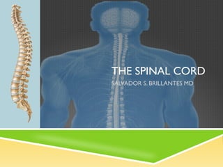
2. Spinal Cord Function
The Spinal Cord is a long, thin
bundle of nervous tissue and
support cells connected to the
brain and located along your
back and neck
The spinal cord receives and
transmits electric signals
throughout the entire body and
then back to the brain
The spinal cord is protected by
the vertebrae, which are bones
running down your back, and
also by cerebral spinal fluid,
which helps to cushion the
nerve tissue
3. EmBRYOLOGY & ANATOMY
Neural tube
18th gestational day
Organization
Butterfly shaped area of gray matter with surrounding white matter
WHITE MATTER
Consist mainly of longitudinal nerve fibers
Ascending and Descending tracts
GRAY MATTER
Groups of nuclei
4. Anatomy
The spinal column is divided into
four areas: Cervical, Thoracic,
Lumbar, and Sacral
Each section contains nerves
that control certain muscles of
your body
Each nerve has its corresponding
vertebrate, with the exception of
C8 which is located between the
C7 and the T1 vertebrates
6. Intervertebral Disc
Fibrocartilaginous joint of the
motion segment
Make up ¼ the length of the
spinal column
Present at levels C2-C3 to L5-S1
Allows compressive, tensile,
and rotational motion
Largest avascular structures in
the body
8. BLOOD SUPPLY OF SPINAL CORD
ANTERIOR SPINAL ARTERY
POSTERIOR SPINAL ARTERY
RADICULAR ARTERIES
THE GREAT RADICULAR
ARTERY OF ADAMKIEWICS
9. ARTERIAL SUPPLY: 3 LONGITUDINAL ARTERIES
1. One anterior spinal artery
• Supplies anterior 2/3’s of the cord
• Covers the length of the cord via the anterior
median fissure
2. Two posterior spinal arteries
• Supplies posterior 1/3 of the cord
• Anastomoses frequently with each other and
with anterior spinal artery
Note: these arteries are only enough to supply the
cervical region of the cord; remaining segments
get there blood supply from the radicular arteries
10. RADICULAR ARTERIES - BRANCHES FROM:
1. VERTEBRAL ARTERY
2. DEEP CERVICAL ARTERY
3. ASCENDING CERVICAL ARTERY
4. POSTERIOR INTERCOSTAL ARTERY
5. LUMBAR ARTERY
6. LATERAL SACRAL ARTERY
*ARTERY OF ADAMKIEWICZ
- Supplies thoracolumbar region and the
lumbosacral enlargement of the cord
- Usually left sided
11. Segmental Arteries
At each vertebral level from T4 to the sacrum, a pair
of SEGMENTAL ARTERIES branches posteriorly from
the aorta to supply blood to the vertebral body, posterior
elements, spinal cord, and costal structures.
Anterior spinal
artery
Spinal
branch
Posterior
branch
Intercostal
artery
Segmental
arteries
Anastomoses
Aorta
12.
13. Venous drainage:
1. Distribution similar to arteries
2. 3 anterior and 3 posterior
spinal sinuses and veins
3. Internal vertebral plexus
• Thin walled and valveless
and surrounds dura mater
4. Path of drainage
• Anterior and posterior spinal
veins venous plexus
vertebral veins ascending
lumbar veins azygous veins
14. SPINAL CORD
1. Extends from foramen magnum of the occipital bone
2. 42-45 cm long
3. Adults L1-L2 disc
4. Occupies only the superior 2/3 of the canal
15. FIVE DIVISIONS OF SPINAL
CORD
1. CERVICAL
2. THORACIC
3. LUMBAR
4. SACRAL
5. COCCYGEAL
1.Enlargement over cervical area – C4-T1
•Forms the brachial plexus and
innervates upper extremit
2.Enlargement over lumbosacral area –
L2-S1
•Forms the lumbar and sacral plexus
and innervates the lower extremity
16. Spinal Nerve Structures
Spinal Cord Foramen
Contained in epidural space magnum
Network of sensory and motor
nerves
Firm, cord-like structure
• Extends from foramen magnum to L1
• Terminates at the conus Conus
medularis medularis
• The cauda equina begins Cauda
below L1 equina
• Filum terminale extends from
conus medularis to the coccyx
17. INEQUALITY OF CORD VS
CANAL:
1. Length and obliquity of
the lower segments
increase due to
increased distance of
cord segments and
vertebral levels
2. Lumbosacral spinal
nerves are the longest
3. Conus medullaris –
tapers into the filum
terminale
4. Cauda equina
18. Meninges
Within the spinal canal, the spinal cord is surrounded by the
EPIDURAL SPACE, filled with fatty tissue, veins, and arteries. The fatty
tissue acts as a shock absorber.
The spinal cord is covered by MENINGES which has three layers.
Pia mater Subarachnoid
space: filled
with CSF
Subdural space
Arachnoid
layer
Dura mater
19. Dura Mater
•Outer most covering
•Long tubular sheath
•Forms dural root sleeve as nerve
exits foramen
Arachnoid Mater
•Avascular intermediate
•Potential space separates it from the
dura mater
Pia Mater
•Innermost covering
•Adheres to spinal cord
20. LAYERS:
1. Extra dural space – fat loose connective tissue; anterior
and posterior plexus of vessels
2. Dura mater
3. Subdural space – only contains a potential space
4. Arachnoid mater
5. Subarachnoid space – CSF and vessels
6. Pia mater
21. Spinal Nerves
Spinal Epidural
cord space
Dura mater and
Arachnoid layers
Dorsal root
Subarachnoid
space
Dorsal root
ganglion
Ventral Peripheral
root nerve
23. Laminae of Rexed - groupings
Laminae I – VI located in the posterior horn
Lamina VII located at the lateral horn
Laminae VIII and IX located at the anterior horn
Lamina X gray substance surrounding the central canal
26. Thank you!
“Out of difficulties…grow miracles’ – malmon buddy
Editor's Notes
Rexed divides the cord into 10 laminae; each lamina extends of the cord, the most dorsal is lamina I, lamina IX is the most ventral, lamina II is also the substantia gelatinosa and is the area of synapse of the spinothalamic tract, lamina IX is the site of cell bodies for the anterior horn motor cells