SlideShare a Scribd company logo
1 of 4
Download to read offline
International Research Journal of Engineering and Technology (IRJET) e-ISSN: 2395-0056
Volume: 05 Issue: 03 | Mar-2018 www.irjet.net p-ISSN: 2395-0072
© 2018, IRJET | Impact Factor value: 6.171 | ISO 9001:2008 Certified Journal | Page 1821
Segmentation of Optic Disc using Bit Plane Technique for Glaucoma
Screening
Kovardhini.K1, Kishore.B2
1PG Scholar, Department of EEE, Dr. Mahalingam College of engineering and technology, India
2Assistant professor, Department of EEE, Dr. Mahalingam College of engineering and technology, India
---------------------------------------------------------------------***---------------------------------------------------------------------
Abstract -It is known that glaucoma, a group ofeyedisease
is the second leading cause of permanent vision impairment.
Glaucoma is caused due to neuro-degeneration of the optic
nerve, which leads to the loss of vision if left untreated. In the
early stages of glaucoma no perceptible symptomsappearand
hence the disease onset is often left unnoticed. As the disease
progresses, vision start to become hazy and eventuallyleadsto
the loss of vision. An approach to automatically segment out
the optic disc and removal of blood vessel from the fundus
image has been presented. The screening system presented
processes and analyses the acquired fundus images. OpticDisc
(OD) segmentation along Optic Cup (OC) fundus image is used
to calculate cup-to-disc-ratio (CDR) which acts one ofthevital
indicators of the disease. The retinal fundus image is acquired
by fundus camera and the proposed system is validated on
public database DRIONS-DB and local dataset.
Key Words: Glaucoma, Fundus images, optic disc and cup,
optic nerve, CDR.
1. INTRODUCTION
Glaucoma is a diseased condition of the retina in
which the optic nerve is damaged and gets into a worse
condition as time progresses. The condition is much often
related to the Intraocular Pressure (IOP) of the eye. The
disease may be due to genetic factor i.e, inherited. Glaucoma
originates from increase in intraocular pressure caused by
aqueous humor, a fluid produced by the eye. In a normaleye,
a balance is maintained as the amount of liquid produced is
equal to the amount of liquid discharged by the eye.
However, in glaucoma the liquid do not flow out of the eye
and increases stress on the eye resulting in the damage of
optic nerve which is responsible for brain and eye
communication. This results more IOP in the eye which
damages the highly sensitive optic nerve causing vision
impairment. It mainly affects the portion inside the optic
disk where the size of the optic cup increases resulting in a
high cup-to-disk ratio. It causes the successive narrowing of
the field of view of affected patients. Therefore diagnosing
glaucoma at initial stage is acutely important for an proper
management of the first-line medical treatment of the
disease. Figure 1 showsa sample retinal image depictingthe
important structures in the eye like blood vessels, fovea,
macula and disk.
Fig 1- Retinal fundus image
One of the most important things to be noticed in glaucoma
is that, one cannot immediately experience the onset of the
disease. By the time the patient starts to experience the
disease, the damage to the retina would already have been
done. Hence it is the required to detect the disease at an
early stage to control the progression of the disease.
Glaucoma hasseveral types, out of which the two maintypes
open angle and close angle glaucoma are of serious concern
and threat. Both the types have high intraocular pressure
inside the eye. Glaucoma can be acquired from birth even.
Wide or Open-angle glaucoma: It is the most common
type, also referred as chronic glaucoma. It occurs due to
partial blockage of drainage canal in which pressure
increasesslowly, as fluid doesnot drain properly.Symptoms
arise from peripheral vision loss and may not be noticed
until central vision is affected.
Angle-closure glaucoma: It is also called acute glaucoma.
Acute angle closure is an uncommon type. It is caused by a
sudden increase in intraocular pressure (IOP) in the eye.
This happens when the drainage canals get blocked, with a
sink smoothing covering the drain. The pressure rises
rapidly leading to loss of vision quickly. In some cases,
complete loss of vision may occur within a day of attack. It
requires immediate medical care.
2. RELATED WORK
Many works in computer vision and image processing
related to glaucoma detection has been proposed in the
literature. Few works are mentioned here. To identity
International Research Journal of Engineering and Technology (IRJET) e-ISSN: 2395-0056
Volume: 05 Issue: 03 | Mar-2018 www.irjet.net p-ISSN: 2395-0072
© 2018, IRJET | Impact Factor value: 6.171 | ISO 9001:2008 Certified Journal | Page 1822
symptoms of glaucoma segmentation of optic disc is
presented [1] which usesthe localimageinformationaround
some point of interest in multi-dimensional feature space
and seems to provide robustness against some variants
around the optic disc.
Segmentation of optic disc and optic cup from color fundus
image is presented [2] where a circular Hough transform is
used. Cup to disc calculation method is presented [3] where
double threshold based approach is used for blood vessel
and background extraction and second threshold for
segmenting the super intensity pixelscontainedbyopticdisc
and optic cup. K-means clustering technique to extract OD
and OC was proposed in [4] and elliptical fitting technique is
used to calculate the CDR. A super-pixel classification based
optic disc and optic cup segmentation[5] was done based on
histograms and center surround statistics method.
A novel OD localization technique [6]horizontal localization
was obtained using scatter degree, by using brightness and
edge gradient vertical location was determined, a
preprocessing step is used to determine the region of
interest in fundus image. Regressive based method [8] and
texture descriptors are used to identify the circle which fits
the OD boundary which is used to detect the OD.
Histogram matching for localizing the OD [7] and average
filter is applied to reduce the noise. OD is extracted by using
a window and threshold was applied on the correlation
function to localize the center of the OD.
3. PROPOSED METHODOLOGY
The optic disc is the primary area where the glaucoma
symptoms occur. The proposed method of Optic Disc
segmentation involves optic disc detection, segmentation
and removal of blood vessel from the retinal fundus image.
The input image will be automatically processed and the
following steps will be made without any manual
intervention as shown in Figure 2.
Fig 2- Flow Diagram of Proposed OD segmentation
The proposed method of glaucoma detection is broadly
classified into four different stages
i) Image Acquisition
ii) Optic disc detection and localization.
iii) Optic disc segmentation.
iv) Blood vessel removal.
a) Digital Fundus image database
The fundus imagesused in this work were collectedfromthe
DRIONS-DB [8] and local database. In this work total of 110
images were used. The inbuilt imaging software of the
funduscamera is used to take picturesof the eye. Theimages
are in JPEG format and had a resolution of 600x 400 of 8
bits/pixel. Fig 3 shows the two samples of the digital fundus
image of normal and glaucoma affected eye.
(a) (b)
Fig 3 Fundus Image (a) Normal (b) Glaucoma
b) Optic Disc Localization
It is known that optic disc is the brightest intensity part in
the fundus image. Optic disc is the slightly elliptical shaped
in nature and it serves as the exit point of the optic nerves.
The strategy adopted here is to detect the bright intensity
object in the fundus image. Optic disc center detection
involves extraction of green channel from the fundus image
because green channel has the maximum intensity feature
needed for optic disc detection [9,10]. Kaiser window and
hat shaped Kaiser Window is passed to run through each of
the rows and columns of the fundus image respectively to
detect the optic disc center with exact co-ordinates.
c) Optic Disk Segmentation
Segmentation of optic disk can be carried out by the way of
locating the disk. Various literatures have shown that the
optic disk can be easily segregated from the red channel of
the fundus RGB image components [11-13]. Optic disc is
segmented from the fundusimage using bit plane technique.
A digital fundus image is segregated into its bit planes. This
isquite useful for analyzing the importance or roleplayedby
every bit of the image and it calculates the sufficiency of
numbers of bits needed in quantizing each pixel. The
database image used has 8 bit representation, hence 6th,
7thand 8th bit plane of the fundus image are considered for
International Research Journal of Engineering and Technology (IRJET) e-ISSN: 2395-0056
Volume: 05 Issue: 03 | Mar-2018 www.irjet.net p-ISSN: 2395-0072
© 2018, IRJET | Impact Factor value: 6.171 | ISO 9001:2008 Certified Journal | Page 1823
the optic disc segmentation. After obtaining the red channel
of the input image, a circular sub image is extracted
depending on the number of rowsand columns in theimage.
It is meant to completely cover the optic disc area. Using
logical operators, it is possible to combine the bit planes to
obtain a common area. The accuracy of this step dependson
locating the bright spot. There will not be any bright pixel in
the OD; if the above process is done correctly else another
common area has to be checked for. If the above passes the
check point correctly, then the final segmentation is
completed, else using another bit plane, common areahasto
be segmented. Similarly using the logical operator, this
common area and the previous common area can be
combined to get the new area. Commonly logical “AND” can
be used. From this newly segmented common area, noisy
pixels have to be removed. Labeling of the objects removes
noise pixels and the objects are selected having the largest
area and roundness. The selected object is nothing but optic
disc. Fig 4 shows the fundus image, circular sub image and
segmented optic disc.
Fig 4 - (a) (b) (c)
(a)Fundus image (b) Circular sub image
(c) Segmented OD
d) Blood Vessel Removal
Blood vessels are considered as noisy pixels in the fundus
image and have to be eliminated to obtain theaccuracyofOD
detection. To remove the noisy pixels from the segmented
optic disc, image in-painting is adopted [14]. The pixel
intensity is considered as a function of distance from the
optic disk center. It is to be noted that as the distance
increases outwards from the OD, the gray pixel values tend
to decrease. Similar intensity levels are observed for all the
pixels that are at equidistant from the center of the optic
disk. Presence of blood vessel is detected, if there is any
deviation from the above. In this process, the disk pixels are
not disturbed. Fig 5 representstheODsub-image,segmented
OD, OD image after blood vessel in painted.
Fig 5 - (a) (b) (c)
(a)OD Sub-image (b) Segmented OD Image
(C) OD image after blood vessel in-painted
4. SEGMENTATION METRICS
Dice metric is quite commonly used to evaluate the
segmentation. It is given by
A B
DM = 2*
A + B
 (1)
A is the segmented image and B is the ground truth image.
A perfect match between the segmented result and the
ground truth one is indicated by a dice mean value of 1 and
no overlap case is indicated by 0 [15]. Another metric that
shows whether the segmented is over segmented or under
segmented is the Relative Area Difference (RAD) mean. It is
given by
A + B
RAD mean = *100
B
(2)
The sign of the RAD mean indicated the metric such that
over segmentation is indicated by positive values and vice –
versa. The proposed system of OD segmentation is tested on
DRIONS-DB and local dataset. The average Dice coefficient
and RAD Mean for the OD segmentation was 0.94and96.9%,
respectively on the local dataset, while obtaining 0.93 and
95% for DRIONS-DB image set.
5. CONCLUSION
The automatic optic disc segmentation technique has been
proposed by using double windowing method. The noisy
blood vessels are removed from segmented optic disc by
using image in-painting technique. The efficiency of the
segmentation results were tested on the DRIONS-DB and
local dataset and their performance is evaluated using two
metrics. As OD segmentation plays a vital role in the
detection of glaucoma, this method can aid in the early
detection or screening of the disease, glaucoma can only be
treated and cannot be prevented.
6. REFERENCES
[1] G. Joshi, J. Sivaswamy, S.R. Krishnadas, Optic disc and
cup segmentation from monocular color fundus images
for glaucoma assessment, IEEE Trans. Med. Imag. 30
(2011, June) 1192–1205
[2] F. Yin, J. Liu, D.W. Kee Wong, N.M. Tan, C. Cheung,
M. Baskaran, T.Y. Wong, Automated segmentation of optic
disc and optic cup in fundus images for glaucoma
diagnosis, in: 25th International Symposium on Computer-
Based Medical System,pp. 1–6, 2012
[3] M.K. Dutta, A.K. Mourya, A. Singh, M. Parthasarathi,
R. Burget, K. Riha, Glaucoma detection by segmenting the
super pixels from fundus color retinal images, in:
International Conference on Medical Imaging, m-Health &
Emerging Communication System, (MEDCOM., 2014) IEEE
Xplore, New York, USA, 2014 Nov.
International Research Journal of Engineering and Technology (IRJET) e-ISSN: 2395-0056
Volume: 05 Issue: 03 | Mar-2018 www.irjet.net p-ISSN: 2395-0072
© 2018, IRJET | Impact Factor value: 6.171 | ISO 9001:2008 Certified Journal | Page 1824
[4] K. Narasimhan and K. Vijayarekha ,“An efficient
automated system for glaucoma detection using fundus
image,” Journal of Theoretical and Applied Information
Technology, vol. 33, no. 1, pp.104–110,2011
[5] J.Cheng,J.Liu,Y.Xuetal.,“Superpixel classification based
optic disc and optic cup segmentation for glaucoma
screening, ”IEEE Transactions on Medical Imaging, vol. 32,
no. 6, pp. 1019–1032, 2013.
[6] D.Zhang,Y.Yi,X.Shang,andY.Peng,“Optic disc localization
by projection with vessel distribution and appearance
characteristics,” in Proceedings of the 21st International
Conference on Pattern Recognition(ICPR’12),pp.3176–
3179,November2012
[7] S.Lu, “Accurate and efficient optic disc detection and
segmentation by a circular transformation, ”IEEE
Transactions on Medical Imaging,vol.30,no.12,pp.2126–
2133,2011.
[8]. http://www.ia.uned.es/~ejcarmona/DRIONS-DB_1.html
[9]. M. Usman Akram Aftab et, al.”Retinal Images: Optic Disk
Localization and Detection”, ProceedingsoftheInternational
Conference Image Analysis and Recognition, pp40-49,2010
[10] C.A.Lupa¸scu,D.Tegolo,andL.DiRosa,“Automated
detection of optic disc location in retinal images,” in
Proceedings of the 21st IEEE International Symposium on
Computer-Based Medical Systems (CBMS’08),pp.1722,
Jyvaskyla, Finland,June2008.
[11] G. Joshi, J. Sivaswamy, S.R. Krishnadas, Optic disc
and cup segmentation from monocular color fundus
images for glaucoma assessment,” IEEE Transactions on
Medical Imaging. Vol 30. pp 1192–1205, 2011
[12] A. A.-H. A.-R. Youssif, A. Z. Ghalwash, and A. A. S. A.-R.
Ghoneim,“Optic disc detection from normalized digital
fundus images by means of a vessels’ direction matched
filter,” IEEE Transactions on Medical Imaging, Vol 27, no 1,
pp.11–18,2008.
[13] J. Cheng, J. Liu, Y. Xu, F. Yin, D.W. Kee Wong, N.-M.
Tan, D. Tao, C.-Y. Cheng, T. Aung, T.Y. Wong, Superpixel
classification based optic disc and optic cup segmentation
for glaucoma screening, IEEE Transactions on Medical
Imaging. Vol 32, pp. 1019–1032, 2013
[14]. M. M. Fraz, P. Remagnino, A. Hoppe et al., “Blood vessel
segmentation methodologies in retinal images—a survey,”
Computer Methods and Programs in Biomedicine, vol. 108,
no. 1, pp. 407–433, 2012.
[15]. F. Yin, J. Liu, D. W. K. Wong et al., “Automated
segmentation of optic disc and optic cupinfundusimagesfor
glaucoma diagnosis,” in Proceedings of the 25th IEEE
International Symposium on Computer-Based Medical
Systems (CBMS '12), pp. 1–6, 2012.

More Related Content

What's hot

Glaucoma Detection from Retinal Images
Glaucoma Detection from Retinal ImagesGlaucoma Detection from Retinal Images
Glaucoma Detection from Retinal Imagesijtsrd
 
Detection of eye disorders through retinal image analysis
Detection of eye disorders through retinal image analysisDetection of eye disorders through retinal image analysis
Detection of eye disorders through retinal image analysisRahul Dey
 
Segmentation of the Blood Vessel and Optic Disc in Retinal Images Using EM Al...
Segmentation of the Blood Vessel and Optic Disc in Retinal Images Using EM Al...Segmentation of the Blood Vessel and Optic Disc in Retinal Images Using EM Al...
Segmentation of the Blood Vessel and Optic Disc in Retinal Images Using EM Al...iosrjce
 
Vessels delineation in retinal 
images using COSFIRE filters
Vessels delineation in retinal 
images using COSFIRE filtersVessels delineation in retinal 
images using COSFIRE filters
Vessels delineation in retinal 
images using COSFIRE filtersNicola Strisciuglio
 
Automatic detection of optic disc and blood vessels from retinal images using...
Automatic detection of optic disc and blood vessels from retinal images using...Automatic detection of optic disc and blood vessels from retinal images using...
Automatic detection of optic disc and blood vessels from retinal images using...eSAT Publishing House
 
Detection of Glaucoma using Optic Disk and Incremental Cup Segmentation from ...
Detection of Glaucoma using Optic Disk and Incremental Cup Segmentation from ...Detection of Glaucoma using Optic Disk and Incremental Cup Segmentation from ...
Detection of Glaucoma using Optic Disk and Incremental Cup Segmentation from ...theijes
 
IRJET- Retinal Health Diagnosis using Image Processing
IRJET- Retinal Health Diagnosis using Image ProcessingIRJET- Retinal Health Diagnosis using Image Processing
IRJET- Retinal Health Diagnosis using Image ProcessingIRJET Journal
 
Automatic Detection of Diabetic Maculopathy from Funduas Images Using Image A...
Automatic Detection of Diabetic Maculopathy from Funduas Images Using Image A...Automatic Detection of Diabetic Maculopathy from Funduas Images Using Image A...
Automatic Detection of Diabetic Maculopathy from Funduas Images Using Image A...Eman Al-dhaher
 
(539011774) 8 th sem glaucoma proj report
(539011774) 8 th sem glaucoma  proj report(539011774) 8 th sem glaucoma  proj report
(539011774) 8 th sem glaucoma proj reportDebangana Dutta
 
IOL power calculation in post-myopic LASIK eyes
IOL power calculation in post-myopic LASIK eyesIOL power calculation in post-myopic LASIK eyes
IOL power calculation in post-myopic LASIK eyesHan Chieh Yu
 
Automatic identification and classification of microaneurysms for detection o...
Automatic identification and classification of microaneurysms for detection o...Automatic identification and classification of microaneurysms for detection o...
Automatic identification and classification of microaneurysms for detection o...eSAT Journals
 
Automated feature extraction for early detection of diabetic retinopathy i
Automated feature extraction for early detection of diabetic retinopathy iAutomated feature extraction for early detection of diabetic retinopathy i
Automated feature extraction for early detection of diabetic retinopathy immanish91
 
IRJET-Optic Disc and Fovea Localization with GUI in Retinal Fundus Images
IRJET-Optic Disc and Fovea Localization with GUI in Retinal Fundus ImagesIRJET-Optic Disc and Fovea Localization with GUI in Retinal Fundus Images
IRJET-Optic Disc and Fovea Localization with GUI in Retinal Fundus ImagesIRJET Journal
 
Review of methods for diabetic retinopathy detection and severity classification
Review of methods for diabetic retinopathy detection and severity classificationReview of methods for diabetic retinopathy detection and severity classification
Review of methods for diabetic retinopathy detection and severity classificationeSAT Journals
 
SUPERPIXEL CLASSIFICATION BASED OPTIC DISC AND OPTIC CUP SEGMENTATION FOR GLA...
SUPERPIXEL CLASSIFICATION BASED OPTIC DISC AND OPTIC CUP SEGMENTATION FOR GLA...SUPERPIXEL CLASSIFICATION BASED OPTIC DISC AND OPTIC CUP SEGMENTATION FOR GLA...
SUPERPIXEL CLASSIFICATION BASED OPTIC DISC AND OPTIC CUP SEGMENTATION FOR GLA...pharmaindexing
 
Performance analysis of retinal image blood vessel segmentation
Performance analysis of retinal image blood vessel segmentationPerformance analysis of retinal image blood vessel segmentation
Performance analysis of retinal image blood vessel segmentationacijjournal
 
Diagnosis of Diabetic Retinopathy
Diagnosis of Diabetic RetinopathyDiagnosis of Diabetic Retinopathy
Diagnosis of Diabetic RetinopathyIJERA Editor
 

What's hot (20)

Glaucoma Detection from Retinal Images
Glaucoma Detection from Retinal ImagesGlaucoma Detection from Retinal Images
Glaucoma Detection from Retinal Images
 
Detection of eye disorders through retinal image analysis
Detection of eye disorders through retinal image analysisDetection of eye disorders through retinal image analysis
Detection of eye disorders through retinal image analysis
 
Segmentation of the Blood Vessel and Optic Disc in Retinal Images Using EM Al...
Segmentation of the Blood Vessel and Optic Disc in Retinal Images Using EM Al...Segmentation of the Blood Vessel and Optic Disc in Retinal Images Using EM Al...
Segmentation of the Blood Vessel and Optic Disc in Retinal Images Using EM Al...
 
Vessels delineation in retinal 
images using COSFIRE filters
Vessels delineation in retinal 
images using COSFIRE filtersVessels delineation in retinal 
images using COSFIRE filters
Vessels delineation in retinal 
images using COSFIRE filters
 
Automatic detection of optic disc and blood vessels from retinal images using...
Automatic detection of optic disc and blood vessels from retinal images using...Automatic detection of optic disc and blood vessels from retinal images using...
Automatic detection of optic disc and blood vessels from retinal images using...
 
Thesis presentation
Thesis presentationThesis presentation
Thesis presentation
 
Detection of Glaucoma using Optic Disk and Incremental Cup Segmentation from ...
Detection of Glaucoma using Optic Disk and Incremental Cup Segmentation from ...Detection of Glaucoma using Optic Disk and Incremental Cup Segmentation from ...
Detection of Glaucoma using Optic Disk and Incremental Cup Segmentation from ...
 
IRJET- Retinal Health Diagnosis using Image Processing
IRJET- Retinal Health Diagnosis using Image ProcessingIRJET- Retinal Health Diagnosis using Image Processing
IRJET- Retinal Health Diagnosis using Image Processing
 
Automatic Detection of Diabetic Maculopathy from Funduas Images Using Image A...
Automatic Detection of Diabetic Maculopathy from Funduas Images Using Image A...Automatic Detection of Diabetic Maculopathy from Funduas Images Using Image A...
Automatic Detection of Diabetic Maculopathy from Funduas Images Using Image A...
 
(539011774) 8 th sem glaucoma proj report
(539011774) 8 th sem glaucoma  proj report(539011774) 8 th sem glaucoma  proj report
(539011774) 8 th sem glaucoma proj report
 
IOL power calculation in post-myopic LASIK eyes
IOL power calculation in post-myopic LASIK eyesIOL power calculation in post-myopic LASIK eyes
IOL power calculation in post-myopic LASIK eyes
 
Automatic identification and classification of microaneurysms for detection o...
Automatic identification and classification of microaneurysms for detection o...Automatic identification and classification of microaneurysms for detection o...
Automatic identification and classification of microaneurysms for detection o...
 
Automated feature extraction for early detection of diabetic retinopathy i
Automated feature extraction for early detection of diabetic retinopathy iAutomated feature extraction for early detection of diabetic retinopathy i
Automated feature extraction for early detection of diabetic retinopathy i
 
IRJET-Optic Disc and Fovea Localization with GUI in Retinal Fundus Images
IRJET-Optic Disc and Fovea Localization with GUI in Retinal Fundus ImagesIRJET-Optic Disc and Fovea Localization with GUI in Retinal Fundus Images
IRJET-Optic Disc and Fovea Localization with GUI in Retinal Fundus Images
 
Review of methods for diabetic retinopathy detection and severity classification
Review of methods for diabetic retinopathy detection and severity classificationReview of methods for diabetic retinopathy detection and severity classification
Review of methods for diabetic retinopathy detection and severity classification
 
SUPERPIXEL CLASSIFICATION BASED OPTIC DISC AND OPTIC CUP SEGMENTATION FOR GLA...
SUPERPIXEL CLASSIFICATION BASED OPTIC DISC AND OPTIC CUP SEGMENTATION FOR GLA...SUPERPIXEL CLASSIFICATION BASED OPTIC DISC AND OPTIC CUP SEGMENTATION FOR GLA...
SUPERPIXEL CLASSIFICATION BASED OPTIC DISC AND OPTIC CUP SEGMENTATION FOR GLA...
 
PROJECT FINAL PPT
PROJECT FINAL PPTPROJECT FINAL PPT
PROJECT FINAL PPT
 
Performance analysis of retinal image blood vessel segmentation
Performance analysis of retinal image blood vessel segmentationPerformance analysis of retinal image blood vessel segmentation
Performance analysis of retinal image blood vessel segmentation
 
Diagnosis of Diabetic Retinopathy
Diagnosis of Diabetic RetinopathyDiagnosis of Diabetic Retinopathy
Diagnosis of Diabetic Retinopathy
 
Ijetcas14 523
Ijetcas14 523Ijetcas14 523
Ijetcas14 523
 

Similar to IRJET- Segmentation of Optic Disc using Bit Plane Technique for Glaucoma Screening

Glaucoma progressiondetection based on Retinal Features.pptx
 Glaucoma progressiondetection based on Retinal Features.pptx Glaucoma progressiondetection based on Retinal Features.pptx
Glaucoma progressiondetection based on Retinal Features.pptxssuser097984
 
IRJET- Early Detection of Glaucoma using Image Processing
IRJET- Early Detection of Glaucoma using Image ProcessingIRJET- Early Detection of Glaucoma using Image Processing
IRJET- Early Detection of Glaucoma using Image ProcessingIRJET Journal
 
FPGA Implementation of Glaucoma Detection using Neural Networks
FPGA Implementation of Glaucoma Detection using Neural NetworksFPGA Implementation of Glaucoma Detection using Neural Networks
FPGA Implementation of Glaucoma Detection using Neural NetworksIRJET Journal
 
IRJET- Performance Analysis of Learning Algorithms for Automated Detection of...
IRJET- Performance Analysis of Learning Algorithms for Automated Detection of...IRJET- Performance Analysis of Learning Algorithms for Automated Detection of...
IRJET- Performance Analysis of Learning Algorithms for Automated Detection of...IRJET Journal
 
FEATURE EXTRACTION FROM RETINAL FUNDUS IMAGES
FEATURE EXTRACTION FROM RETINAL FUNDUS IMAGESFEATURE EXTRACTION FROM RETINAL FUNDUS IMAGES
FEATURE EXTRACTION FROM RETINAL FUNDUS IMAGESIRJET Journal
 
3c31fd00-8ac4-4e1e-82c6-ee6f24a54210-492.pptx
3c31fd00-8ac4-4e1e-82c6-ee6f24a54210-492.pptx3c31fd00-8ac4-4e1e-82c6-ee6f24a54210-492.pptx
3c31fd00-8ac4-4e1e-82c6-ee6f24a54210-492.pptxsajalmenon99
 
Detection of Macular Edema by using Various Techniques of Feature Extraction ...
Detection of Macular Edema by using Various Techniques of Feature Extraction ...Detection of Macular Edema by using Various Techniques of Feature Extraction ...
Detection of Macular Edema by using Various Techniques of Feature Extraction ...IRJET Journal
 
Optic Disk and Retinal Vesssel Segmentation in Fundus Images
Optic Disk and Retinal Vesssel Segmentation in Fundus ImagesOptic Disk and Retinal Vesssel Segmentation in Fundus Images
Optic Disk and Retinal Vesssel Segmentation in Fundus ImagesIRJET Journal
 
IRJET- Automatic Detection of Diabetic Retinopathy using R-CNN
IRJET- Automatic Detection of Diabetic Retinopathy using R-CNNIRJET- Automatic Detection of Diabetic Retinopathy using R-CNN
IRJET- Automatic Detection of Diabetic Retinopathy using R-CNNIRJET Journal
 
Diagnosis of Diabetic Retinopathy by Detection of Microneurysm And Exudates
Diagnosis of Diabetic Retinopathy by Detection of Microneurysm And ExudatesDiagnosis of Diabetic Retinopathy by Detection of Microneurysm And Exudates
Diagnosis of Diabetic Retinopathy by Detection of Microneurysm And ExudatesIRJET Journal
 
IRJET- An Efficient Techniques in Digital Image Processing to Detect Glau...
IRJET-  	  An Efficient Techniques in Digital Image Processing to Detect Glau...IRJET-  	  An Efficient Techniques in Digital Image Processing to Detect Glau...
IRJET- An Efficient Techniques in Digital Image Processing to Detect Glau...IRJET Journal
 
Optic Disc and Macula Localization from Retinal Optical Coherence Tomography ...
Optic Disc and Macula Localization from Retinal Optical Coherence Tomography ...Optic Disc and Macula Localization from Retinal Optical Coherence Tomography ...
Optic Disc and Macula Localization from Retinal Optical Coherence Tomography ...IJECEIAES
 
Ieee smc99 hci-eyegaze
Ieee smc99 hci-eyegazeIeee smc99 hci-eyegaze
Ieee smc99 hci-eyegazeAyusha Patnaik
 
An Approach for the Detection of Vascular Abnormalities in Diabetic Retinopathy
An Approach for the Detection of Vascular Abnormalities in Diabetic RetinopathyAn Approach for the Detection of Vascular Abnormalities in Diabetic Retinopathy
An Approach for the Detection of Vascular Abnormalities in Diabetic Retinopathyijdmtaiir
 
IRJET- Survey based on Detection of Optic Disc in Retinal Images using Segmen...
IRJET- Survey based on Detection of Optic Disc in Retinal Images using Segmen...IRJET- Survey based on Detection of Optic Disc in Retinal Images using Segmen...
IRJET- Survey based on Detection of Optic Disc in Retinal Images using Segmen...IRJET Journal
 
An Automatic ROI of The Fundus Photography
An Automatic ROI of The Fundus Photography An Automatic ROI of The Fundus Photography
An Automatic ROI of The Fundus Photography IJECEIAES
 
Driver Alert Control & Accident Prevention System
Driver Alert Control & Accident Prevention SystemDriver Alert Control & Accident Prevention System
Driver Alert Control & Accident Prevention SystemIRJET Journal
 
IRJET- Blood Vessel Segmentation in Retinal Images using Matlab
IRJET- Blood Vessel Segmentation in Retinal Images using MatlabIRJET- Blood Vessel Segmentation in Retinal Images using Matlab
IRJET- Blood Vessel Segmentation in Retinal Images using MatlabIRJET Journal
 
Study on Glaucoma Detection Using CNN
Study on Glaucoma Detection Using CNNStudy on Glaucoma Detection Using CNN
Study on Glaucoma Detection Using CNNIRJET Journal
 

Similar to IRJET- Segmentation of Optic Disc using Bit Plane Technique for Glaucoma Screening (20)

Glaucoma progressiondetection based on Retinal Features.pptx
 Glaucoma progressiondetection based on Retinal Features.pptx Glaucoma progressiondetection based on Retinal Features.pptx
Glaucoma progressiondetection based on Retinal Features.pptx
 
IRJET- Early Detection of Glaucoma using Image Processing
IRJET- Early Detection of Glaucoma using Image ProcessingIRJET- Early Detection of Glaucoma using Image Processing
IRJET- Early Detection of Glaucoma using Image Processing
 
FPGA Implementation of Glaucoma Detection using Neural Networks
FPGA Implementation of Glaucoma Detection using Neural NetworksFPGA Implementation of Glaucoma Detection using Neural Networks
FPGA Implementation of Glaucoma Detection using Neural Networks
 
IRJET- Performance Analysis of Learning Algorithms for Automated Detection of...
IRJET- Performance Analysis of Learning Algorithms for Automated Detection of...IRJET- Performance Analysis of Learning Algorithms for Automated Detection of...
IRJET- Performance Analysis of Learning Algorithms for Automated Detection of...
 
FEATURE EXTRACTION FROM RETINAL FUNDUS IMAGES
FEATURE EXTRACTION FROM RETINAL FUNDUS IMAGESFEATURE EXTRACTION FROM RETINAL FUNDUS IMAGES
FEATURE EXTRACTION FROM RETINAL FUNDUS IMAGES
 
20120130406019
2012013040601920120130406019
20120130406019
 
3c31fd00-8ac4-4e1e-82c6-ee6f24a54210-492.pptx
3c31fd00-8ac4-4e1e-82c6-ee6f24a54210-492.pptx3c31fd00-8ac4-4e1e-82c6-ee6f24a54210-492.pptx
3c31fd00-8ac4-4e1e-82c6-ee6f24a54210-492.pptx
 
Detection of Macular Edema by using Various Techniques of Feature Extraction ...
Detection of Macular Edema by using Various Techniques of Feature Extraction ...Detection of Macular Edema by using Various Techniques of Feature Extraction ...
Detection of Macular Edema by using Various Techniques of Feature Extraction ...
 
Optic Disk and Retinal Vesssel Segmentation in Fundus Images
Optic Disk and Retinal Vesssel Segmentation in Fundus ImagesOptic Disk and Retinal Vesssel Segmentation in Fundus Images
Optic Disk and Retinal Vesssel Segmentation in Fundus Images
 
IRJET- Automatic Detection of Diabetic Retinopathy using R-CNN
IRJET- Automatic Detection of Diabetic Retinopathy using R-CNNIRJET- Automatic Detection of Diabetic Retinopathy using R-CNN
IRJET- Automatic Detection of Diabetic Retinopathy using R-CNN
 
Diagnosis of Diabetic Retinopathy by Detection of Microneurysm And Exudates
Diagnosis of Diabetic Retinopathy by Detection of Microneurysm And ExudatesDiagnosis of Diabetic Retinopathy by Detection of Microneurysm And Exudates
Diagnosis of Diabetic Retinopathy by Detection of Microneurysm And Exudates
 
IRJET- An Efficient Techniques in Digital Image Processing to Detect Glau...
IRJET-  	  An Efficient Techniques in Digital Image Processing to Detect Glau...IRJET-  	  An Efficient Techniques in Digital Image Processing to Detect Glau...
IRJET- An Efficient Techniques in Digital Image Processing to Detect Glau...
 
Optic Disc and Macula Localization from Retinal Optical Coherence Tomography ...
Optic Disc and Macula Localization from Retinal Optical Coherence Tomography ...Optic Disc and Macula Localization from Retinal Optical Coherence Tomography ...
Optic Disc and Macula Localization from Retinal Optical Coherence Tomography ...
 
Ieee smc99 hci-eyegaze
Ieee smc99 hci-eyegazeIeee smc99 hci-eyegaze
Ieee smc99 hci-eyegaze
 
An Approach for the Detection of Vascular Abnormalities in Diabetic Retinopathy
An Approach for the Detection of Vascular Abnormalities in Diabetic RetinopathyAn Approach for the Detection of Vascular Abnormalities in Diabetic Retinopathy
An Approach for the Detection of Vascular Abnormalities in Diabetic Retinopathy
 
IRJET- Survey based on Detection of Optic Disc in Retinal Images using Segmen...
IRJET- Survey based on Detection of Optic Disc in Retinal Images using Segmen...IRJET- Survey based on Detection of Optic Disc in Retinal Images using Segmen...
IRJET- Survey based on Detection of Optic Disc in Retinal Images using Segmen...
 
An Automatic ROI of The Fundus Photography
An Automatic ROI of The Fundus Photography An Automatic ROI of The Fundus Photography
An Automatic ROI of The Fundus Photography
 
Driver Alert Control & Accident Prevention System
Driver Alert Control & Accident Prevention SystemDriver Alert Control & Accident Prevention System
Driver Alert Control & Accident Prevention System
 
IRJET- Blood Vessel Segmentation in Retinal Images using Matlab
IRJET- Blood Vessel Segmentation in Retinal Images using MatlabIRJET- Blood Vessel Segmentation in Retinal Images using Matlab
IRJET- Blood Vessel Segmentation in Retinal Images using Matlab
 
Study on Glaucoma Detection Using CNN
Study on Glaucoma Detection Using CNNStudy on Glaucoma Detection Using CNN
Study on Glaucoma Detection Using CNN
 

More from IRJET Journal

TUNNELING IN HIMALAYAS WITH NATM METHOD: A SPECIAL REFERENCES TO SUNGAL TUNNE...
TUNNELING IN HIMALAYAS WITH NATM METHOD: A SPECIAL REFERENCES TO SUNGAL TUNNE...TUNNELING IN HIMALAYAS WITH NATM METHOD: A SPECIAL REFERENCES TO SUNGAL TUNNE...
TUNNELING IN HIMALAYAS WITH NATM METHOD: A SPECIAL REFERENCES TO SUNGAL TUNNE...IRJET Journal
 
STUDY THE EFFECT OF RESPONSE REDUCTION FACTOR ON RC FRAMED STRUCTURE
STUDY THE EFFECT OF RESPONSE REDUCTION FACTOR ON RC FRAMED STRUCTURESTUDY THE EFFECT OF RESPONSE REDUCTION FACTOR ON RC FRAMED STRUCTURE
STUDY THE EFFECT OF RESPONSE REDUCTION FACTOR ON RC FRAMED STRUCTUREIRJET Journal
 
A COMPARATIVE ANALYSIS OF RCC ELEMENT OF SLAB WITH STARK STEEL (HYSD STEEL) A...
A COMPARATIVE ANALYSIS OF RCC ELEMENT OF SLAB WITH STARK STEEL (HYSD STEEL) A...A COMPARATIVE ANALYSIS OF RCC ELEMENT OF SLAB WITH STARK STEEL (HYSD STEEL) A...
A COMPARATIVE ANALYSIS OF RCC ELEMENT OF SLAB WITH STARK STEEL (HYSD STEEL) A...IRJET Journal
 
Effect of Camber and Angles of Attack on Airfoil Characteristics
Effect of Camber and Angles of Attack on Airfoil CharacteristicsEffect of Camber and Angles of Attack on Airfoil Characteristics
Effect of Camber and Angles of Attack on Airfoil CharacteristicsIRJET Journal
 
A Review on the Progress and Challenges of Aluminum-Based Metal Matrix Compos...
A Review on the Progress and Challenges of Aluminum-Based Metal Matrix Compos...A Review on the Progress and Challenges of Aluminum-Based Metal Matrix Compos...
A Review on the Progress and Challenges of Aluminum-Based Metal Matrix Compos...IRJET Journal
 
Dynamic Urban Transit Optimization: A Graph Neural Network Approach for Real-...
Dynamic Urban Transit Optimization: A Graph Neural Network Approach for Real-...Dynamic Urban Transit Optimization: A Graph Neural Network Approach for Real-...
Dynamic Urban Transit Optimization: A Graph Neural Network Approach for Real-...IRJET Journal
 
Structural Analysis and Design of Multi-Storey Symmetric and Asymmetric Shape...
Structural Analysis and Design of Multi-Storey Symmetric and Asymmetric Shape...Structural Analysis and Design of Multi-Storey Symmetric and Asymmetric Shape...
Structural Analysis and Design of Multi-Storey Symmetric and Asymmetric Shape...IRJET Journal
 
A Review of “Seismic Response of RC Structures Having Plan and Vertical Irreg...
A Review of “Seismic Response of RC Structures Having Plan and Vertical Irreg...A Review of “Seismic Response of RC Structures Having Plan and Vertical Irreg...
A Review of “Seismic Response of RC Structures Having Plan and Vertical Irreg...IRJET Journal
 
A REVIEW ON MACHINE LEARNING IN ADAS
A REVIEW ON MACHINE LEARNING IN ADASA REVIEW ON MACHINE LEARNING IN ADAS
A REVIEW ON MACHINE LEARNING IN ADASIRJET Journal
 
Long Term Trend Analysis of Precipitation and Temperature for Asosa district,...
Long Term Trend Analysis of Precipitation and Temperature for Asosa district,...Long Term Trend Analysis of Precipitation and Temperature for Asosa district,...
Long Term Trend Analysis of Precipitation and Temperature for Asosa district,...IRJET Journal
 
P.E.B. Framed Structure Design and Analysis Using STAAD Pro
P.E.B. Framed Structure Design and Analysis Using STAAD ProP.E.B. Framed Structure Design and Analysis Using STAAD Pro
P.E.B. Framed Structure Design and Analysis Using STAAD ProIRJET Journal
 
A Review on Innovative Fiber Integration for Enhanced Reinforcement of Concre...
A Review on Innovative Fiber Integration for Enhanced Reinforcement of Concre...A Review on Innovative Fiber Integration for Enhanced Reinforcement of Concre...
A Review on Innovative Fiber Integration for Enhanced Reinforcement of Concre...IRJET Journal
 
Survey Paper on Cloud-Based Secured Healthcare System
Survey Paper on Cloud-Based Secured Healthcare SystemSurvey Paper on Cloud-Based Secured Healthcare System
Survey Paper on Cloud-Based Secured Healthcare SystemIRJET Journal
 
Review on studies and research on widening of existing concrete bridges
Review on studies and research on widening of existing concrete bridgesReview on studies and research on widening of existing concrete bridges
Review on studies and research on widening of existing concrete bridgesIRJET Journal
 
React based fullstack edtech web application
React based fullstack edtech web applicationReact based fullstack edtech web application
React based fullstack edtech web applicationIRJET Journal
 
A Comprehensive Review of Integrating IoT and Blockchain Technologies in the ...
A Comprehensive Review of Integrating IoT and Blockchain Technologies in the ...A Comprehensive Review of Integrating IoT and Blockchain Technologies in the ...
A Comprehensive Review of Integrating IoT and Blockchain Technologies in the ...IRJET Journal
 
A REVIEW ON THE PERFORMANCE OF COCONUT FIBRE REINFORCED CONCRETE.
A REVIEW ON THE PERFORMANCE OF COCONUT FIBRE REINFORCED CONCRETE.A REVIEW ON THE PERFORMANCE OF COCONUT FIBRE REINFORCED CONCRETE.
A REVIEW ON THE PERFORMANCE OF COCONUT FIBRE REINFORCED CONCRETE.IRJET Journal
 
Optimizing Business Management Process Workflows: The Dynamic Influence of Mi...
Optimizing Business Management Process Workflows: The Dynamic Influence of Mi...Optimizing Business Management Process Workflows: The Dynamic Influence of Mi...
Optimizing Business Management Process Workflows: The Dynamic Influence of Mi...IRJET Journal
 
Multistoried and Multi Bay Steel Building Frame by using Seismic Design
Multistoried and Multi Bay Steel Building Frame by using Seismic DesignMultistoried and Multi Bay Steel Building Frame by using Seismic Design
Multistoried and Multi Bay Steel Building Frame by using Seismic DesignIRJET Journal
 
Cost Optimization of Construction Using Plastic Waste as a Sustainable Constr...
Cost Optimization of Construction Using Plastic Waste as a Sustainable Constr...Cost Optimization of Construction Using Plastic Waste as a Sustainable Constr...
Cost Optimization of Construction Using Plastic Waste as a Sustainable Constr...IRJET Journal
 

More from IRJET Journal (20)

TUNNELING IN HIMALAYAS WITH NATM METHOD: A SPECIAL REFERENCES TO SUNGAL TUNNE...
TUNNELING IN HIMALAYAS WITH NATM METHOD: A SPECIAL REFERENCES TO SUNGAL TUNNE...TUNNELING IN HIMALAYAS WITH NATM METHOD: A SPECIAL REFERENCES TO SUNGAL TUNNE...
TUNNELING IN HIMALAYAS WITH NATM METHOD: A SPECIAL REFERENCES TO SUNGAL TUNNE...
 
STUDY THE EFFECT OF RESPONSE REDUCTION FACTOR ON RC FRAMED STRUCTURE
STUDY THE EFFECT OF RESPONSE REDUCTION FACTOR ON RC FRAMED STRUCTURESTUDY THE EFFECT OF RESPONSE REDUCTION FACTOR ON RC FRAMED STRUCTURE
STUDY THE EFFECT OF RESPONSE REDUCTION FACTOR ON RC FRAMED STRUCTURE
 
A COMPARATIVE ANALYSIS OF RCC ELEMENT OF SLAB WITH STARK STEEL (HYSD STEEL) A...
A COMPARATIVE ANALYSIS OF RCC ELEMENT OF SLAB WITH STARK STEEL (HYSD STEEL) A...A COMPARATIVE ANALYSIS OF RCC ELEMENT OF SLAB WITH STARK STEEL (HYSD STEEL) A...
A COMPARATIVE ANALYSIS OF RCC ELEMENT OF SLAB WITH STARK STEEL (HYSD STEEL) A...
 
Effect of Camber and Angles of Attack on Airfoil Characteristics
Effect of Camber and Angles of Attack on Airfoil CharacteristicsEffect of Camber and Angles of Attack on Airfoil Characteristics
Effect of Camber and Angles of Attack on Airfoil Characteristics
 
A Review on the Progress and Challenges of Aluminum-Based Metal Matrix Compos...
A Review on the Progress and Challenges of Aluminum-Based Metal Matrix Compos...A Review on the Progress and Challenges of Aluminum-Based Metal Matrix Compos...
A Review on the Progress and Challenges of Aluminum-Based Metal Matrix Compos...
 
Dynamic Urban Transit Optimization: A Graph Neural Network Approach for Real-...
Dynamic Urban Transit Optimization: A Graph Neural Network Approach for Real-...Dynamic Urban Transit Optimization: A Graph Neural Network Approach for Real-...
Dynamic Urban Transit Optimization: A Graph Neural Network Approach for Real-...
 
Structural Analysis and Design of Multi-Storey Symmetric and Asymmetric Shape...
Structural Analysis and Design of Multi-Storey Symmetric and Asymmetric Shape...Structural Analysis and Design of Multi-Storey Symmetric and Asymmetric Shape...
Structural Analysis and Design of Multi-Storey Symmetric and Asymmetric Shape...
 
A Review of “Seismic Response of RC Structures Having Plan and Vertical Irreg...
A Review of “Seismic Response of RC Structures Having Plan and Vertical Irreg...A Review of “Seismic Response of RC Structures Having Plan and Vertical Irreg...
A Review of “Seismic Response of RC Structures Having Plan and Vertical Irreg...
 
A REVIEW ON MACHINE LEARNING IN ADAS
A REVIEW ON MACHINE LEARNING IN ADASA REVIEW ON MACHINE LEARNING IN ADAS
A REVIEW ON MACHINE LEARNING IN ADAS
 
Long Term Trend Analysis of Precipitation and Temperature for Asosa district,...
Long Term Trend Analysis of Precipitation and Temperature for Asosa district,...Long Term Trend Analysis of Precipitation and Temperature for Asosa district,...
Long Term Trend Analysis of Precipitation and Temperature for Asosa district,...
 
P.E.B. Framed Structure Design and Analysis Using STAAD Pro
P.E.B. Framed Structure Design and Analysis Using STAAD ProP.E.B. Framed Structure Design and Analysis Using STAAD Pro
P.E.B. Framed Structure Design and Analysis Using STAAD Pro
 
A Review on Innovative Fiber Integration for Enhanced Reinforcement of Concre...
A Review on Innovative Fiber Integration for Enhanced Reinforcement of Concre...A Review on Innovative Fiber Integration for Enhanced Reinforcement of Concre...
A Review on Innovative Fiber Integration for Enhanced Reinforcement of Concre...
 
Survey Paper on Cloud-Based Secured Healthcare System
Survey Paper on Cloud-Based Secured Healthcare SystemSurvey Paper on Cloud-Based Secured Healthcare System
Survey Paper on Cloud-Based Secured Healthcare System
 
Review on studies and research on widening of existing concrete bridges
Review on studies and research on widening of existing concrete bridgesReview on studies and research on widening of existing concrete bridges
Review on studies and research on widening of existing concrete bridges
 
React based fullstack edtech web application
React based fullstack edtech web applicationReact based fullstack edtech web application
React based fullstack edtech web application
 
A Comprehensive Review of Integrating IoT and Blockchain Technologies in the ...
A Comprehensive Review of Integrating IoT and Blockchain Technologies in the ...A Comprehensive Review of Integrating IoT and Blockchain Technologies in the ...
A Comprehensive Review of Integrating IoT and Blockchain Technologies in the ...
 
A REVIEW ON THE PERFORMANCE OF COCONUT FIBRE REINFORCED CONCRETE.
A REVIEW ON THE PERFORMANCE OF COCONUT FIBRE REINFORCED CONCRETE.A REVIEW ON THE PERFORMANCE OF COCONUT FIBRE REINFORCED CONCRETE.
A REVIEW ON THE PERFORMANCE OF COCONUT FIBRE REINFORCED CONCRETE.
 
Optimizing Business Management Process Workflows: The Dynamic Influence of Mi...
Optimizing Business Management Process Workflows: The Dynamic Influence of Mi...Optimizing Business Management Process Workflows: The Dynamic Influence of Mi...
Optimizing Business Management Process Workflows: The Dynamic Influence of Mi...
 
Multistoried and Multi Bay Steel Building Frame by using Seismic Design
Multistoried and Multi Bay Steel Building Frame by using Seismic DesignMultistoried and Multi Bay Steel Building Frame by using Seismic Design
Multistoried and Multi Bay Steel Building Frame by using Seismic Design
 
Cost Optimization of Construction Using Plastic Waste as a Sustainable Constr...
Cost Optimization of Construction Using Plastic Waste as a Sustainable Constr...Cost Optimization of Construction Using Plastic Waste as a Sustainable Constr...
Cost Optimization of Construction Using Plastic Waste as a Sustainable Constr...
 

Recently uploaded

The Most Attractive Pune Call Girls Budhwar Peth 8250192130 Will You Miss Thi...
The Most Attractive Pune Call Girls Budhwar Peth 8250192130 Will You Miss Thi...The Most Attractive Pune Call Girls Budhwar Peth 8250192130 Will You Miss Thi...
The Most Attractive Pune Call Girls Budhwar Peth 8250192130 Will You Miss Thi...ranjana rawat
 
Porous Ceramics seminar and technical writing
Porous Ceramics seminar and technical writingPorous Ceramics seminar and technical writing
Porous Ceramics seminar and technical writingrakeshbaidya232001
 
Sheet Pile Wall Design and Construction: A Practical Guide for Civil Engineer...
Sheet Pile Wall Design and Construction: A Practical Guide for Civil Engineer...Sheet Pile Wall Design and Construction: A Practical Guide for Civil Engineer...
Sheet Pile Wall Design and Construction: A Practical Guide for Civil Engineer...Dr.Costas Sachpazis
 
Processing & Properties of Floor and Wall Tiles.pptx
Processing & Properties of Floor and Wall Tiles.pptxProcessing & Properties of Floor and Wall Tiles.pptx
Processing & Properties of Floor and Wall Tiles.pptxpranjaldaimarysona
 
Introduction to Multiple Access Protocol.pptx
Introduction to Multiple Access Protocol.pptxIntroduction to Multiple Access Protocol.pptx
Introduction to Multiple Access Protocol.pptxupamatechverse
 
College Call Girls Nashik Nehal 7001305949 Independent Escort Service Nashik
College Call Girls Nashik Nehal 7001305949 Independent Escort Service NashikCollege Call Girls Nashik Nehal 7001305949 Independent Escort Service Nashik
College Call Girls Nashik Nehal 7001305949 Independent Escort Service NashikCall Girls in Nagpur High Profile
 
SPICE PARK APR2024 ( 6,793 SPICE Models )
SPICE PARK APR2024 ( 6,793 SPICE Models )SPICE PARK APR2024 ( 6,793 SPICE Models )
SPICE PARK APR2024 ( 6,793 SPICE Models )Tsuyoshi Horigome
 
VIP Call Girls Service Hitech City Hyderabad Call +91-8250192130
VIP Call Girls Service Hitech City Hyderabad Call +91-8250192130VIP Call Girls Service Hitech City Hyderabad Call +91-8250192130
VIP Call Girls Service Hitech City Hyderabad Call +91-8250192130Suhani Kapoor
 
What are the advantages and disadvantages of membrane structures.pptx
What are the advantages and disadvantages of membrane structures.pptxWhat are the advantages and disadvantages of membrane structures.pptx
What are the advantages and disadvantages of membrane structures.pptxwendy cai
 
VIP Call Girls Service Kondapur Hyderabad Call +91-8250192130
VIP Call Girls Service Kondapur Hyderabad Call +91-8250192130VIP Call Girls Service Kondapur Hyderabad Call +91-8250192130
VIP Call Girls Service Kondapur Hyderabad Call +91-8250192130Suhani Kapoor
 
the ladakh protest in leh ladakh 2024 sonam wangchuk.pptx
the ladakh protest in leh ladakh 2024 sonam wangchuk.pptxthe ladakh protest in leh ladakh 2024 sonam wangchuk.pptx
the ladakh protest in leh ladakh 2024 sonam wangchuk.pptxhumanexperienceaaa
 
Structural Analysis and Design of Foundations: A Comprehensive Handbook for S...
Structural Analysis and Design of Foundations: A Comprehensive Handbook for S...Structural Analysis and Design of Foundations: A Comprehensive Handbook for S...
Structural Analysis and Design of Foundations: A Comprehensive Handbook for S...Dr.Costas Sachpazis
 
HARDNESS, FRACTURE TOUGHNESS AND STRENGTH OF CERAMICS
HARDNESS, FRACTURE TOUGHNESS AND STRENGTH OF CERAMICSHARDNESS, FRACTURE TOUGHNESS AND STRENGTH OF CERAMICS
HARDNESS, FRACTURE TOUGHNESS AND STRENGTH OF CERAMICSRajkumarAkumalla
 
Call Girls in Nagpur Suman Call 7001035870 Meet With Nagpur Escorts
Call Girls in Nagpur Suman Call 7001035870 Meet With Nagpur EscortsCall Girls in Nagpur Suman Call 7001035870 Meet With Nagpur Escorts
Call Girls in Nagpur Suman Call 7001035870 Meet With Nagpur EscortsCall Girls in Nagpur High Profile
 
Coefficient of Thermal Expansion and their Importance.pptx
Coefficient of Thermal Expansion and their Importance.pptxCoefficient of Thermal Expansion and their Importance.pptx
Coefficient of Thermal Expansion and their Importance.pptxAsutosh Ranjan
 

Recently uploaded (20)

The Most Attractive Pune Call Girls Budhwar Peth 8250192130 Will You Miss Thi...
The Most Attractive Pune Call Girls Budhwar Peth 8250192130 Will You Miss Thi...The Most Attractive Pune Call Girls Budhwar Peth 8250192130 Will You Miss Thi...
The Most Attractive Pune Call Girls Budhwar Peth 8250192130 Will You Miss Thi...
 
Porous Ceramics seminar and technical writing
Porous Ceramics seminar and technical writingPorous Ceramics seminar and technical writing
Porous Ceramics seminar and technical writing
 
Sheet Pile Wall Design and Construction: A Practical Guide for Civil Engineer...
Sheet Pile Wall Design and Construction: A Practical Guide for Civil Engineer...Sheet Pile Wall Design and Construction: A Practical Guide for Civil Engineer...
Sheet Pile Wall Design and Construction: A Practical Guide for Civil Engineer...
 
Processing & Properties of Floor and Wall Tiles.pptx
Processing & Properties of Floor and Wall Tiles.pptxProcessing & Properties of Floor and Wall Tiles.pptx
Processing & Properties of Floor and Wall Tiles.pptx
 
Introduction to Multiple Access Protocol.pptx
Introduction to Multiple Access Protocol.pptxIntroduction to Multiple Access Protocol.pptx
Introduction to Multiple Access Protocol.pptx
 
★ CALL US 9953330565 ( HOT Young Call Girls In Badarpur delhi NCR
★ CALL US 9953330565 ( HOT Young Call Girls In Badarpur delhi NCR★ CALL US 9953330565 ( HOT Young Call Girls In Badarpur delhi NCR
★ CALL US 9953330565 ( HOT Young Call Girls In Badarpur delhi NCR
 
DJARUM4D - SLOT GACOR ONLINE | SLOT DEMO ONLINE
DJARUM4D - SLOT GACOR ONLINE | SLOT DEMO ONLINEDJARUM4D - SLOT GACOR ONLINE | SLOT DEMO ONLINE
DJARUM4D - SLOT GACOR ONLINE | SLOT DEMO ONLINE
 
College Call Girls Nashik Nehal 7001305949 Independent Escort Service Nashik
College Call Girls Nashik Nehal 7001305949 Independent Escort Service NashikCollege Call Girls Nashik Nehal 7001305949 Independent Escort Service Nashik
College Call Girls Nashik Nehal 7001305949 Independent Escort Service Nashik
 
SPICE PARK APR2024 ( 6,793 SPICE Models )
SPICE PARK APR2024 ( 6,793 SPICE Models )SPICE PARK APR2024 ( 6,793 SPICE Models )
SPICE PARK APR2024 ( 6,793 SPICE Models )
 
VIP Call Girls Service Hitech City Hyderabad Call +91-8250192130
VIP Call Girls Service Hitech City Hyderabad Call +91-8250192130VIP Call Girls Service Hitech City Hyderabad Call +91-8250192130
VIP Call Girls Service Hitech City Hyderabad Call +91-8250192130
 
What are the advantages and disadvantages of membrane structures.pptx
What are the advantages and disadvantages of membrane structures.pptxWhat are the advantages and disadvantages of membrane structures.pptx
What are the advantages and disadvantages of membrane structures.pptx
 
Exploring_Network_Security_with_JA3_by_Rakesh Seal.pptx
Exploring_Network_Security_with_JA3_by_Rakesh Seal.pptxExploring_Network_Security_with_JA3_by_Rakesh Seal.pptx
Exploring_Network_Security_with_JA3_by_Rakesh Seal.pptx
 
VIP Call Girls Service Kondapur Hyderabad Call +91-8250192130
VIP Call Girls Service Kondapur Hyderabad Call +91-8250192130VIP Call Girls Service Kondapur Hyderabad Call +91-8250192130
VIP Call Girls Service Kondapur Hyderabad Call +91-8250192130
 
Call Us -/9953056974- Call Girls In Vikaspuri-/- Delhi NCR
Call Us -/9953056974- Call Girls In Vikaspuri-/- Delhi NCRCall Us -/9953056974- Call Girls In Vikaspuri-/- Delhi NCR
Call Us -/9953056974- Call Girls In Vikaspuri-/- Delhi NCR
 
the ladakh protest in leh ladakh 2024 sonam wangchuk.pptx
the ladakh protest in leh ladakh 2024 sonam wangchuk.pptxthe ladakh protest in leh ladakh 2024 sonam wangchuk.pptx
the ladakh protest in leh ladakh 2024 sonam wangchuk.pptx
 
Structural Analysis and Design of Foundations: A Comprehensive Handbook for S...
Structural Analysis and Design of Foundations: A Comprehensive Handbook for S...Structural Analysis and Design of Foundations: A Comprehensive Handbook for S...
Structural Analysis and Design of Foundations: A Comprehensive Handbook for S...
 
HARDNESS, FRACTURE TOUGHNESS AND STRENGTH OF CERAMICS
HARDNESS, FRACTURE TOUGHNESS AND STRENGTH OF CERAMICSHARDNESS, FRACTURE TOUGHNESS AND STRENGTH OF CERAMICS
HARDNESS, FRACTURE TOUGHNESS AND STRENGTH OF CERAMICS
 
Call Girls in Nagpur Suman Call 7001035870 Meet With Nagpur Escorts
Call Girls in Nagpur Suman Call 7001035870 Meet With Nagpur EscortsCall Girls in Nagpur Suman Call 7001035870 Meet With Nagpur Escorts
Call Girls in Nagpur Suman Call 7001035870 Meet With Nagpur Escorts
 
Roadmap to Membership of RICS - Pathways and Routes
Roadmap to Membership of RICS - Pathways and RoutesRoadmap to Membership of RICS - Pathways and Routes
Roadmap to Membership of RICS - Pathways and Routes
 
Coefficient of Thermal Expansion and their Importance.pptx
Coefficient of Thermal Expansion and their Importance.pptxCoefficient of Thermal Expansion and their Importance.pptx
Coefficient of Thermal Expansion and their Importance.pptx
 

IRJET- Segmentation of Optic Disc using Bit Plane Technique for Glaucoma Screening

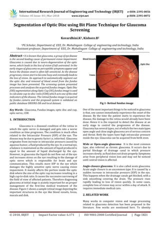
  • 1. International Research Journal of Engineering and Technology (IRJET) e-ISSN: 2395-0056 Volume: 05 Issue: 03 | Mar-2018 www.irjet.net p-ISSN: 2395-0072 © 2018, IRJET | Impact Factor value: 6.171 | ISO 9001:2008 Certified Journal | Page 1821 Segmentation of Optic Disc using Bit Plane Technique for Glaucoma Screening Kovardhini.K1, Kishore.B2 1PG Scholar, Department of EEE, Dr. Mahalingam College of engineering and technology, India 2Assistant professor, Department of EEE, Dr. Mahalingam College of engineering and technology, India ---------------------------------------------------------------------***--------------------------------------------------------------------- Abstract -It is known that glaucoma, a group ofeyedisease is the second leading cause of permanent vision impairment. Glaucoma is caused due to neuro-degeneration of the optic nerve, which leads to the loss of vision if left untreated. In the early stages of glaucoma no perceptible symptomsappearand hence the disease onset is often left unnoticed. As the disease progresses, vision start to become hazy and eventuallyleadsto the loss of vision. An approach to automatically segment out the optic disc and removal of blood vessel from the fundus image has been presented. The screening system presented processes and analyses the acquired fundus images. OpticDisc (OD) segmentation along Optic Cup (OC) fundus image is used to calculate cup-to-disc-ratio (CDR) which acts one ofthevital indicators of the disease. The retinal fundus image is acquired by fundus camera and the proposed system is validated on public database DRIONS-DB and local dataset. Key Words: Glaucoma, Fundus images, optic disc and cup, optic nerve, CDR. 1. INTRODUCTION Glaucoma is a diseased condition of the retina in which the optic nerve is damaged and gets into a worse condition as time progresses. The condition is much often related to the Intraocular Pressure (IOP) of the eye. The disease may be due to genetic factor i.e, inherited. Glaucoma originates from increase in intraocular pressure caused by aqueous humor, a fluid produced by the eye. In a normaleye, a balance is maintained as the amount of liquid produced is equal to the amount of liquid discharged by the eye. However, in glaucoma the liquid do not flow out of the eye and increases stress on the eye resulting in the damage of optic nerve which is responsible for brain and eye communication. This results more IOP in the eye which damages the highly sensitive optic nerve causing vision impairment. It mainly affects the portion inside the optic disk where the size of the optic cup increases resulting in a high cup-to-disk ratio. It causes the successive narrowing of the field of view of affected patients. Therefore diagnosing glaucoma at initial stage is acutely important for an proper management of the first-line medical treatment of the disease. Figure 1 showsa sample retinal image depictingthe important structures in the eye like blood vessels, fovea, macula and disk. Fig 1- Retinal fundus image One of the most important things to be noticed in glaucoma is that, one cannot immediately experience the onset of the disease. By the time the patient starts to experience the disease, the damage to the retina would already have been done. Hence it is the required to detect the disease at an early stage to control the progression of the disease. Glaucoma hasseveral types, out of which the two maintypes open angle and close angle glaucoma are of serious concern and threat. Both the types have high intraocular pressure inside the eye. Glaucoma can be acquired from birth even. Wide or Open-angle glaucoma: It is the most common type, also referred as chronic glaucoma. It occurs due to partial blockage of drainage canal in which pressure increasesslowly, as fluid doesnot drain properly.Symptoms arise from peripheral vision loss and may not be noticed until central vision is affected. Angle-closure glaucoma: It is also called acute glaucoma. Acute angle closure is an uncommon type. It is caused by a sudden increase in intraocular pressure (IOP) in the eye. This happens when the drainage canals get blocked, with a sink smoothing covering the drain. The pressure rises rapidly leading to loss of vision quickly. In some cases, complete loss of vision may occur within a day of attack. It requires immediate medical care. 2. RELATED WORK Many works in computer vision and image processing related to glaucoma detection has been proposed in the literature. Few works are mentioned here. To identity
  • 2. International Research Journal of Engineering and Technology (IRJET) e-ISSN: 2395-0056 Volume: 05 Issue: 03 | Mar-2018 www.irjet.net p-ISSN: 2395-0072 © 2018, IRJET | Impact Factor value: 6.171 | ISO 9001:2008 Certified Journal | Page 1822 symptoms of glaucoma segmentation of optic disc is presented [1] which usesthe localimageinformationaround some point of interest in multi-dimensional feature space and seems to provide robustness against some variants around the optic disc. Segmentation of optic disc and optic cup from color fundus image is presented [2] where a circular Hough transform is used. Cup to disc calculation method is presented [3] where double threshold based approach is used for blood vessel and background extraction and second threshold for segmenting the super intensity pixelscontainedbyopticdisc and optic cup. K-means clustering technique to extract OD and OC was proposed in [4] and elliptical fitting technique is used to calculate the CDR. A super-pixel classification based optic disc and optic cup segmentation[5] was done based on histograms and center surround statistics method. A novel OD localization technique [6]horizontal localization was obtained using scatter degree, by using brightness and edge gradient vertical location was determined, a preprocessing step is used to determine the region of interest in fundus image. Regressive based method [8] and texture descriptors are used to identify the circle which fits the OD boundary which is used to detect the OD. Histogram matching for localizing the OD [7] and average filter is applied to reduce the noise. OD is extracted by using a window and threshold was applied on the correlation function to localize the center of the OD. 3. PROPOSED METHODOLOGY The optic disc is the primary area where the glaucoma symptoms occur. The proposed method of Optic Disc segmentation involves optic disc detection, segmentation and removal of blood vessel from the retinal fundus image. The input image will be automatically processed and the following steps will be made without any manual intervention as shown in Figure 2. Fig 2- Flow Diagram of Proposed OD segmentation The proposed method of glaucoma detection is broadly classified into four different stages i) Image Acquisition ii) Optic disc detection and localization. iii) Optic disc segmentation. iv) Blood vessel removal. a) Digital Fundus image database The fundus imagesused in this work were collectedfromthe DRIONS-DB [8] and local database. In this work total of 110 images were used. The inbuilt imaging software of the funduscamera is used to take picturesof the eye. Theimages are in JPEG format and had a resolution of 600x 400 of 8 bits/pixel. Fig 3 shows the two samples of the digital fundus image of normal and glaucoma affected eye. (a) (b) Fig 3 Fundus Image (a) Normal (b) Glaucoma b) Optic Disc Localization It is known that optic disc is the brightest intensity part in the fundus image. Optic disc is the slightly elliptical shaped in nature and it serves as the exit point of the optic nerves. The strategy adopted here is to detect the bright intensity object in the fundus image. Optic disc center detection involves extraction of green channel from the fundus image because green channel has the maximum intensity feature needed for optic disc detection [9,10]. Kaiser window and hat shaped Kaiser Window is passed to run through each of the rows and columns of the fundus image respectively to detect the optic disc center with exact co-ordinates. c) Optic Disk Segmentation Segmentation of optic disk can be carried out by the way of locating the disk. Various literatures have shown that the optic disk can be easily segregated from the red channel of the fundus RGB image components [11-13]. Optic disc is segmented from the fundusimage using bit plane technique. A digital fundus image is segregated into its bit planes. This isquite useful for analyzing the importance or roleplayedby every bit of the image and it calculates the sufficiency of numbers of bits needed in quantizing each pixel. The database image used has 8 bit representation, hence 6th, 7thand 8th bit plane of the fundus image are considered for
  • 3. International Research Journal of Engineering and Technology (IRJET) e-ISSN: 2395-0056 Volume: 05 Issue: 03 | Mar-2018 www.irjet.net p-ISSN: 2395-0072 © 2018, IRJET | Impact Factor value: 6.171 | ISO 9001:2008 Certified Journal | Page 1823 the optic disc segmentation. After obtaining the red channel of the input image, a circular sub image is extracted depending on the number of rowsand columns in theimage. It is meant to completely cover the optic disc area. Using logical operators, it is possible to combine the bit planes to obtain a common area. The accuracy of this step dependson locating the bright spot. There will not be any bright pixel in the OD; if the above process is done correctly else another common area has to be checked for. If the above passes the check point correctly, then the final segmentation is completed, else using another bit plane, common areahasto be segmented. Similarly using the logical operator, this common area and the previous common area can be combined to get the new area. Commonly logical “AND” can be used. From this newly segmented common area, noisy pixels have to be removed. Labeling of the objects removes noise pixels and the objects are selected having the largest area and roundness. The selected object is nothing but optic disc. Fig 4 shows the fundus image, circular sub image and segmented optic disc. Fig 4 - (a) (b) (c) (a)Fundus image (b) Circular sub image (c) Segmented OD d) Blood Vessel Removal Blood vessels are considered as noisy pixels in the fundus image and have to be eliminated to obtain theaccuracyofOD detection. To remove the noisy pixels from the segmented optic disc, image in-painting is adopted [14]. The pixel intensity is considered as a function of distance from the optic disk center. It is to be noted that as the distance increases outwards from the OD, the gray pixel values tend to decrease. Similar intensity levels are observed for all the pixels that are at equidistant from the center of the optic disk. Presence of blood vessel is detected, if there is any deviation from the above. In this process, the disk pixels are not disturbed. Fig 5 representstheODsub-image,segmented OD, OD image after blood vessel in painted. Fig 5 - (a) (b) (c) (a)OD Sub-image (b) Segmented OD Image (C) OD image after blood vessel in-painted 4. SEGMENTATION METRICS Dice metric is quite commonly used to evaluate the segmentation. It is given by A B DM = 2* A + B  (1) A is the segmented image and B is the ground truth image. A perfect match between the segmented result and the ground truth one is indicated by a dice mean value of 1 and no overlap case is indicated by 0 [15]. Another metric that shows whether the segmented is over segmented or under segmented is the Relative Area Difference (RAD) mean. It is given by A + B RAD mean = *100 B (2) The sign of the RAD mean indicated the metric such that over segmentation is indicated by positive values and vice – versa. The proposed system of OD segmentation is tested on DRIONS-DB and local dataset. The average Dice coefficient and RAD Mean for the OD segmentation was 0.94and96.9%, respectively on the local dataset, while obtaining 0.93 and 95% for DRIONS-DB image set. 5. CONCLUSION The automatic optic disc segmentation technique has been proposed by using double windowing method. The noisy blood vessels are removed from segmented optic disc by using image in-painting technique. The efficiency of the segmentation results were tested on the DRIONS-DB and local dataset and their performance is evaluated using two metrics. As OD segmentation plays a vital role in the detection of glaucoma, this method can aid in the early detection or screening of the disease, glaucoma can only be treated and cannot be prevented. 6. REFERENCES [1] G. Joshi, J. Sivaswamy, S.R. Krishnadas, Optic disc and cup segmentation from monocular color fundus images for glaucoma assessment, IEEE Trans. Med. Imag. 30 (2011, June) 1192–1205 [2] F. Yin, J. Liu, D.W. Kee Wong, N.M. Tan, C. Cheung, M. Baskaran, T.Y. Wong, Automated segmentation of optic disc and optic cup in fundus images for glaucoma diagnosis, in: 25th International Symposium on Computer- Based Medical System,pp. 1–6, 2012 [3] M.K. Dutta, A.K. Mourya, A. Singh, M. Parthasarathi, R. Burget, K. Riha, Glaucoma detection by segmenting the super pixels from fundus color retinal images, in: International Conference on Medical Imaging, m-Health & Emerging Communication System, (MEDCOM., 2014) IEEE Xplore, New York, USA, 2014 Nov.
  • 4. International Research Journal of Engineering and Technology (IRJET) e-ISSN: 2395-0056 Volume: 05 Issue: 03 | Mar-2018 www.irjet.net p-ISSN: 2395-0072 © 2018, IRJET | Impact Factor value: 6.171 | ISO 9001:2008 Certified Journal | Page 1824 [4] K. Narasimhan and K. Vijayarekha ,“An efficient automated system for glaucoma detection using fundus image,” Journal of Theoretical and Applied Information Technology, vol. 33, no. 1, pp.104–110,2011 [5] J.Cheng,J.Liu,Y.Xuetal.,“Superpixel classification based optic disc and optic cup segmentation for glaucoma screening, ”IEEE Transactions on Medical Imaging, vol. 32, no. 6, pp. 1019–1032, 2013. [6] D.Zhang,Y.Yi,X.Shang,andY.Peng,“Optic disc localization by projection with vessel distribution and appearance characteristics,” in Proceedings of the 21st International Conference on Pattern Recognition(ICPR’12),pp.3176– 3179,November2012 [7] S.Lu, “Accurate and efficient optic disc detection and segmentation by a circular transformation, ”IEEE Transactions on Medical Imaging,vol.30,no.12,pp.2126– 2133,2011. [8]. http://www.ia.uned.es/~ejcarmona/DRIONS-DB_1.html [9]. M. Usman Akram Aftab et, al.”Retinal Images: Optic Disk Localization and Detection”, ProceedingsoftheInternational Conference Image Analysis and Recognition, pp40-49,2010 [10] C.A.Lupa¸scu,D.Tegolo,andL.DiRosa,“Automated detection of optic disc location in retinal images,” in Proceedings of the 21st IEEE International Symposium on Computer-Based Medical Systems (CBMS’08),pp.1722, Jyvaskyla, Finland,June2008. [11] G. Joshi, J. Sivaswamy, S.R. Krishnadas, Optic disc and cup segmentation from monocular color fundus images for glaucoma assessment,” IEEE Transactions on Medical Imaging. Vol 30. pp 1192–1205, 2011 [12] A. A.-H. A.-R. Youssif, A. Z. Ghalwash, and A. A. S. A.-R. Ghoneim,“Optic disc detection from normalized digital fundus images by means of a vessels’ direction matched filter,” IEEE Transactions on Medical Imaging, Vol 27, no 1, pp.11–18,2008. [13] J. Cheng, J. Liu, Y. Xu, F. Yin, D.W. Kee Wong, N.-M. Tan, D. Tao, C.-Y. Cheng, T. Aung, T.Y. Wong, Superpixel classification based optic disc and optic cup segmentation for glaucoma screening, IEEE Transactions on Medical Imaging. Vol 32, pp. 1019–1032, 2013 [14]. M. M. Fraz, P. Remagnino, A. Hoppe et al., “Blood vessel segmentation methodologies in retinal images—a survey,” Computer Methods and Programs in Biomedicine, vol. 108, no. 1, pp. 407–433, 2012. [15]. F. Yin, J. Liu, D. W. K. Wong et al., “Automated segmentation of optic disc and optic cupinfundusimagesfor glaucoma diagnosis,” in Proceedings of the 25th IEEE International Symposium on Computer-Based Medical Systems (CBMS '12), pp. 1–6, 2012.