SlideShare a Scribd company logo
1 of 5
Download to read offline
International Research Journal of Engineering and Technology (IRJET) e-ISSN: 2395-0056
Volume: 09 Issue: 05 | May 2022 www.irjet.net p-ISSN: 2395-0072
© 2022, IRJET | Impact Factor value: 7.529 | ISO 9001:2008 Certified Journal | Page 3300
FEATURE EXTRACTION FROM RETINAL FUNDUS IMAGES
A. ARUL AFRIN MARY1, I. MUTHUMANI2, V. SELVAKUMAR3
1Student, Dept. of ECE, GCE, Tirunelveli, Tamil Nadu, India
2Professor, Dept. of ECE, GCE, Tirunelveli, Tamil Nadu, India
3Assistant Professor, Dept. of ECE, GCE, Tirunelveli, Tamil Nadu, India
---------------------------------------------------------------------***---------------------------------------------------------------------
Abstract - COVID-19 pandemic has brought around
changes in human life and work habits due to lockdown and
has resulted in an increase in the frequency of numerous
conditions like Diabetes. One such side effect of Diabetes is
Diabetic Retinopathy. The proposed method aims to detect
the optic disc, blood vessels and exudates in the retinal
fundus images. Optic disc is detected using Histogram
Normalization technique. Blood vessels are detected using
Kirsch’s template and Morphological Operations. Exudates
appear as small white dots on the retinal fundus images
which are detected using Morphological Operations. Retinal
fundus images for testing were collected from the standard
diabetic retinopathy database DIARETDB0 and DIARETDB1.
From the results, it is found that the area of the extracted
optic disc, blood vessels and exudates are more accurate
than the state-of-the-art methods.
Key Words: Fundus Images, Optic Disc, Blood Vessels,
Exudates, Morphological Operations.
1. INTRODUCTION
Human eye is a sense receptor that reacts to light
and permits vision with the assistance of the
photoreceptor cells (rod and cone cells) present in it. Rod
cell is helpful for vision during night time or in dim light
and this vision is named as scotopic vision. Cone cells
facilitate vision during daytime or in bright light and this
vision is termed as photopic vision. Internal surface of the
eye is the fundus which incorporates macula, retina, optic
disc and fovea and can be examined by ophthalmoscope
and/or fundus photography. Optic disc also known as
optic nerve head is the point where the optic nerve
connects to the brain. Optic disc is additionally referred to
as blind spot since it doesn’t embody any photoreceptor
cells. Common eye disorders are cataract, night blindness,
glaucoma, Diabetic Retinopathy.
Cataracts may occur either due to an eye injury or
excessive exposure to actinic radiations. Nyctalopia also
called night-blindness is a condition where the affected
person is unable to see during night time or in dim light. It
may also occur due to the deficiency of Vitamin-A.
Glaucoma is a group of eye conditions which damages the
optic nerve caused by an abnormally high pressure within
the eye.
The first stage known as non-proliferative
diabetic retinopathy (NPDR) in which the affected person
has no symptoms and can be detected by fundus
examination either by direct opthalmoscope or indirect
ophthalmoscope by a trained oculist or optometrist.
The second stage known as proliferative diabetic
retinopathy (PDR) in which abnormal new blood vessels
(neovascularization) are formed at the back of the eye.
Upon rupturing, these blood vessels leak fluid which
causes deposition of lipids within the eye. These deposits
are termed as exudates which are the primary clinical
signs of DR, and they appear as small white dots on the
retinal fundus images. These exudates are classified into
two sorts as:
 Hard exudates – They’re deep yellow and don’t
have a precise margin.
 Soft exudates – They don’t have a definite
boundary and are also known as cotton wool
spots.
The number of exudates is employed to point the
severity of the disease. Hard and soft exudates are
conjointly known as bright lesions.
Fig -1: Normal Fundus Image vs. DR affected Fundus
Image
Insulin secreted by the duct gland helps glucose
from food get into the cells to be used for energy. Diabetes
is a disease that occurs when the blood glucose is just too
high which could cause many other diseases. One such
disease is Diabetic Retinopathy. Diabetic Retinopathy (DR)
is a medical condition which damages the eye’s retina due
to diabetes which is a leading cause of blindness in several
developed countries. Diabetic retinopathy has no early
warning signs. Diabetic Retinopathy affected fundus
images can be characterized by the presence of either
exudates (or) microaneurysms (or) hemorrhages (or) all
looking on the severity of the disease.
International Research Journal of Engineering and Technology (IRJET) e-ISSN: 2395-0056
Volume: 09 Issue: 05 | May 2022 www.irjet.net p-ISSN: 2395-0072
© 2022, IRJET | Impact Factor value: 7.529 | ISO 9001:2008 Certified Journal | Page 3301
Microaneurysm is an eye condition in which tiny
red dots encircled by yellow rings are found within the eye
as a result of vascular leakage. Retinal hemorrhage refers
to abnormal bleeding within the delicate blood vessels of
the retina whose symptoms range from the undetectable
to severe vision issues. Vision problems are often
temporary, but in some instances, they’ll be permanent.
Microaneurysms and hemorrhages are jointly known as
red lesions.
2. PROPOSED METHODOLOGY
The detection of optic disc, blood vessels and
exudates is a major problem in the automatic processing
of retinal images. The segmentation of exudates is
essential in differentiating the normal eye from the DR
affected eye. In this work fundus image of the retina is
used. MATLAB is used for implementing the process.
2.1 Dataset
The retinal fundus images for testing were taken
from the standard publicly available Diabetic Retinopathy
database DIARETDB0 and DIARETDB1.
2.2 Preprocessing
Preprocessing includes resizing of the retinal
fundus images and choosing the appropriate color
component from the RGB image.
The retinal fundus images collected for testing
were of different resolutions. In order to maintain a
uniform resolution, all the images collected were resized
to 512x512 pixels. The resized retinal fundus images were
in RGB colour space. The contents in the blue colour
channel of the image are of low contrast whereas the red
colour channel of the image is noisy. The optic disc, blood
vessels and exudates has good contrast in the green colour
channel of the image. Hence the green channel is chosen
for the analysis of the retinal fundus images.
2.3 Image Enhancement
CLAHE (Contrast-Limited Adaptive Histogram
Equalization) algorithm is applied for noise reduction by
increasing the contrast and filtering of the image.
Histogram equalization is used for enhancing the contrast
of the image. It removes the background for the resulted
image by converting the background to white color (every
pixel outside the white board changed to white color).
2.4 Exudates Detection
The proposed technique for the detection of
exudates in the retinal fundus images is implemented
using morphological top- and bottom-hat filtering
operations.
The flow diagram for exudates extraction in the
retinal fundus images is shown in fig. 2
Fig -2: Flow Diagram for Exudates Extraction
The exudates from the retinal fundus images are
detected by applying the morphological bottom-hat
filtering on the extracted green channel of the fundus
image with a disc shaped structuring element. Bottom-hat
filtering subtracts the original image from the
morphologically closed image. Then morphological top-hat
filtering is applied on the extracted green channel of the
fundus image with a disc shaped structuring element. Top-
hat filtering subtracts the morphologically opened image
from the original image. Then the bottom-hat filtered
image is subtracted from the top-hat filtered image. Then a
binary image is obtained by converting the resultant
image with a threshold value i.e., the pixels with
luminance value greater than the threshold value are
replaced by value 1 (white) and the remaining pixels are
replaced by a value 0 (black). The white pixels correspond
to the exudates pixels in the original image.
2.5 Blood Vessels Detection
The proposed technique for the detection of blood
vessels in the retinal fundus images is implemented using
Kirsch’s templates.
The flow diagram for blood vessel extraction in
retinal fundus images is shown in fig. 3
Fig -3: Flow Diagram for Blood Vessels Extraction
The blood vessels in the retinal fundus images
were detected by applying a 2-dimensional digital finite
impulse response (FIR) on the bottom-hat transformed
The flow diagram for exudates feature extraction in the retinal fundus images is shown in fig. 3
Fig. 3 Flow Diagram for Exudates Extraction
Retinal Fundus Images
Extract Green Channel
Extracted ExudatePixels
Convert to Binary Image
Morphological Bottom Hat
Morphological Top Hat
The flow diagram for blood vessels feature extraction in the retinal fundus images is shown in fig. 4
Fig. 4 Flow Diagram for Blood Vessels Extraction
Retinal Fundus Images
Extract GreenChannel
Extracted Blood Vessel
Pixels
Threshold Image
Morphological Bottom Hat
FIR Filter
International Research Journal of Engineering and Technology (IRJET) e-ISSN: 2395-0056
Volume: 09 Issue: 05 | May 2022 www.irjet.net p-ISSN: 2395-0072
© 2022, IRJET | Impact Factor value: 7.529 | ISO 9001:2008 Certified Journal | Page 3302
image and the Kirsch’s template co - efficients. Then a
threshold is applied on the 2-D FIR filtered image to
extract the pixels of the blood vessels from the filtered
image. The pixels greater than the threshold value
correspond to the blood vessel pixels.
2.6 Optic Disc Detection
The proposed technique for the detection of optic
disc in the retinal fundus images is implemented using
histogram conversion and normalization of histogram.
The flow diagram for optic disc extraction in
retinal fundus images is shown in fig. 4
Fig -4: Flow Diagram for Optic Disc Extraction
In image processing, normalization is a process
used for changing the range of intensity values of the
pixels in the image. It is also called histogram stretching or
contrast stretching. Histogram normalization enhances the
fine details within an image. Then a threshold is applied on
the normalized histogram to find the pixels of the optic
disc from the normalized histogram. The pixels greater
than the threshold value correspond to the optic disc
pixels.
3. RESULT
3.1 Retinal Fundus Image
Fundus Image consists of RGB Channel which has
unique feature.
Fig -5: Normal Retinal
Fundus Image
Fig -6: DR affected Retinal
Fundus Image
3.2 Detected Optic Disc
Fig. 7, 8 shows the detected optic edges disc in
normal and DR affected fundus images.
Fig -7: Detected Optic Disc
in a Normal Retinal Fundus
Image
Fig -8: Detected Optic Disc
in a DR affected Retinal
Fundus Image
3.3 Detected Blood Vessels
Fig. 9, 10 shows the detected blood vessels in
normal and DR affected fundus images.
Fig -9: Detected Blood
Vessels in a Normal Retinal
Fundus Image
Fig -10: Detected Blood
Vessels in a DR affected
Retinal Fundus Image
3.4 Detected Exudates
Fig. 11, 12 shows the detected exudates in normal
and DR affected fundus images.
Fig -11: Detected Exudates
in a Normal Retinal Fundus
Image
Fig -12: Detected Exudates
in a DR affected Retinal
Fundus Image
The flow diagram for optic disc feature extractioninthe retinal fundus images is shown in fig. 3
Fig. 3 Flow Diagram for Optic Disc Extraction
Retinal Fundus Images
Extract GreenChannel
Extracted Optic Disc Pixels
Threshold Image
Contrast Enhancement
Normalization of Histogram
International Research Journal of Engineering and Technology (IRJET) e-ISSN: 2395-0056
Volume: 09 Issue: 05 | May 2022 www.irjet.net p-ISSN: 2395-0072
© 2022, IRJET | Impact Factor value: 7.529 | ISO 9001:2008 Certified Journal | Page 3303
From fig.11, it is observed that the normal retinal
fundus images do not have any exudates. The yellow dots
in the above figure (fig. 12) shows the detected exudates.
3.5 Area of the Detected Features
Fig. 13, 14 shows the area of the extracted
features from the retinal fundus image.
Fig -13: Area of the
Extracted Features in a
Normal Retinal Fundus
Image
Fig -14: Area of the
Extracted Features in a DR
affected Retinal Fundus
Image
4. CONCLUSION
In this work, features like optic disc, blood vessels
and exudates in the human retinal fundus images were
extracted. Retinal fundus images including both normal
and Diabetic Retinopathy affected images were taken from
the publicly available standard Diabetic Retinopathy
Databases like DIARETDB0 and DIARETDB1 for testing.
Optic Disc in the retinal fundus images were detected
using the Histogram Normalization technique. Blood
Vessels in the retinal fundus images were detected using
Kirsch’s templates and Morphological Operations.
Exudates in the retinal fundus images were detected using
the Morphological Operations. The area of the extracted
features was also measured. The extraction of all the
above three features and the area of these extracted
features were performed using MATLAB R2018a. From
the extracted exudates and its area, it was observed that
the area of the exudates was zero in the normal retinal
fundus images and greater than zero in the case of
Diabetic Retinopathy affected retinal fundus images. From
this observation, it is inferred that if the area of the
exudates is greater than zero, then the corresponding
retinal fundus image can be classified as a Diabetic
Retinopathy affected retinal fundus image and if the area
of the exudates is equal to zero, then the corresponding
retinal fundus image can be classified as a normal retinal
fundus image.
REFERENCES
[1] Meindert Niemeijer, Bram van Ginneken, Joes
Staal, Maria S. A. Suttorp-Schulten, Michael D.
Abramoff, “Automatic Detection of Red Lesions in
Digital Color Fundus Photographs,” IEEE
Transactions on Medical Imaging, Vol. 24, No. 5,
pp. 584-592, May 2005.
[2] Dongbo Zhang, Xiong Li, Xinyu Shang, Yao Yi,
Yaonan Wang, “Robust Hemorrhage Detection in
Diabetic Retinopathy Detection,” pp. 209-213,
2011 IEEE.
[3] Balint Antal, Andras Hajdu, “An Ensemble-Based
System for Microaneurysms Detection and
Diabetic Retinopathy Grading,” IEEE Transactions
on Biomedical Engineering, Vol. 59, 6, pp. 1720-
1726, June 2012.
[4] Sohini Roychowdhury, Dara D. Koozekanani,
Keshab K. Parthi, “Screening Fundus Images for
Diabetic Retinopathy,” pp. 1641-1645, 2012 IEEE.
[5] M. Usman Akram, Shehzad Khalid, Shoab A. Khan,
“Identification and classification of
microaneurysms for early detection of diabetic
retinopathy,” ELSEVIER - Pattern Recognition 46
(2013), pp. 107-116.
[6] L. Giancardo, F. Meriaudeau, T. P. Karnowski, K.W.
Tobin, E. Chaum, “Validation of Microaneurysm-
based Diabetic Retinopathy Screening across
Retinal Fundus Datasets,” pp. 125-130, 2013 IEEE.
[7] Sundararaj Wifred Franklin, Samuelnadar Edwar
Rajan, “Diagnosis of Diabetic Retinopathy by
employing Image Processing Techniques to detect
Exudates in retinal images,” IET Image Process,
2014 Vol. 8, Iss. 10, pp. 601 – 609.
[8] R. S. Deshmukh, “Analysis of Diabetic Retinopathy
by Automatic Detection of Exudates,”
International Journal of Electronics and
Communication Engineering (IJECET), Vol. 5, Iss.
12, December (2014), pp. 266 – 275.
[9] ManojKumar S B, Manjunath R, Dr. H S Sheshadri,
“Feature extraction from the Fundus Images for
the diagnosis of diabetic retinopathy,”
International Conference on Emerging Research
in Electronics, Computer Science and Technology,
pp. 240-245, 2015 IEEE.
[10] Sudeshna Sil Kar, Santi P. Maity, “Automatic
Detection of Retinal Lesions for Screening of
Diabetic Retinopathy,” IEEE Transactions on
Biomedical Engineering, pp. 1-9, 2016 IEEE.
[11] Karan Bhatia, Shikar Arora, Ravi Tomar,
“Diagnosis of Diabetic Retinopathy Using Machine
Learning Classification Algorithm,” 2016 2nd
International Research Journal of Engineering and Technology (IRJET) e-ISSN: 2395-0056
Volume: 09 Issue: 05 | May 2022 www.irjet.net p-ISSN: 2395-0072
© 2022, IRJET | Impact Factor value: 7.529 | ISO 9001:2008 Certified Journal | Page 3304
International Conference on Next Generation
Computing Technologies (NGCT-2016), pp. 347-
351, 2016 IEEE.
[12] Sarni Suhaila Rahim, Vasile Palade, James
Shuttleworth, Christiana Jayne, “Automatic
screening and classification of diabetic
retinopathy and maculopathy using fuzzy image
processing,” Brain Informatics (2016) 3: 249 –
267.
[13] Dilip Singh Sisodia, Shruti Nair, Pooja Khobragde,
“Diabetic Retinal Fundus Images: Preprocessing
and Feature Extraction For Early Detection of
Diabetic Retinopathy,” Biomedical and
Pharmacology Journal, Vol. 10(2), pp. 615-626
(2017).
[14] Wei Zhou, Chendong Wu, Dali Chen, Zhenzhu
Wang, Yugen Yi, Wenyou Du, “Automatic
Microaneurysm Detection of Diabetic Retinopathy
in Fundus Images,” pp. 7453-7458, 2017 IEEE.
[15] Shuang Yu, Di Xiao, Yogesan Kanagasingam,
“Exudate Detection for Diabetic Retinopathy with
Convolutional Neural Networks,” pp. 1744-1747,
2017 IEEE.
[16] Wei Zhou, Chengdong Wu, Xiaosheng Yu,
“Computer Aided Diagnosis for Diabetic
Retinopathy based on Fundus Image,”
Proceedings of the 37th Chinese Control
Conference 2018, pp. 9214-9219.
[17] Zihao Wang, Ke-Jia Chen, Lingli Zhang, “A R-CNN
Based Approach for Microaneurysm Detection in
Retinal Fundus Images,” – 2019.
[18] Muhammad Mateen, Junhao Wen, Nasrullah
Nasrullah, Song Sun, Shaukat Hayat, “Exudate
Detection for Diabetic Retinopathy using
Pretrained Convolutional Neural Networks,”
Hindawi Complexity, Vol. 2020, pp. 1-11.
[19] DIARETDB0 and DIARETDB1 Database.
[20] Software Package, “MATLAB (2018a)”.

More Related Content

Similar to FEATURE EXTRACTION FROM RETINAL FUNDUS IMAGES

Glaucoma Detection from Retinal Images
Glaucoma Detection from Retinal ImagesGlaucoma Detection from Retinal Images
Glaucoma Detection from Retinal Images
ijtsrd
 
A review on implementation of algorithms for detection of diabetic retinopathy
A review on implementation of algorithms for detection of diabetic retinopathyA review on implementation of algorithms for detection of diabetic retinopathy
A review on implementation of algorithms for detection of diabetic retinopathy
eSAT Journals
 

Similar to FEATURE EXTRACTION FROM RETINAL FUNDUS IMAGES (20)

Detection and Grading of Diabetic Maculopathy Automatically in Digital Retina...
Detection and Grading of Diabetic Maculopathy Automatically in Digital Retina...Detection and Grading of Diabetic Maculopathy Automatically in Digital Retina...
Detection and Grading of Diabetic Maculopathy Automatically in Digital Retina...
 
IRJET- Retinal Health Diagnosis using Image Processing
IRJET- Retinal Health Diagnosis using Image ProcessingIRJET- Retinal Health Diagnosis using Image Processing
IRJET- Retinal Health Diagnosis using Image Processing
 
Diagnosis of Diabetic Retinopathy
Diagnosis of Diabetic RetinopathyDiagnosis of Diabetic Retinopathy
Diagnosis of Diabetic Retinopathy
 
CATARACT DISEASE DETECTION AND CLASSIFICATION USING RETINAL IMAGE MODEL
CATARACT DISEASE DETECTION AND CLASSIFICATION USING RETINAL IMAGE MODELCATARACT DISEASE DETECTION AND CLASSIFICATION USING RETINAL IMAGE MODEL
CATARACT DISEASE DETECTION AND CLASSIFICATION USING RETINAL IMAGE MODEL
 
IRJET- Early Detection of Glaucoma using Image Processing
IRJET- Early Detection of Glaucoma using Image ProcessingIRJET- Early Detection of Glaucoma using Image Processing
IRJET- Early Detection of Glaucoma using Image Processing
 
IRJET- Approach for Diabetic Retinopathy Analysis using Artificial Neural Net...
IRJET- Approach for Diabetic Retinopathy Analysis using Artificial Neural Net...IRJET- Approach for Diabetic Retinopathy Analysis using Artificial Neural Net...
IRJET- Approach for Diabetic Retinopathy Analysis using Artificial Neural Net...
 
SVM based CSR disease detection for OCT and Fundus Imaging
SVM based CSR disease detection for OCT and Fundus ImagingSVM based CSR disease detection for OCT and Fundus Imaging
SVM based CSR disease detection for OCT and Fundus Imaging
 
IRJET- Study on Segmentation and Classification Techniques for Automatic ...
IRJET-  	  Study on Segmentation and Classification Techniques for Automatic ...IRJET-  	  Study on Segmentation and Classification Techniques for Automatic ...
IRJET- Study on Segmentation and Classification Techniques for Automatic ...
 
IRJET- Retinal Structure Segmentation using Adaptive Fuzzy Thresholding
IRJET- Retinal Structure Segmentation using Adaptive Fuzzy ThresholdingIRJET- Retinal Structure Segmentation using Adaptive Fuzzy Thresholding
IRJET- Retinal Structure Segmentation using Adaptive Fuzzy Thresholding
 
Blood vessel segmentation in fundus images
Blood vessel segmentation in fundus imagesBlood vessel segmentation in fundus images
Blood vessel segmentation in fundus images
 
IRJET -An Automatated Learning Approach for Detection of Diabetic Retinopathy...
IRJET -An Automatated Learning Approach for Detection of Diabetic Retinopathy...IRJET -An Automatated Learning Approach for Detection of Diabetic Retinopathy...
IRJET -An Automatated Learning Approach for Detection of Diabetic Retinopathy...
 
Glaucoma Detection from Retinal Images
Glaucoma Detection from Retinal ImagesGlaucoma Detection from Retinal Images
Glaucoma Detection from Retinal Images
 
IRJET -Analysis of Ophthalmic System Applications using Signal Processing
IRJET -Analysis of Ophthalmic System Applications using Signal ProcessingIRJET -Analysis of Ophthalmic System Applications using Signal Processing
IRJET -Analysis of Ophthalmic System Applications using Signal Processing
 
Glaucoma Screening Using Novel Evaluated CNN Architecture: An Automated Appro...
Glaucoma Screening Using Novel Evaluated CNN Architecture: An Automated Appro...Glaucoma Screening Using Novel Evaluated CNN Architecture: An Automated Appro...
Glaucoma Screening Using Novel Evaluated CNN Architecture: An Automated Appro...
 
V01761152156
V01761152156V01761152156
V01761152156
 
Automated Detection of Microaneurysm, Hard Exudates, and Cotton Wool Spots in...
Automated Detection of Microaneurysm, Hard Exudates, and Cotton Wool Spots in...Automated Detection of Microaneurysm, Hard Exudates, and Cotton Wool Spots in...
Automated Detection of Microaneurysm, Hard Exudates, and Cotton Wool Spots in...
 
IRJET- Automatic Detection of Diabetic Retinopathy using R-CNN
IRJET- Automatic Detection of Diabetic Retinopathy using R-CNNIRJET- Automatic Detection of Diabetic Retinopathy using R-CNN
IRJET- Automatic Detection of Diabetic Retinopathy using R-CNN
 
Diagnosis of Diabetic Retinopathy by Detection of Microneurysm And Exudates
Diagnosis of Diabetic Retinopathy by Detection of Microneurysm And ExudatesDiagnosis of Diabetic Retinopathy by Detection of Microneurysm And Exudates
Diagnosis of Diabetic Retinopathy by Detection of Microneurysm And Exudates
 
A review on implementation of algorithms for detection of diabetic retinopathy
A review on implementation of algorithms for detection of diabetic retinopathyA review on implementation of algorithms for detection of diabetic retinopathy
A review on implementation of algorithms for detection of diabetic retinopathy
 
A review on implementation of algorithms for detection of diabetic retinopathy
A review on implementation of algorithms for detection of diabetic retinopathyA review on implementation of algorithms for detection of diabetic retinopathy
A review on implementation of algorithms for detection of diabetic retinopathy
 

More from IRJET Journal

More from IRJET Journal (20)

TUNNELING IN HIMALAYAS WITH NATM METHOD: A SPECIAL REFERENCES TO SUNGAL TUNNE...
TUNNELING IN HIMALAYAS WITH NATM METHOD: A SPECIAL REFERENCES TO SUNGAL TUNNE...TUNNELING IN HIMALAYAS WITH NATM METHOD: A SPECIAL REFERENCES TO SUNGAL TUNNE...
TUNNELING IN HIMALAYAS WITH NATM METHOD: A SPECIAL REFERENCES TO SUNGAL TUNNE...
 
STUDY THE EFFECT OF RESPONSE REDUCTION FACTOR ON RC FRAMED STRUCTURE
STUDY THE EFFECT OF RESPONSE REDUCTION FACTOR ON RC FRAMED STRUCTURESTUDY THE EFFECT OF RESPONSE REDUCTION FACTOR ON RC FRAMED STRUCTURE
STUDY THE EFFECT OF RESPONSE REDUCTION FACTOR ON RC FRAMED STRUCTURE
 
A COMPARATIVE ANALYSIS OF RCC ELEMENT OF SLAB WITH STARK STEEL (HYSD STEEL) A...
A COMPARATIVE ANALYSIS OF RCC ELEMENT OF SLAB WITH STARK STEEL (HYSD STEEL) A...A COMPARATIVE ANALYSIS OF RCC ELEMENT OF SLAB WITH STARK STEEL (HYSD STEEL) A...
A COMPARATIVE ANALYSIS OF RCC ELEMENT OF SLAB WITH STARK STEEL (HYSD STEEL) A...
 
Effect of Camber and Angles of Attack on Airfoil Characteristics
Effect of Camber and Angles of Attack on Airfoil CharacteristicsEffect of Camber and Angles of Attack on Airfoil Characteristics
Effect of Camber and Angles of Attack on Airfoil Characteristics
 
A Review on the Progress and Challenges of Aluminum-Based Metal Matrix Compos...
A Review on the Progress and Challenges of Aluminum-Based Metal Matrix Compos...A Review on the Progress and Challenges of Aluminum-Based Metal Matrix Compos...
A Review on the Progress and Challenges of Aluminum-Based Metal Matrix Compos...
 
Dynamic Urban Transit Optimization: A Graph Neural Network Approach for Real-...
Dynamic Urban Transit Optimization: A Graph Neural Network Approach for Real-...Dynamic Urban Transit Optimization: A Graph Neural Network Approach for Real-...
Dynamic Urban Transit Optimization: A Graph Neural Network Approach for Real-...
 
Structural Analysis and Design of Multi-Storey Symmetric and Asymmetric Shape...
Structural Analysis and Design of Multi-Storey Symmetric and Asymmetric Shape...Structural Analysis and Design of Multi-Storey Symmetric and Asymmetric Shape...
Structural Analysis and Design of Multi-Storey Symmetric and Asymmetric Shape...
 
A Review of “Seismic Response of RC Structures Having Plan and Vertical Irreg...
A Review of “Seismic Response of RC Structures Having Plan and Vertical Irreg...A Review of “Seismic Response of RC Structures Having Plan and Vertical Irreg...
A Review of “Seismic Response of RC Structures Having Plan and Vertical Irreg...
 
A REVIEW ON MACHINE LEARNING IN ADAS
A REVIEW ON MACHINE LEARNING IN ADASA REVIEW ON MACHINE LEARNING IN ADAS
A REVIEW ON MACHINE LEARNING IN ADAS
 
Long Term Trend Analysis of Precipitation and Temperature for Asosa district,...
Long Term Trend Analysis of Precipitation and Temperature for Asosa district,...Long Term Trend Analysis of Precipitation and Temperature for Asosa district,...
Long Term Trend Analysis of Precipitation and Temperature for Asosa district,...
 
P.E.B. Framed Structure Design and Analysis Using STAAD Pro
P.E.B. Framed Structure Design and Analysis Using STAAD ProP.E.B. Framed Structure Design and Analysis Using STAAD Pro
P.E.B. Framed Structure Design and Analysis Using STAAD Pro
 
A Review on Innovative Fiber Integration for Enhanced Reinforcement of Concre...
A Review on Innovative Fiber Integration for Enhanced Reinforcement of Concre...A Review on Innovative Fiber Integration for Enhanced Reinforcement of Concre...
A Review on Innovative Fiber Integration for Enhanced Reinforcement of Concre...
 
Survey Paper on Cloud-Based Secured Healthcare System
Survey Paper on Cloud-Based Secured Healthcare SystemSurvey Paper on Cloud-Based Secured Healthcare System
Survey Paper on Cloud-Based Secured Healthcare System
 
Review on studies and research on widening of existing concrete bridges
Review on studies and research on widening of existing concrete bridgesReview on studies and research on widening of existing concrete bridges
Review on studies and research on widening of existing concrete bridges
 
React based fullstack edtech web application
React based fullstack edtech web applicationReact based fullstack edtech web application
React based fullstack edtech web application
 
A Comprehensive Review of Integrating IoT and Blockchain Technologies in the ...
A Comprehensive Review of Integrating IoT and Blockchain Technologies in the ...A Comprehensive Review of Integrating IoT and Blockchain Technologies in the ...
A Comprehensive Review of Integrating IoT and Blockchain Technologies in the ...
 
A REVIEW ON THE PERFORMANCE OF COCONUT FIBRE REINFORCED CONCRETE.
A REVIEW ON THE PERFORMANCE OF COCONUT FIBRE REINFORCED CONCRETE.A REVIEW ON THE PERFORMANCE OF COCONUT FIBRE REINFORCED CONCRETE.
A REVIEW ON THE PERFORMANCE OF COCONUT FIBRE REINFORCED CONCRETE.
 
Optimizing Business Management Process Workflows: The Dynamic Influence of Mi...
Optimizing Business Management Process Workflows: The Dynamic Influence of Mi...Optimizing Business Management Process Workflows: The Dynamic Influence of Mi...
Optimizing Business Management Process Workflows: The Dynamic Influence of Mi...
 
Multistoried and Multi Bay Steel Building Frame by using Seismic Design
Multistoried and Multi Bay Steel Building Frame by using Seismic DesignMultistoried and Multi Bay Steel Building Frame by using Seismic Design
Multistoried and Multi Bay Steel Building Frame by using Seismic Design
 
Cost Optimization of Construction Using Plastic Waste as a Sustainable Constr...
Cost Optimization of Construction Using Plastic Waste as a Sustainable Constr...Cost Optimization of Construction Using Plastic Waste as a Sustainable Constr...
Cost Optimization of Construction Using Plastic Waste as a Sustainable Constr...
 

Recently uploaded

VIP Call Girls Palanpur 7001035870 Whatsapp Number, 24/07 Booking
VIP Call Girls Palanpur 7001035870 Whatsapp Number, 24/07 BookingVIP Call Girls Palanpur 7001035870 Whatsapp Number, 24/07 Booking
VIP Call Girls Palanpur 7001035870 Whatsapp Number, 24/07 Booking
dharasingh5698
 
Call Now ≽ 9953056974 ≼🔝 Call Girls In New Ashok Nagar ≼🔝 Delhi door step de...
Call Now ≽ 9953056974 ≼🔝 Call Girls In New Ashok Nagar  ≼🔝 Delhi door step de...Call Now ≽ 9953056974 ≼🔝 Call Girls In New Ashok Nagar  ≼🔝 Delhi door step de...
Call Now ≽ 9953056974 ≼🔝 Call Girls In New Ashok Nagar ≼🔝 Delhi door step de...
9953056974 Low Rate Call Girls In Saket, Delhi NCR
 
Integrated Test Rig For HTFE-25 - Neometrix
Integrated Test Rig For HTFE-25 - NeometrixIntegrated Test Rig For HTFE-25 - Neometrix
Integrated Test Rig For HTFE-25 - Neometrix
Neometrix_Engineering_Pvt_Ltd
 
VIP Call Girls Ankleshwar 7001035870 Whatsapp Number, 24/07 Booking
VIP Call Girls Ankleshwar 7001035870 Whatsapp Number, 24/07 BookingVIP Call Girls Ankleshwar 7001035870 Whatsapp Number, 24/07 Booking
VIP Call Girls Ankleshwar 7001035870 Whatsapp Number, 24/07 Booking
dharasingh5698
 

Recently uploaded (20)

UNIT - IV - Air Compressors and its Performance
UNIT - IV - Air Compressors and its PerformanceUNIT - IV - Air Compressors and its Performance
UNIT - IV - Air Compressors and its Performance
 
COST-EFFETIVE and Energy Efficient BUILDINGS ptx
COST-EFFETIVE  and Energy Efficient BUILDINGS ptxCOST-EFFETIVE  and Energy Efficient BUILDINGS ptx
COST-EFFETIVE and Energy Efficient BUILDINGS ptx
 
VIP Call Girls Palanpur 7001035870 Whatsapp Number, 24/07 Booking
VIP Call Girls Palanpur 7001035870 Whatsapp Number, 24/07 BookingVIP Call Girls Palanpur 7001035870 Whatsapp Number, 24/07 Booking
VIP Call Girls Palanpur 7001035870 Whatsapp Number, 24/07 Booking
 
FEA Based Level 3 Assessment of Deformed Tanks with Fluid Induced Loads
FEA Based Level 3 Assessment of Deformed Tanks with Fluid Induced LoadsFEA Based Level 3 Assessment of Deformed Tanks with Fluid Induced Loads
FEA Based Level 3 Assessment of Deformed Tanks with Fluid Induced Loads
 
(INDIRA) Call Girl Bhosari Call Now 8617697112 Bhosari Escorts 24x7
(INDIRA) Call Girl Bhosari Call Now 8617697112 Bhosari Escorts 24x7(INDIRA) Call Girl Bhosari Call Now 8617697112 Bhosari Escorts 24x7
(INDIRA) Call Girl Bhosari Call Now 8617697112 Bhosari Escorts 24x7
 
chapter 5.pptx: drainage and irrigation engineering
chapter 5.pptx: drainage and irrigation engineeringchapter 5.pptx: drainage and irrigation engineering
chapter 5.pptx: drainage and irrigation engineering
 
VIP Model Call Girls Kothrud ( Pune ) Call ON 8005736733 Starting From 5K to ...
VIP Model Call Girls Kothrud ( Pune ) Call ON 8005736733 Starting From 5K to ...VIP Model Call Girls Kothrud ( Pune ) Call ON 8005736733 Starting From 5K to ...
VIP Model Call Girls Kothrud ( Pune ) Call ON 8005736733 Starting From 5K to ...
 
Call Now ≽ 9953056974 ≼🔝 Call Girls In New Ashok Nagar ≼🔝 Delhi door step de...
Call Now ≽ 9953056974 ≼🔝 Call Girls In New Ashok Nagar  ≼🔝 Delhi door step de...Call Now ≽ 9953056974 ≼🔝 Call Girls In New Ashok Nagar  ≼🔝 Delhi door step de...
Call Now ≽ 9953056974 ≼🔝 Call Girls In New Ashok Nagar ≼🔝 Delhi door step de...
 
22-prompt engineering noted slide shown.pdf
22-prompt engineering noted slide shown.pdf22-prompt engineering noted slide shown.pdf
22-prompt engineering noted slide shown.pdf
 
Minimum and Maximum Modes of microprocessor 8086
Minimum and Maximum Modes of microprocessor 8086Minimum and Maximum Modes of microprocessor 8086
Minimum and Maximum Modes of microprocessor 8086
 
Thermal Engineering Unit - I & II . ppt
Thermal Engineering  Unit - I & II . pptThermal Engineering  Unit - I & II . ppt
Thermal Engineering Unit - I & II . ppt
 
Employee leave management system project.
Employee leave management system project.Employee leave management system project.
Employee leave management system project.
 
KubeKraft presentation @CloudNativeHooghly
KubeKraft presentation @CloudNativeHooghlyKubeKraft presentation @CloudNativeHooghly
KubeKraft presentation @CloudNativeHooghly
 
Unit 2- Effective stress & Permeability.pdf
Unit 2- Effective stress & Permeability.pdfUnit 2- Effective stress & Permeability.pdf
Unit 2- Effective stress & Permeability.pdf
 
Generative AI or GenAI technology based PPT
Generative AI or GenAI technology based PPTGenerative AI or GenAI technology based PPT
Generative AI or GenAI technology based PPT
 
Water Industry Process Automation & Control Monthly - April 2024
Water Industry Process Automation & Control Monthly - April 2024Water Industry Process Automation & Control Monthly - April 2024
Water Industry Process Automation & Control Monthly - April 2024
 
Integrated Test Rig For HTFE-25 - Neometrix
Integrated Test Rig For HTFE-25 - NeometrixIntegrated Test Rig For HTFE-25 - Neometrix
Integrated Test Rig For HTFE-25 - Neometrix
 
Block diagram reduction techniques in control systems.ppt
Block diagram reduction techniques in control systems.pptBlock diagram reduction techniques in control systems.ppt
Block diagram reduction techniques in control systems.ppt
 
VIP Call Girls Ankleshwar 7001035870 Whatsapp Number, 24/07 Booking
VIP Call Girls Ankleshwar 7001035870 Whatsapp Number, 24/07 BookingVIP Call Girls Ankleshwar 7001035870 Whatsapp Number, 24/07 Booking
VIP Call Girls Ankleshwar 7001035870 Whatsapp Number, 24/07 Booking
 
Call Girls Pimpri Chinchwad Call Me 7737669865 Budget Friendly No Advance Boo...
Call Girls Pimpri Chinchwad Call Me 7737669865 Budget Friendly No Advance Boo...Call Girls Pimpri Chinchwad Call Me 7737669865 Budget Friendly No Advance Boo...
Call Girls Pimpri Chinchwad Call Me 7737669865 Budget Friendly No Advance Boo...
 

FEATURE EXTRACTION FROM RETINAL FUNDUS IMAGES

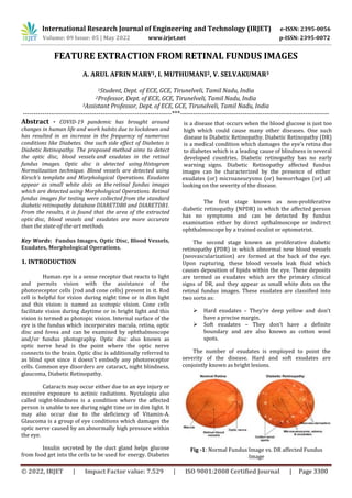
  • 1. International Research Journal of Engineering and Technology (IRJET) e-ISSN: 2395-0056 Volume: 09 Issue: 05 | May 2022 www.irjet.net p-ISSN: 2395-0072 © 2022, IRJET | Impact Factor value: 7.529 | ISO 9001:2008 Certified Journal | Page 3300 FEATURE EXTRACTION FROM RETINAL FUNDUS IMAGES A. ARUL AFRIN MARY1, I. MUTHUMANI2, V. SELVAKUMAR3 1Student, Dept. of ECE, GCE, Tirunelveli, Tamil Nadu, India 2Professor, Dept. of ECE, GCE, Tirunelveli, Tamil Nadu, India 3Assistant Professor, Dept. of ECE, GCE, Tirunelveli, Tamil Nadu, India ---------------------------------------------------------------------***--------------------------------------------------------------------- Abstract - COVID-19 pandemic has brought around changes in human life and work habits due to lockdown and has resulted in an increase in the frequency of numerous conditions like Diabetes. One such side effect of Diabetes is Diabetic Retinopathy. The proposed method aims to detect the optic disc, blood vessels and exudates in the retinal fundus images. Optic disc is detected using Histogram Normalization technique. Blood vessels are detected using Kirsch’s template and Morphological Operations. Exudates appear as small white dots on the retinal fundus images which are detected using Morphological Operations. Retinal fundus images for testing were collected from the standard diabetic retinopathy database DIARETDB0 and DIARETDB1. From the results, it is found that the area of the extracted optic disc, blood vessels and exudates are more accurate than the state-of-the-art methods. Key Words: Fundus Images, Optic Disc, Blood Vessels, Exudates, Morphological Operations. 1. INTRODUCTION Human eye is a sense receptor that reacts to light and permits vision with the assistance of the photoreceptor cells (rod and cone cells) present in it. Rod cell is helpful for vision during night time or in dim light and this vision is named as scotopic vision. Cone cells facilitate vision during daytime or in bright light and this vision is termed as photopic vision. Internal surface of the eye is the fundus which incorporates macula, retina, optic disc and fovea and can be examined by ophthalmoscope and/or fundus photography. Optic disc also known as optic nerve head is the point where the optic nerve connects to the brain. Optic disc is additionally referred to as blind spot since it doesn’t embody any photoreceptor cells. Common eye disorders are cataract, night blindness, glaucoma, Diabetic Retinopathy. Cataracts may occur either due to an eye injury or excessive exposure to actinic radiations. Nyctalopia also called night-blindness is a condition where the affected person is unable to see during night time or in dim light. It may also occur due to the deficiency of Vitamin-A. Glaucoma is a group of eye conditions which damages the optic nerve caused by an abnormally high pressure within the eye. The first stage known as non-proliferative diabetic retinopathy (NPDR) in which the affected person has no symptoms and can be detected by fundus examination either by direct opthalmoscope or indirect ophthalmoscope by a trained oculist or optometrist. The second stage known as proliferative diabetic retinopathy (PDR) in which abnormal new blood vessels (neovascularization) are formed at the back of the eye. Upon rupturing, these blood vessels leak fluid which causes deposition of lipids within the eye. These deposits are termed as exudates which are the primary clinical signs of DR, and they appear as small white dots on the retinal fundus images. These exudates are classified into two sorts as:  Hard exudates – They’re deep yellow and don’t have a precise margin.  Soft exudates – They don’t have a definite boundary and are also known as cotton wool spots. The number of exudates is employed to point the severity of the disease. Hard and soft exudates are conjointly known as bright lesions. Fig -1: Normal Fundus Image vs. DR affected Fundus Image Insulin secreted by the duct gland helps glucose from food get into the cells to be used for energy. Diabetes is a disease that occurs when the blood glucose is just too high which could cause many other diseases. One such disease is Diabetic Retinopathy. Diabetic Retinopathy (DR) is a medical condition which damages the eye’s retina due to diabetes which is a leading cause of blindness in several developed countries. Diabetic retinopathy has no early warning signs. Diabetic Retinopathy affected fundus images can be characterized by the presence of either exudates (or) microaneurysms (or) hemorrhages (or) all looking on the severity of the disease.
  • 2. International Research Journal of Engineering and Technology (IRJET) e-ISSN: 2395-0056 Volume: 09 Issue: 05 | May 2022 www.irjet.net p-ISSN: 2395-0072 © 2022, IRJET | Impact Factor value: 7.529 | ISO 9001:2008 Certified Journal | Page 3301 Microaneurysm is an eye condition in which tiny red dots encircled by yellow rings are found within the eye as a result of vascular leakage. Retinal hemorrhage refers to abnormal bleeding within the delicate blood vessels of the retina whose symptoms range from the undetectable to severe vision issues. Vision problems are often temporary, but in some instances, they’ll be permanent. Microaneurysms and hemorrhages are jointly known as red lesions. 2. PROPOSED METHODOLOGY The detection of optic disc, blood vessels and exudates is a major problem in the automatic processing of retinal images. The segmentation of exudates is essential in differentiating the normal eye from the DR affected eye. In this work fundus image of the retina is used. MATLAB is used for implementing the process. 2.1 Dataset The retinal fundus images for testing were taken from the standard publicly available Diabetic Retinopathy database DIARETDB0 and DIARETDB1. 2.2 Preprocessing Preprocessing includes resizing of the retinal fundus images and choosing the appropriate color component from the RGB image. The retinal fundus images collected for testing were of different resolutions. In order to maintain a uniform resolution, all the images collected were resized to 512x512 pixels. The resized retinal fundus images were in RGB colour space. The contents in the blue colour channel of the image are of low contrast whereas the red colour channel of the image is noisy. The optic disc, blood vessels and exudates has good contrast in the green colour channel of the image. Hence the green channel is chosen for the analysis of the retinal fundus images. 2.3 Image Enhancement CLAHE (Contrast-Limited Adaptive Histogram Equalization) algorithm is applied for noise reduction by increasing the contrast and filtering of the image. Histogram equalization is used for enhancing the contrast of the image. It removes the background for the resulted image by converting the background to white color (every pixel outside the white board changed to white color). 2.4 Exudates Detection The proposed technique for the detection of exudates in the retinal fundus images is implemented using morphological top- and bottom-hat filtering operations. The flow diagram for exudates extraction in the retinal fundus images is shown in fig. 2 Fig -2: Flow Diagram for Exudates Extraction The exudates from the retinal fundus images are detected by applying the morphological bottom-hat filtering on the extracted green channel of the fundus image with a disc shaped structuring element. Bottom-hat filtering subtracts the original image from the morphologically closed image. Then morphological top-hat filtering is applied on the extracted green channel of the fundus image with a disc shaped structuring element. Top- hat filtering subtracts the morphologically opened image from the original image. Then the bottom-hat filtered image is subtracted from the top-hat filtered image. Then a binary image is obtained by converting the resultant image with a threshold value i.e., the pixels with luminance value greater than the threshold value are replaced by value 1 (white) and the remaining pixels are replaced by a value 0 (black). The white pixels correspond to the exudates pixels in the original image. 2.5 Blood Vessels Detection The proposed technique for the detection of blood vessels in the retinal fundus images is implemented using Kirsch’s templates. The flow diagram for blood vessel extraction in retinal fundus images is shown in fig. 3 Fig -3: Flow Diagram for Blood Vessels Extraction The blood vessels in the retinal fundus images were detected by applying a 2-dimensional digital finite impulse response (FIR) on the bottom-hat transformed The flow diagram for exudates feature extraction in the retinal fundus images is shown in fig. 3 Fig. 3 Flow Diagram for Exudates Extraction Retinal Fundus Images Extract Green Channel Extracted ExudatePixels Convert to Binary Image Morphological Bottom Hat Morphological Top Hat The flow diagram for blood vessels feature extraction in the retinal fundus images is shown in fig. 4 Fig. 4 Flow Diagram for Blood Vessels Extraction Retinal Fundus Images Extract GreenChannel Extracted Blood Vessel Pixels Threshold Image Morphological Bottom Hat FIR Filter
  • 3. International Research Journal of Engineering and Technology (IRJET) e-ISSN: 2395-0056 Volume: 09 Issue: 05 | May 2022 www.irjet.net p-ISSN: 2395-0072 © 2022, IRJET | Impact Factor value: 7.529 | ISO 9001:2008 Certified Journal | Page 3302 image and the Kirsch’s template co - efficients. Then a threshold is applied on the 2-D FIR filtered image to extract the pixels of the blood vessels from the filtered image. The pixels greater than the threshold value correspond to the blood vessel pixels. 2.6 Optic Disc Detection The proposed technique for the detection of optic disc in the retinal fundus images is implemented using histogram conversion and normalization of histogram. The flow diagram for optic disc extraction in retinal fundus images is shown in fig. 4 Fig -4: Flow Diagram for Optic Disc Extraction In image processing, normalization is a process used for changing the range of intensity values of the pixels in the image. It is also called histogram stretching or contrast stretching. Histogram normalization enhances the fine details within an image. Then a threshold is applied on the normalized histogram to find the pixels of the optic disc from the normalized histogram. The pixels greater than the threshold value correspond to the optic disc pixels. 3. RESULT 3.1 Retinal Fundus Image Fundus Image consists of RGB Channel which has unique feature. Fig -5: Normal Retinal Fundus Image Fig -6: DR affected Retinal Fundus Image 3.2 Detected Optic Disc Fig. 7, 8 shows the detected optic edges disc in normal and DR affected fundus images. Fig -7: Detected Optic Disc in a Normal Retinal Fundus Image Fig -8: Detected Optic Disc in a DR affected Retinal Fundus Image 3.3 Detected Blood Vessels Fig. 9, 10 shows the detected blood vessels in normal and DR affected fundus images. Fig -9: Detected Blood Vessels in a Normal Retinal Fundus Image Fig -10: Detected Blood Vessels in a DR affected Retinal Fundus Image 3.4 Detected Exudates Fig. 11, 12 shows the detected exudates in normal and DR affected fundus images. Fig -11: Detected Exudates in a Normal Retinal Fundus Image Fig -12: Detected Exudates in a DR affected Retinal Fundus Image The flow diagram for optic disc feature extractioninthe retinal fundus images is shown in fig. 3 Fig. 3 Flow Diagram for Optic Disc Extraction Retinal Fundus Images Extract GreenChannel Extracted Optic Disc Pixels Threshold Image Contrast Enhancement Normalization of Histogram
  • 4. International Research Journal of Engineering and Technology (IRJET) e-ISSN: 2395-0056 Volume: 09 Issue: 05 | May 2022 www.irjet.net p-ISSN: 2395-0072 © 2022, IRJET | Impact Factor value: 7.529 | ISO 9001:2008 Certified Journal | Page 3303 From fig.11, it is observed that the normal retinal fundus images do not have any exudates. The yellow dots in the above figure (fig. 12) shows the detected exudates. 3.5 Area of the Detected Features Fig. 13, 14 shows the area of the extracted features from the retinal fundus image. Fig -13: Area of the Extracted Features in a Normal Retinal Fundus Image Fig -14: Area of the Extracted Features in a DR affected Retinal Fundus Image 4. CONCLUSION In this work, features like optic disc, blood vessels and exudates in the human retinal fundus images were extracted. Retinal fundus images including both normal and Diabetic Retinopathy affected images were taken from the publicly available standard Diabetic Retinopathy Databases like DIARETDB0 and DIARETDB1 for testing. Optic Disc in the retinal fundus images were detected using the Histogram Normalization technique. Blood Vessels in the retinal fundus images were detected using Kirsch’s templates and Morphological Operations. Exudates in the retinal fundus images were detected using the Morphological Operations. The area of the extracted features was also measured. The extraction of all the above three features and the area of these extracted features were performed using MATLAB R2018a. From the extracted exudates and its area, it was observed that the area of the exudates was zero in the normal retinal fundus images and greater than zero in the case of Diabetic Retinopathy affected retinal fundus images. From this observation, it is inferred that if the area of the exudates is greater than zero, then the corresponding retinal fundus image can be classified as a Diabetic Retinopathy affected retinal fundus image and if the area of the exudates is equal to zero, then the corresponding retinal fundus image can be classified as a normal retinal fundus image. REFERENCES [1] Meindert Niemeijer, Bram van Ginneken, Joes Staal, Maria S. A. Suttorp-Schulten, Michael D. Abramoff, “Automatic Detection of Red Lesions in Digital Color Fundus Photographs,” IEEE Transactions on Medical Imaging, Vol. 24, No. 5, pp. 584-592, May 2005. [2] Dongbo Zhang, Xiong Li, Xinyu Shang, Yao Yi, Yaonan Wang, “Robust Hemorrhage Detection in Diabetic Retinopathy Detection,” pp. 209-213, 2011 IEEE. [3] Balint Antal, Andras Hajdu, “An Ensemble-Based System for Microaneurysms Detection and Diabetic Retinopathy Grading,” IEEE Transactions on Biomedical Engineering, Vol. 59, 6, pp. 1720- 1726, June 2012. [4] Sohini Roychowdhury, Dara D. Koozekanani, Keshab K. Parthi, “Screening Fundus Images for Diabetic Retinopathy,” pp. 1641-1645, 2012 IEEE. [5] M. Usman Akram, Shehzad Khalid, Shoab A. Khan, “Identification and classification of microaneurysms for early detection of diabetic retinopathy,” ELSEVIER - Pattern Recognition 46 (2013), pp. 107-116. [6] L. Giancardo, F. Meriaudeau, T. P. Karnowski, K.W. Tobin, E. Chaum, “Validation of Microaneurysm- based Diabetic Retinopathy Screening across Retinal Fundus Datasets,” pp. 125-130, 2013 IEEE. [7] Sundararaj Wifred Franklin, Samuelnadar Edwar Rajan, “Diagnosis of Diabetic Retinopathy by employing Image Processing Techniques to detect Exudates in retinal images,” IET Image Process, 2014 Vol. 8, Iss. 10, pp. 601 – 609. [8] R. S. Deshmukh, “Analysis of Diabetic Retinopathy by Automatic Detection of Exudates,” International Journal of Electronics and Communication Engineering (IJECET), Vol. 5, Iss. 12, December (2014), pp. 266 – 275. [9] ManojKumar S B, Manjunath R, Dr. H S Sheshadri, “Feature extraction from the Fundus Images for the diagnosis of diabetic retinopathy,” International Conference on Emerging Research in Electronics, Computer Science and Technology, pp. 240-245, 2015 IEEE. [10] Sudeshna Sil Kar, Santi P. Maity, “Automatic Detection of Retinal Lesions for Screening of Diabetic Retinopathy,” IEEE Transactions on Biomedical Engineering, pp. 1-9, 2016 IEEE. [11] Karan Bhatia, Shikar Arora, Ravi Tomar, “Diagnosis of Diabetic Retinopathy Using Machine Learning Classification Algorithm,” 2016 2nd
  • 5. International Research Journal of Engineering and Technology (IRJET) e-ISSN: 2395-0056 Volume: 09 Issue: 05 | May 2022 www.irjet.net p-ISSN: 2395-0072 © 2022, IRJET | Impact Factor value: 7.529 | ISO 9001:2008 Certified Journal | Page 3304 International Conference on Next Generation Computing Technologies (NGCT-2016), pp. 347- 351, 2016 IEEE. [12] Sarni Suhaila Rahim, Vasile Palade, James Shuttleworth, Christiana Jayne, “Automatic screening and classification of diabetic retinopathy and maculopathy using fuzzy image processing,” Brain Informatics (2016) 3: 249 – 267. [13] Dilip Singh Sisodia, Shruti Nair, Pooja Khobragde, “Diabetic Retinal Fundus Images: Preprocessing and Feature Extraction For Early Detection of Diabetic Retinopathy,” Biomedical and Pharmacology Journal, Vol. 10(2), pp. 615-626 (2017). [14] Wei Zhou, Chendong Wu, Dali Chen, Zhenzhu Wang, Yugen Yi, Wenyou Du, “Automatic Microaneurysm Detection of Diabetic Retinopathy in Fundus Images,” pp. 7453-7458, 2017 IEEE. [15] Shuang Yu, Di Xiao, Yogesan Kanagasingam, “Exudate Detection for Diabetic Retinopathy with Convolutional Neural Networks,” pp. 1744-1747, 2017 IEEE. [16] Wei Zhou, Chengdong Wu, Xiaosheng Yu, “Computer Aided Diagnosis for Diabetic Retinopathy based on Fundus Image,” Proceedings of the 37th Chinese Control Conference 2018, pp. 9214-9219. [17] Zihao Wang, Ke-Jia Chen, Lingli Zhang, “A R-CNN Based Approach for Microaneurysm Detection in Retinal Fundus Images,” – 2019. [18] Muhammad Mateen, Junhao Wen, Nasrullah Nasrullah, Song Sun, Shaukat Hayat, “Exudate Detection for Diabetic Retinopathy using Pretrained Convolutional Neural Networks,” Hindawi Complexity, Vol. 2020, pp. 1-11. [19] DIARETDB0 and DIARETDB1 Database. [20] Software Package, “MATLAB (2018a)”.