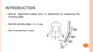
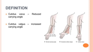
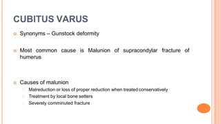
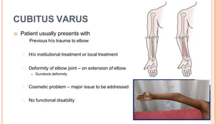
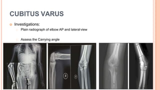
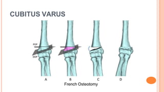
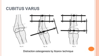
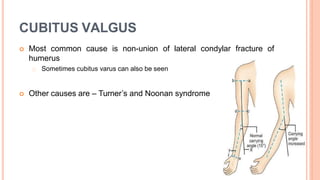
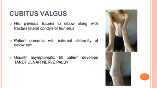
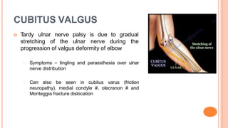
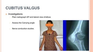
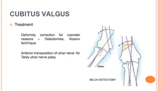
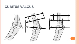
CUBITUS VARUS AND
CUBITUS VALGUS
Dr. Ponnilavan
Senior Resident
INTRODUCTION
 Normal alignment of elbow joint is determined by measuring the
Carrying angle
 Normal carrying angle – 5 to 15 deg
 More in females than in males
DEFINITION
 Cubitus varus – Reduced
carrying angle
 Cubitus valgus – increased
carrying angle
CUBITUS VARUS
 Synonyms – Gunstock deformity
 Most common cause is Malunion of supracondylar
humerus
fracture of
 Causes of malunion
Malreduction or loss of proper reduction when treated conservatively
Treatment by local bone setters
Severely comminuted fracture
CUBITUS VARUS
 Patient usually presents with
Previous h/o trauma to elbow
H/o institutional treatment or local treatment
Deformity of elbow joint – on extension of elbow
 Gunstock deformity
Cosmetic problem – major issue to be addressed
No functional disability
CUBITUS VARUS
 Investigations:
Plain radiograph of elbow AP and lateral view
Assess the Carrying angle
CUBITUS VARUS
 Treatment – cubitus varus has no tendency for spontaneous
correction by remodelling. Hence has to corrected surgically
 Mostly done for cosmetic reasons
 Options available are:
Hemiepiphysiodesis and growth alteration – in patients with medial growth
arrest and progressive deformity
Corrective osteotomies – Commonly done
Distraction osteogenesis by Ilizarov technique
CUBITUS VARUS
 Corrective osteotomies
Lateral closing wedge osteotomy (French osteotomy)
Medial open wedge osteotomy – stretches ulnar nerve
Dome osteotomy
Others – Step cut, Pentalateral and Oblique osteotomy
CUBITUS VARUS
French Osteotomy
CUBITUS VARUS
Distraction osteogenesis by Ilizarov technique
CUBITUS VALGUS
 Most common cause is non-union of lateral condylar fracture of
humerus
Sometimes cubitus varus can also be seen
 Other causes are – Turner’s and Noonan syndrome
CUBITUS VALGUS
 H/o previous trauma to elbow
fracture lateral condyle of humerus
along with
 Patient presents with external deformity of
elbow joint
 Usually asymptomatic till patient develops
TARDY ULNAR NERVE PALSY
CUBITUS VALGUS
 T
ardy ulnar nerve palsy is due to gradual
stretching of the ulnar nerve during the
progression of valgus deformity of elbow
Symptoms – tingling and paraesthesia over ulnar
nerve distribution
Can also be seen in cubitus varus (friction
neuropathy), medial condyle #, olecranon # and
Monteggia fracture dislocation
CUBITUS VALGUS
 Investigations
Plain radiograph AP and lateral view of elbow
Assess the Carrying angle
Nerve conduction studies
CUBITUS VALGUS
 Treatment
Deformity correction for cosmetic
reasons – Osteotomies, Ilizarov
technique
Anterior transposition of ulnar nerve for
Tardy ulnar nerve palsy
MILCH OSTEOTOMY
CUBITUS VALGUS
T
H
A
N
KY
O
U

Cubitus varus and valgus

  • 1.
    CUBITUS VARUS AND CUBITUSVALGUS Dr. Ponnilavan Senior Resident
  • 2.
    INTRODUCTION  Normal alignmentof elbow joint is determined by measuring the Carrying angle  Normal carrying angle – 5 to 15 deg  More in females than in males
  • 3.
    DEFINITION  Cubitus varus– Reduced carrying angle  Cubitus valgus – increased carrying angle
  • 4.
    CUBITUS VARUS  Synonyms– Gunstock deformity  Most common cause is Malunion of supracondylar humerus fracture of  Causes of malunion Malreduction or loss of proper reduction when treated conservatively Treatment by local bone setters Severely comminuted fracture
  • 5.
    CUBITUS VARUS  Patientusually presents with Previous h/o trauma to elbow H/o institutional treatment or local treatment Deformity of elbow joint – on extension of elbow  Gunstock deformity Cosmetic problem – major issue to be addressed No functional disability
  • 6.
    CUBITUS VARUS  Investigations: Plainradiograph of elbow AP and lateral view Assess the Carrying angle
  • 7.
    CUBITUS VARUS  Treatment– cubitus varus has no tendency for spontaneous correction by remodelling. Hence has to corrected surgically  Mostly done for cosmetic reasons  Options available are: Hemiepiphysiodesis and growth alteration – in patients with medial growth arrest and progressive deformity Corrective osteotomies – Commonly done Distraction osteogenesis by Ilizarov technique
  • 8.
    CUBITUS VARUS  Correctiveosteotomies Lateral closing wedge osteotomy (French osteotomy) Medial open wedge osteotomy – stretches ulnar nerve Dome osteotomy Others – Step cut, Pentalateral and Oblique osteotomy
  • 9.
  • 10.
  • 11.
    CUBITUS VALGUS  Mostcommon cause is non-union of lateral condylar fracture of humerus Sometimes cubitus varus can also be seen  Other causes are – Turner’s and Noonan syndrome
  • 12.
    CUBITUS VALGUS  H/oprevious trauma to elbow fracture lateral condyle of humerus along with  Patient presents with external deformity of elbow joint  Usually asymptomatic till patient develops TARDY ULNAR NERVE PALSY
  • 13.
    CUBITUS VALGUS  T ardyulnar nerve palsy is due to gradual stretching of the ulnar nerve during the progression of valgus deformity of elbow Symptoms – tingling and paraesthesia over ulnar nerve distribution Can also be seen in cubitus varus (friction neuropathy), medial condyle #, olecranon # and Monteggia fracture dislocation
  • 14.
    CUBITUS VALGUS  Investigations Plainradiograph AP and lateral view of elbow Assess the Carrying angle Nerve conduction studies
  • 15.
    CUBITUS VALGUS  Treatment Deformitycorrection for cosmetic reasons – Osteotomies, Ilizarov technique Anterior transposition of ulnar nerve for Tardy ulnar nerve palsy MILCH OSTEOTOMY
  • 16.
  • 17.