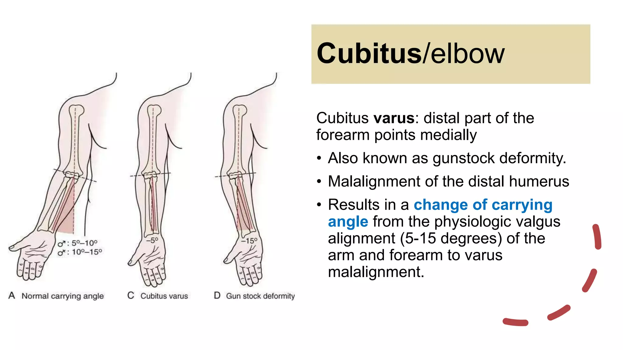
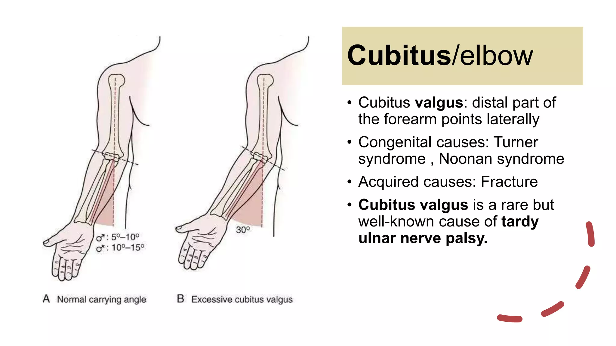
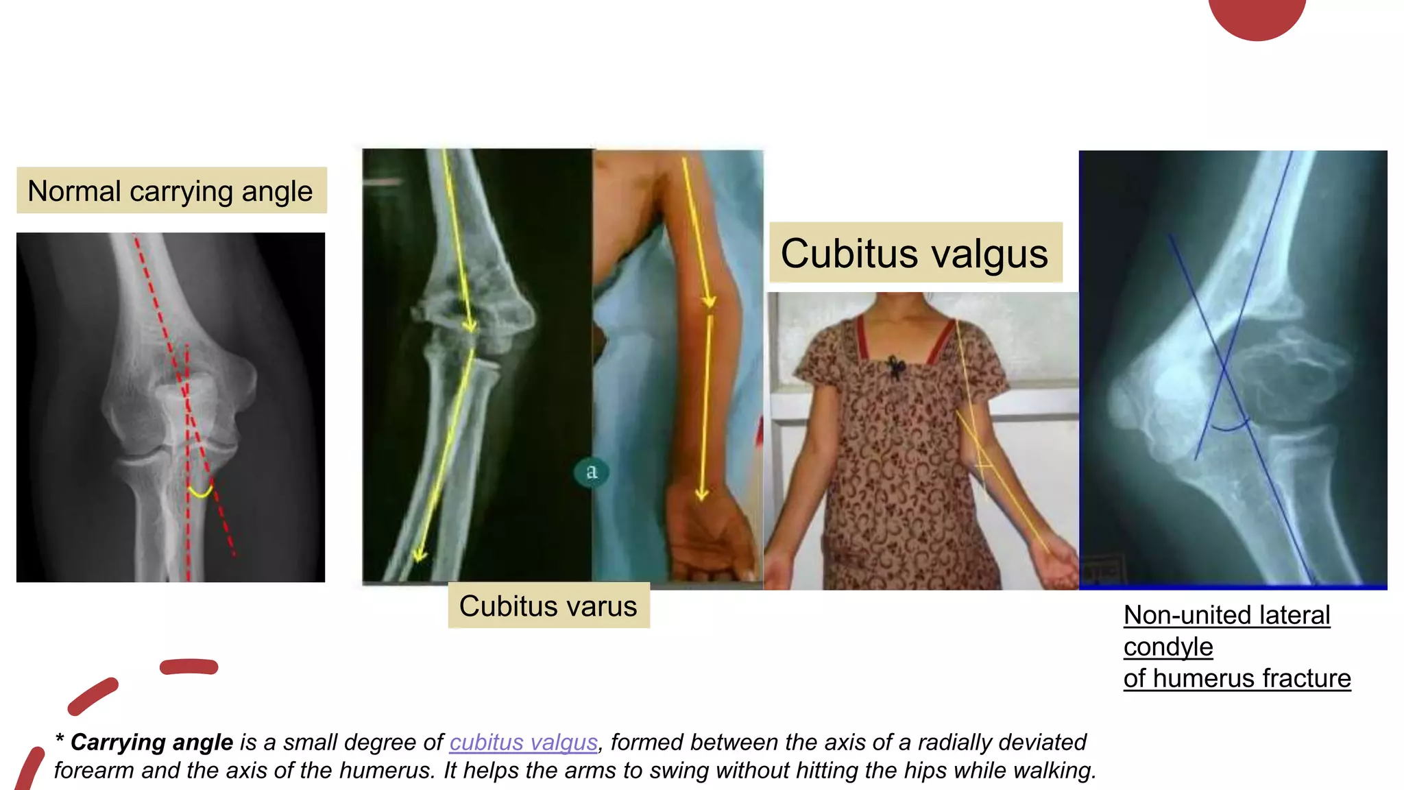
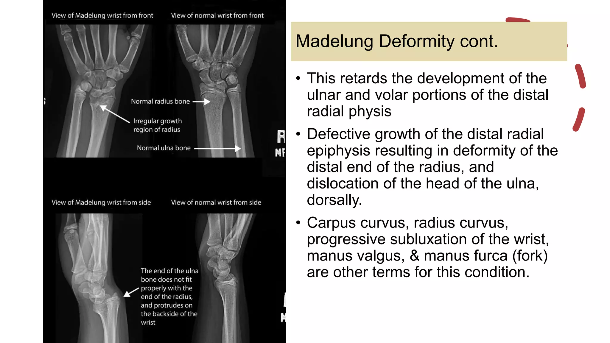
SlideShare a Scribd company logo
Terminologies & deformities:
• Varus and valgus
• Hallux Valgus/Varus
• Coxa Vara/Coxa Vaiga
• Genu Varum/Valgum
• Cubitus Varus/Valgus
• Madelung Deformity
• Bunion
• Osteotomy
By Dhanulekha
Varus & valgus
• The terms valgus and varus refer to angulation (or bowing)
within the shaft of a bone or at a joint. It is determined by the
distal part of the bone forming the joint being more medial or
lateral than it should be.
• Valgus = The distal part is more lateral/goes outwards
• Varus = The distal part is more medial/goes inwards
Coxa/
Hip
Knee/
Genu
Hallux
/Toe
Heel Foot/
Pes
Cubitus/
Elbow
Manus
/Hand
Wrist
Coxa/hip
• Relation of the femoral shaft to the femoral head and neck
• Coxa vara: Angle between the head and neck of the femur and
its shaft is decreased, <120 degrees.
• Coxa valga: There is an increased angle between the femoral
head and neck and femoral shaft.
Coxa/hip
Laterally
Medially
• Depends on the direction of
distal part of the tibia
• Genu varum:
Medial misalignment of the knee
• Genu valgum:
Lateral misalignment of the knee
Genu/knee
Hallux/Big toe
• The hallux usually moves in the transverse plane.
• Hallux varus:
Characterized by medial deviation of great toe relative to the 1st metatarsal
bone.
Hallux valgus / Bunion
• A bunion is a localized painful swelling at the
base of the big toe. The joint is enlarged (due to
new bone formation) and the toe is often
misaligned. It is frequently associated with
inflammation.
• It can be related to inflammation of the nearby
bursa (bursitis) or degenerative joint disease
(osteoarthritis)
• It is characterized by medial deviation of the
first metatarsal and lateral deviation rotation of
the hallux, with associated joint subluxation.
Cubitus/elbow
Cubitus varus: distal part of the
forearm points medially
• Also known as gunstock deformity.
• Malalignment of the distal humerus
• Results in a change of carrying
angle from the physiologic valgus
alignment (5-15 degrees) of the
arm and forearm to varus
malalignment.
Cubitus/elbow
• Cubitus valgus: distal part of
the forearm points laterally
• Congenital causes: Turner
syndrome , Noonan syndrome
• Acquired causes: Fracture
• Cubitus valgus is a rare but
well-known cause of tardy
ulnar nerve palsy.
Cubitus varus
Cubitus valgus
Non-united lateral
condyle
of humerus fracture
Normal carrying angle
* Carrying angle is a small degree of cubitus valgus, formed between the axis of a radially deviated
forearm and the axis of the humerus. It helps the arms to swing without hitting the hips while walking.
Madelung Deformity
• It can be bilateral in 50-66% of patients.
• Rare congenital deformity and does not
usually manifest until 10-14 years.
• It may also be seen as an acquired
consequence of trauma to the growth plate,
e.g. Salter-Harris fracture (type V).
• Growth disturbance of volar-ulnar distal
radial physis
• This retards the development of the
ulnar and volar portions of the distal
radial physis
• Defective growth of the distal radial
epiphysis resulting in deformity of the
distal end of the radius, and
dislocation of the head of the ulna,
dorsally.
• Carpus curvus, radius curvus,
progressive subluxation of the wrist,
manus valgus, & manus furca (fork)
are other terms for this condition.
Madelung Deformity cont.
Osteotomy
• An osteotomy is a surgical operation whereby a bone is cut to
shorten or lengthen it or to change its alignment.
• It is sometimes performed to correct a hallux valgus, or to
straighten a bone that has healed crookedly following a fracture.
It is also used to correct a coxa vara, genu valgum, and genu
varum.
References
1) Maheshwari, J. and Mhaskar, V., 2015. Essential Orthopaedics. 5th ed. The Health Science
Publisher.
2) Jones, J., 2020. Coxa Valga | Radiology Reference Article | Radiopaedia.Org. [online]
Radiopaedia.org. Available at: https://radiopaedia.org/articles/coxa-valga
3) Lamberti, P., 2020. Madelung Deformity: Background, Anatomy, Pathophysiology. [online]
Emedicine.medscape.com. Available at: https://emedicine.medscape.com/article/1260002-
overview
4) Luijkx, T., 2020. Valgus Vs Varus | Radiology Reference Article | Radiopaedia.Org. [online]
Radiopaedia.org. Available at: https://radiopaedia.org/articles/valgus-vs-varus-1
Thank you

More Related Content

What's hot

Coxa vara
Coxa varaCoxa vara
Coxa vara
manoj das
 
CTEV UG
CTEV UGCTEV UG
CTEV UG
HETA PATEL
 
Congenital talipes equinovarus (club foot/ctev) ppt by Dr Pratik
Congenital talipes equinovarus (club foot/ctev) ppt by Dr PratikCongenital talipes equinovarus (club foot/ctev) ppt by Dr Pratik
Congenital talipes equinovarus (club foot/ctev) ppt by Dr Pratik
Dr. Pratik Agarwal
 
Congenital coxa vara
Congenital coxa varaCongenital coxa vara
Congenital coxa vara
Libin Thomas
 
MCL,LCL & ALL injuries of the knee
MCL,LCL & ALL injuries of the knee MCL,LCL & ALL injuries of the knee
MCL,LCL & ALL injuries of the knee
Mohamed Abulsoud
 
Pffd
PffdPffd
Pffd
Dr-fadikh
 
Dislocation of patella
Dislocation of patellaDislocation of patella
Dislocation of patellaorthoprince
 
Sprengel deformity
Sprengel deformitySprengel deformity
Sprengel deformity
Joydeep Mallik
 
Pes planus
Pes planusPes planus
Pes planus
Dr. Anurag Mittal
 
Proximal humerus fractures
Proximal humerus fracturesProximal humerus fractures
Proximal humerus fractures
MONTHER ALKHAWLANY
 
Clinical Examination of the Hip
Clinical Examination of the HipClinical Examination of the Hip
Clinical Examination of the Hiporthoprince
 
Femoroacetabular impingement
Femoroacetabular impingementFemoroacetabular impingement
Femoroacetabular impingement
Advanced Physiotherapy
 
Congenital vertical talus
Congenital vertical talusCongenital vertical talus
Congenital vertical talus
Joydeep Mandal
 
Slipped Capital Femoral Epiphysis (SCFE)
Slipped Capital Femoral Epiphysis (SCFE)Slipped Capital Femoral Epiphysis (SCFE)
Slipped Capital Femoral Epiphysis (SCFE)
Sijan Bhattachan
 
Non union neck of femur
Non union neck of femurNon union neck of femur
Non union neck of femur
ramachandra reddy
 
Flat foot By Dr.Mahbub
Flat foot By Dr.MahbubFlat foot By Dr.Mahbub
Flat foot By Dr.Mahbub
dr_mhb21
 
Painful Arc Syndrome.pptx
Painful Arc Syndrome.pptxPainful Arc Syndrome.pptx
Painful Arc Syndrome.pptx
Prashant Badiger
 
Fracture of talus ppt
Fracture of talus pptFracture of talus ppt
Fracture of talus ppt
sunil kumar daha
 
Radiographic evaluation of Paediatric elbow injury
Radiographic evaluation of Paediatric elbow injury Radiographic evaluation of Paediatric elbow injury
Radiographic evaluation of Paediatric elbow injury
saikat ghosh
 

What's hot (20)

Coxa vara
Coxa varaCoxa vara
Coxa vara
 
CTEV UG
CTEV UGCTEV UG
CTEV UG
 
Congenital talipes equinovarus (club foot/ctev) ppt by Dr Pratik
Congenital talipes equinovarus (club foot/ctev) ppt by Dr PratikCongenital talipes equinovarus (club foot/ctev) ppt by Dr Pratik
Congenital talipes equinovarus (club foot/ctev) ppt by Dr Pratik
 
Congenital coxa vara
Congenital coxa varaCongenital coxa vara
Congenital coxa vara
 
MCL,LCL & ALL injuries of the knee
MCL,LCL & ALL injuries of the knee MCL,LCL & ALL injuries of the knee
MCL,LCL & ALL injuries of the knee
 
Pffd
PffdPffd
Pffd
 
Dislocation of patella
Dislocation of patellaDislocation of patella
Dislocation of patella
 
Sprengel deformity
Sprengel deformitySprengel deformity
Sprengel deformity
 
Pes planus
Pes planusPes planus
Pes planus
 
Proximal humerus fractures
Proximal humerus fracturesProximal humerus fractures
Proximal humerus fractures
 
Clinical Examination of the Hip
Clinical Examination of the HipClinical Examination of the Hip
Clinical Examination of the Hip
 
Femoroacetabular impingement
Femoroacetabular impingementFemoroacetabular impingement
Femoroacetabular impingement
 
Congenital vertical talus
Congenital vertical talusCongenital vertical talus
Congenital vertical talus
 
Slipped Capital Femoral Epiphysis (SCFE)
Slipped Capital Femoral Epiphysis (SCFE)Slipped Capital Femoral Epiphysis (SCFE)
Slipped Capital Femoral Epiphysis (SCFE)
 
Non union neck of femur
Non union neck of femurNon union neck of femur
Non union neck of femur
 
Flat foot By Dr.Mahbub
Flat foot By Dr.MahbubFlat foot By Dr.Mahbub
Flat foot By Dr.Mahbub
 
Painful Arc Syndrome.pptx
Painful Arc Syndrome.pptxPainful Arc Syndrome.pptx
Painful Arc Syndrome.pptx
 
Fracture of talus ppt
Fracture of talus pptFracture of talus ppt
Fracture of talus ppt
 
Radiographic evaluation of Paediatric elbow injury
Radiographic evaluation of Paediatric elbow injury Radiographic evaluation of Paediatric elbow injury
Radiographic evaluation of Paediatric elbow injury
 
Femoroacetabular impingement
Femoroacetabular impingementFemoroacetabular impingement
Femoroacetabular impingement
 

Similar to Student seminar valgus & varus

Coxavaradnbid 110424114226-phpapp02
Coxavaradnbid 110424114226-phpapp02Coxavaradnbid 110424114226-phpapp02
Coxavaradnbid 110424114226-phpapp02Abdelwahab Hussein
 
Common deformities, deviations, and injuries of ankle and foot
Common deformities, deviations, and injuries of ankle and footCommon deformities, deviations, and injuries of ankle and foot
Common deformities, deviations, and injuries of ankle and foot
Dr Vicky Kasundra
 
Cubitus varus and valgus
Cubitus varus and valgusCubitus varus and valgus
Cubitus varus and valgus
BipulBorthakur
 
Cavus foot
Cavus footCavus foot
Cavus foot
Dr. Anurag Mittal
 
Lower limb deformities.pptx
Lower limb deformities.pptxLower limb deformities.pptx
Lower limb deformities.pptx
IrfanNashad1
 
hallux valgus.pptx
hallux valgus.pptxhallux valgus.pptx
hallux valgus.pptx
sonalidas935894
 
The knee in cerebral palsy
The knee in cerebral palsy The knee in cerebral palsy
The knee in cerebral palsy
Libin Thomas
 
Pathomechanics of Ankle joint
Pathomechanics of Ankle joint Pathomechanics of Ankle joint
Pathomechanics of Ankle joint
shanmugimadhavan
 
Deformities related to cerebral palsy and their orthotic
Deformities related to cerebral palsy and their orthoticDeformities related to cerebral palsy and their orthotic
Deformities related to cerebral palsy and their orthotic
Ainaa Khan
 
Hip joint
Hip jointHip joint
Hip joint
mariamanwar010
 
Hallux valgus.pptx
Hallux valgus.pptxHallux valgus.pptx
Hallux valgus.pptx
Siwaporn Khureerung
 
Congenital and Acquired Deformities around Elbow.pptx
Congenital and Acquired  Deformities around Elbow.pptxCongenital and Acquired  Deformities around Elbow.pptx
Congenital and Acquired Deformities around Elbow.pptx
Arun Upreti
 
Wrist Joint
Wrist JointWrist Joint
Wrist Joint
Shweta Kotwani
 
Cubitus varus and valgus
Cubitus varus and valgusCubitus varus and valgus
Cubitus varus and valgus
Ponnilavan Ponz
 
Coxa Vara, Genu VArum & Valgum. Under Gradts.
Coxa Vara, Genu VArum & Valgum. Under Gradts.Coxa Vara, Genu VArum & Valgum. Under Gradts.
Coxa Vara, Genu VArum & Valgum. Under Gradts.
Sarthy Velayutham
 
Flat foot and Cavus foot
 Flat foot and Cavus foot Flat foot and Cavus foot
Flat foot and Cavus foot
Dr Thouseef Abdul Majeed
 
COXA VARA AND COXA VALGA, DEVLOPMENTAL COXA VARA.pptx
COXA VARA AND COXA VALGA, DEVLOPMENTAL COXA VARA.pptxCOXA VARA AND COXA VALGA, DEVLOPMENTAL COXA VARA.pptx
COXA VARA AND COXA VALGA, DEVLOPMENTAL COXA VARA.pptx
KisanNepali
 
SPORTS INJURIES OF ANKLE AND FOOT original.pptx
SPORTS INJURIES OF ANKLE AND FOOT original.pptxSPORTS INJURIES OF ANKLE AND FOOT original.pptx
SPORTS INJURIES OF ANKLE AND FOOT original.pptx
MeghaPrakash9
 
Cubitus varus deformity
Cubitus varus deformityCubitus varus deformity
Cubitus varus deformity
ramachandra reddy
 

Similar to Student seminar valgus & varus (20)

Coxavaradnbid 110424114226-phpapp02
Coxavaradnbid 110424114226-phpapp02Coxavaradnbid 110424114226-phpapp02
Coxavaradnbid 110424114226-phpapp02
 
Common deformities, deviations, and injuries of ankle and foot
Common deformities, deviations, and injuries of ankle and footCommon deformities, deviations, and injuries of ankle and foot
Common deformities, deviations, and injuries of ankle and foot
 
Cubitus varus and valgus
Cubitus varus and valgusCubitus varus and valgus
Cubitus varus and valgus
 
Cavus foot
Cavus footCavus foot
Cavus foot
 
Lower limb deformities.pptx
Lower limb deformities.pptxLower limb deformities.pptx
Lower limb deformities.pptx
 
hallux valgus.pptx
hallux valgus.pptxhallux valgus.pptx
hallux valgus.pptx
 
The knee in cerebral palsy
The knee in cerebral palsy The knee in cerebral palsy
The knee in cerebral palsy
 
Pathomechanics of Ankle joint
Pathomechanics of Ankle joint Pathomechanics of Ankle joint
Pathomechanics of Ankle joint
 
Deformities related to cerebral palsy and their orthotic
Deformities related to cerebral palsy and their orthoticDeformities related to cerebral palsy and their orthotic
Deformities related to cerebral palsy and their orthotic
 
Cubitus valgus varus
Cubitus valgus varusCubitus valgus varus
Cubitus valgus varus
 
Hip joint
Hip jointHip joint
Hip joint
 
Hallux valgus.pptx
Hallux valgus.pptxHallux valgus.pptx
Hallux valgus.pptx
 
Congenital and Acquired Deformities around Elbow.pptx
Congenital and Acquired  Deformities around Elbow.pptxCongenital and Acquired  Deformities around Elbow.pptx
Congenital and Acquired Deformities around Elbow.pptx
 
Wrist Joint
Wrist JointWrist Joint
Wrist Joint
 
Cubitus varus and valgus
Cubitus varus and valgusCubitus varus and valgus
Cubitus varus and valgus
 
Coxa Vara, Genu VArum & Valgum. Under Gradts.
Coxa Vara, Genu VArum & Valgum. Under Gradts.Coxa Vara, Genu VArum & Valgum. Under Gradts.
Coxa Vara, Genu VArum & Valgum. Under Gradts.
 
Flat foot and Cavus foot
 Flat foot and Cavus foot Flat foot and Cavus foot
Flat foot and Cavus foot
 
COXA VARA AND COXA VALGA, DEVLOPMENTAL COXA VARA.pptx
COXA VARA AND COXA VALGA, DEVLOPMENTAL COXA VARA.pptxCOXA VARA AND COXA VALGA, DEVLOPMENTAL COXA VARA.pptx
COXA VARA AND COXA VALGA, DEVLOPMENTAL COXA VARA.pptx
 
SPORTS INJURIES OF ANKLE AND FOOT original.pptx
SPORTS INJURIES OF ANKLE AND FOOT original.pptxSPORTS INJURIES OF ANKLE AND FOOT original.pptx
SPORTS INJURIES OF ANKLE AND FOOT original.pptx
 
Cubitus varus deformity
Cubitus varus deformityCubitus varus deformity
Cubitus varus deformity
 

Recently uploaded

Top-Vitamin-Supplement-Brands-in-India List
Top-Vitamin-Supplement-Brands-in-India ListTop-Vitamin-Supplement-Brands-in-India List
Top-Vitamin-Supplement-Brands-in-India List
SwisschemDerma
 
Cardiac Assessment for B.sc Nursing Student.pdf
Cardiac Assessment for B.sc Nursing Student.pdfCardiac Assessment for B.sc Nursing Student.pdf
Cardiac Assessment for B.sc Nursing Student.pdf
shivalingatalekar1
 
Sex determination from mandible pelvis and skull
Sex determination from mandible pelvis and skullSex determination from mandible pelvis and skull
Sex determination from mandible pelvis and skull
ShashankRoodkee
 
Cervical & Brachial Plexus By Dr. RIG.pptx
Cervical & Brachial Plexus By Dr. RIG.pptxCervical & Brachial Plexus By Dr. RIG.pptx
Cervical & Brachial Plexus By Dr. RIG.pptx
Dr. Rabia Inam Gandapore
 
Ozempic: Preoperative Management of Patients on GLP-1 Receptor Agonists
Ozempic: Preoperative Management of Patients on GLP-1 Receptor Agonists  Ozempic: Preoperative Management of Patients on GLP-1 Receptor Agonists
Ozempic: Preoperative Management of Patients on GLP-1 Receptor Agonists
Saeid Safari
 
How STIs Influence the Development of Pelvic Inflammatory Disease.pptx
How STIs Influence the Development of Pelvic Inflammatory Disease.pptxHow STIs Influence the Development of Pelvic Inflammatory Disease.pptx
How STIs Influence the Development of Pelvic Inflammatory Disease.pptx
FFragrant
 
Integrating Ayurveda into Parkinson’s Management: A Holistic Approach
Integrating Ayurveda into Parkinson’s Management: A Holistic ApproachIntegrating Ayurveda into Parkinson’s Management: A Holistic Approach
Integrating Ayurveda into Parkinson’s Management: A Holistic Approach
Ayurveda ForAll
 
Local Advanced Lung Cancer: Artificial Intelligence, Synergetics, Complex Sys...
Local Advanced Lung Cancer: Artificial Intelligence, Synergetics, Complex Sys...Local Advanced Lung Cancer: Artificial Intelligence, Synergetics, Complex Sys...
Local Advanced Lung Cancer: Artificial Intelligence, Synergetics, Complex Sys...
Oleg Kshivets
 
BRACHYTHERAPY OVERVIEW AND APPLICATORS
BRACHYTHERAPY OVERVIEW  AND  APPLICATORSBRACHYTHERAPY OVERVIEW  AND  APPLICATORS
BRACHYTHERAPY OVERVIEW AND APPLICATORS
Krishan Murari
 
Netter's Atlas of Human Anatomy 7.ed.pdf
Netter's Atlas of Human Anatomy 7.ed.pdfNetter's Atlas of Human Anatomy 7.ed.pdf
Netter's Atlas of Human Anatomy 7.ed.pdf
BrissaOrtiz3
 
SURGICAL ANATOMY OF THE RETROPERITONEUM, ADRENALS, KIDNEYS AND URETERS.pptx
SURGICAL ANATOMY OF THE RETROPERITONEUM, ADRENALS, KIDNEYS AND URETERS.pptxSURGICAL ANATOMY OF THE RETROPERITONEUM, ADRENALS, KIDNEYS AND URETERS.pptx
SURGICAL ANATOMY OF THE RETROPERITONEUM, ADRENALS, KIDNEYS AND URETERS.pptx
Bright Chipili
 
Knee anatomy and clinical tests 2024.pdf
Knee anatomy and clinical tests 2024.pdfKnee anatomy and clinical tests 2024.pdf
Knee anatomy and clinical tests 2024.pdf
vimalpl1234
 
Colonic and anorectal physiology with surgical implications
Colonic and anorectal physiology with surgical implicationsColonic and anorectal physiology with surgical implications
Colonic and anorectal physiology with surgical implications
Dr Maria Tamanna
 
The Electrocardiogram - Physiologic Principles
The Electrocardiogram - Physiologic PrinciplesThe Electrocardiogram - Physiologic Principles
The Electrocardiogram - Physiologic Principles
MedicoseAcademics
 
NVBDCP.pptx Nation vector borne disease control program
NVBDCP.pptx Nation vector borne disease control programNVBDCP.pptx Nation vector borne disease control program
NVBDCP.pptx Nation vector borne disease control program
Sapna Thakur
 
Hemodialysis: Chapter 4, Dialysate Circuit - Dr.Gawad
Hemodialysis: Chapter 4, Dialysate Circuit - Dr.GawadHemodialysis: Chapter 4, Dialysate Circuit - Dr.Gawad
Hemodialysis: Chapter 4, Dialysate Circuit - Dr.Gawad
NephroTube - Dr.Gawad
 
Thyroid Gland- Gross Anatomy by Dr. Rabia Inam Gandapore.pptx
Thyroid Gland- Gross Anatomy by Dr. Rabia Inam Gandapore.pptxThyroid Gland- Gross Anatomy by Dr. Rabia Inam Gandapore.pptx
Thyroid Gland- Gross Anatomy by Dr. Rabia Inam Gandapore.pptx
Dr. Rabia Inam Gandapore
 
ANATOMY AND PHYSIOLOGY OF URINARY SYSTEM.pptx
ANATOMY AND PHYSIOLOGY OF URINARY SYSTEM.pptxANATOMY AND PHYSIOLOGY OF URINARY SYSTEM.pptx
ANATOMY AND PHYSIOLOGY OF URINARY SYSTEM.pptx
Swetaba Besh
 
Role of Mukta Pishti in the Management of Hyperthyroidism
Role of Mukta Pishti in the Management of HyperthyroidismRole of Mukta Pishti in the Management of Hyperthyroidism
Role of Mukta Pishti in the Management of Hyperthyroidism
Dr. Jyothirmai Paindla
 
Pictures of Superficial & Deep Fascia.ppt.pdf
Pictures of Superficial & Deep Fascia.ppt.pdfPictures of Superficial & Deep Fascia.ppt.pdf
Pictures of Superficial & Deep Fascia.ppt.pdf
Dr. Rabia Inam Gandapore
 

Recently uploaded (20)

Top-Vitamin-Supplement-Brands-in-India List
Top-Vitamin-Supplement-Brands-in-India ListTop-Vitamin-Supplement-Brands-in-India List
Top-Vitamin-Supplement-Brands-in-India List
 
Cardiac Assessment for B.sc Nursing Student.pdf
Cardiac Assessment for B.sc Nursing Student.pdfCardiac Assessment for B.sc Nursing Student.pdf
Cardiac Assessment for B.sc Nursing Student.pdf
 
Sex determination from mandible pelvis and skull
Sex determination from mandible pelvis and skullSex determination from mandible pelvis and skull
Sex determination from mandible pelvis and skull
 
Cervical & Brachial Plexus By Dr. RIG.pptx
Cervical & Brachial Plexus By Dr. RIG.pptxCervical & Brachial Plexus By Dr. RIG.pptx
Cervical & Brachial Plexus By Dr. RIG.pptx
 
Ozempic: Preoperative Management of Patients on GLP-1 Receptor Agonists
Ozempic: Preoperative Management of Patients on GLP-1 Receptor Agonists  Ozempic: Preoperative Management of Patients on GLP-1 Receptor Agonists
Ozempic: Preoperative Management of Patients on GLP-1 Receptor Agonists
 
How STIs Influence the Development of Pelvic Inflammatory Disease.pptx
How STIs Influence the Development of Pelvic Inflammatory Disease.pptxHow STIs Influence the Development of Pelvic Inflammatory Disease.pptx
How STIs Influence the Development of Pelvic Inflammatory Disease.pptx
 
Integrating Ayurveda into Parkinson’s Management: A Holistic Approach
Integrating Ayurveda into Parkinson’s Management: A Holistic ApproachIntegrating Ayurveda into Parkinson’s Management: A Holistic Approach
Integrating Ayurveda into Parkinson’s Management: A Holistic Approach
 
Local Advanced Lung Cancer: Artificial Intelligence, Synergetics, Complex Sys...
Local Advanced Lung Cancer: Artificial Intelligence, Synergetics, Complex Sys...Local Advanced Lung Cancer: Artificial Intelligence, Synergetics, Complex Sys...
Local Advanced Lung Cancer: Artificial Intelligence, Synergetics, Complex Sys...
 
BRACHYTHERAPY OVERVIEW AND APPLICATORS
BRACHYTHERAPY OVERVIEW  AND  APPLICATORSBRACHYTHERAPY OVERVIEW  AND  APPLICATORS
BRACHYTHERAPY OVERVIEW AND APPLICATORS
 
Netter's Atlas of Human Anatomy 7.ed.pdf
Netter's Atlas of Human Anatomy 7.ed.pdfNetter's Atlas of Human Anatomy 7.ed.pdf
Netter's Atlas of Human Anatomy 7.ed.pdf
 
SURGICAL ANATOMY OF THE RETROPERITONEUM, ADRENALS, KIDNEYS AND URETERS.pptx
SURGICAL ANATOMY OF THE RETROPERITONEUM, ADRENALS, KIDNEYS AND URETERS.pptxSURGICAL ANATOMY OF THE RETROPERITONEUM, ADRENALS, KIDNEYS AND URETERS.pptx
SURGICAL ANATOMY OF THE RETROPERITONEUM, ADRENALS, KIDNEYS AND URETERS.pptx
 
Knee anatomy and clinical tests 2024.pdf
Knee anatomy and clinical tests 2024.pdfKnee anatomy and clinical tests 2024.pdf
Knee anatomy and clinical tests 2024.pdf
 
Colonic and anorectal physiology with surgical implications
Colonic and anorectal physiology with surgical implicationsColonic and anorectal physiology with surgical implications
Colonic and anorectal physiology with surgical implications
 
The Electrocardiogram - Physiologic Principles
The Electrocardiogram - Physiologic PrinciplesThe Electrocardiogram - Physiologic Principles
The Electrocardiogram - Physiologic Principles
 
NVBDCP.pptx Nation vector borne disease control program
NVBDCP.pptx Nation vector borne disease control programNVBDCP.pptx Nation vector borne disease control program
NVBDCP.pptx Nation vector borne disease control program
 
Hemodialysis: Chapter 4, Dialysate Circuit - Dr.Gawad
Hemodialysis: Chapter 4, Dialysate Circuit - Dr.GawadHemodialysis: Chapter 4, Dialysate Circuit - Dr.Gawad
Hemodialysis: Chapter 4, Dialysate Circuit - Dr.Gawad
 
Thyroid Gland- Gross Anatomy by Dr. Rabia Inam Gandapore.pptx
Thyroid Gland- Gross Anatomy by Dr. Rabia Inam Gandapore.pptxThyroid Gland- Gross Anatomy by Dr. Rabia Inam Gandapore.pptx
Thyroid Gland- Gross Anatomy by Dr. Rabia Inam Gandapore.pptx
 
ANATOMY AND PHYSIOLOGY OF URINARY SYSTEM.pptx
ANATOMY AND PHYSIOLOGY OF URINARY SYSTEM.pptxANATOMY AND PHYSIOLOGY OF URINARY SYSTEM.pptx
ANATOMY AND PHYSIOLOGY OF URINARY SYSTEM.pptx
 
Role of Mukta Pishti in the Management of Hyperthyroidism
Role of Mukta Pishti in the Management of HyperthyroidismRole of Mukta Pishti in the Management of Hyperthyroidism
Role of Mukta Pishti in the Management of Hyperthyroidism
 
Pictures of Superficial & Deep Fascia.ppt.pdf
Pictures of Superficial & Deep Fascia.ppt.pdfPictures of Superficial & Deep Fascia.ppt.pdf
Pictures of Superficial & Deep Fascia.ppt.pdf
 

Student seminar valgus & varus

  • 1. Terminologies & deformities: • Varus and valgus • Hallux Valgus/Varus • Coxa Vara/Coxa Vaiga • Genu Varum/Valgum • Cubitus Varus/Valgus • Madelung Deformity • Bunion • Osteotomy By Dhanulekha
  • 2. Varus & valgus • The terms valgus and varus refer to angulation (or bowing) within the shaft of a bone or at a joint. It is determined by the distal part of the bone forming the joint being more medial or lateral than it should be. • Valgus = The distal part is more lateral/goes outwards • Varus = The distal part is more medial/goes inwards Coxa/ Hip Knee/ Genu Hallux /Toe Heel Foot/ Pes Cubitus/ Elbow Manus /Hand Wrist
  • 3. Coxa/hip • Relation of the femoral shaft to the femoral head and neck • Coxa vara: Angle between the head and neck of the femur and its shaft is decreased, <120 degrees. • Coxa valga: There is an increased angle between the femoral head and neck and femoral shaft.
  • 5. • Depends on the direction of distal part of the tibia • Genu varum: Medial misalignment of the knee • Genu valgum: Lateral misalignment of the knee Genu/knee
  • 6.
  • 7. Hallux/Big toe • The hallux usually moves in the transverse plane. • Hallux varus: Characterized by medial deviation of great toe relative to the 1st metatarsal bone.
  • 8. Hallux valgus / Bunion • A bunion is a localized painful swelling at the base of the big toe. The joint is enlarged (due to new bone formation) and the toe is often misaligned. It is frequently associated with inflammation. • It can be related to inflammation of the nearby bursa (bursitis) or degenerative joint disease (osteoarthritis) • It is characterized by medial deviation of the first metatarsal and lateral deviation rotation of the hallux, with associated joint subluxation.
  • 9. Cubitus/elbow Cubitus varus: distal part of the forearm points medially • Also known as gunstock deformity. • Malalignment of the distal humerus • Results in a change of carrying angle from the physiologic valgus alignment (5-15 degrees) of the arm and forearm to varus malalignment.
  • 10. Cubitus/elbow • Cubitus valgus: distal part of the forearm points laterally • Congenital causes: Turner syndrome , Noonan syndrome • Acquired causes: Fracture • Cubitus valgus is a rare but well-known cause of tardy ulnar nerve palsy.
  • 11. Cubitus varus Cubitus valgus Non-united lateral condyle of humerus fracture Normal carrying angle * Carrying angle is a small degree of cubitus valgus, formed between the axis of a radially deviated forearm and the axis of the humerus. It helps the arms to swing without hitting the hips while walking.
  • 12. Madelung Deformity • It can be bilateral in 50-66% of patients. • Rare congenital deformity and does not usually manifest until 10-14 years. • It may also be seen as an acquired consequence of trauma to the growth plate, e.g. Salter-Harris fracture (type V). • Growth disturbance of volar-ulnar distal radial physis
  • 13. • This retards the development of the ulnar and volar portions of the distal radial physis • Defective growth of the distal radial epiphysis resulting in deformity of the distal end of the radius, and dislocation of the head of the ulna, dorsally. • Carpus curvus, radius curvus, progressive subluxation of the wrist, manus valgus, & manus furca (fork) are other terms for this condition. Madelung Deformity cont.
  • 14. Osteotomy • An osteotomy is a surgical operation whereby a bone is cut to shorten or lengthen it or to change its alignment. • It is sometimes performed to correct a hallux valgus, or to straighten a bone that has healed crookedly following a fracture. It is also used to correct a coxa vara, genu valgum, and genu varum.
  • 15.
  • 16. References 1) Maheshwari, J. and Mhaskar, V., 2015. Essential Orthopaedics. 5th ed. The Health Science Publisher. 2) Jones, J., 2020. Coxa Valga | Radiology Reference Article | Radiopaedia.Org. [online] Radiopaedia.org. Available at: https://radiopaedia.org/articles/coxa-valga 3) Lamberti, P., 2020. Madelung Deformity: Background, Anatomy, Pathophysiology. [online] Emedicine.medscape.com. Available at: https://emedicine.medscape.com/article/1260002- overview 4) Luijkx, T., 2020. Valgus Vs Varus | Radiology Reference Article | Radiopaedia.Org. [online] Radiopaedia.org. Available at: https://radiopaedia.org/articles/valgus-vs-varus-1 Thank you

Editor's Notes

  1. Valgus - A deformity in which the angulation of the part is away from the midline of the body. Varus - A deformity in which the angulation of the part is toward the midline of the body. The terms valgus and varus refer to angulation (or bowing) within the shaft of a bone or at a joint. It is determined by the distal part OF THE BONE FORMING THE JOINT being more medial or lateral than it should be. Whenever the distal part is more lateral, it is called valgus. Whenever the distal part is more medial, it is called varus. Therefore, when the apex of a joint points medially, the deformity, if any, would be called valgus, as the distal part points laterally. It is important to bear in mind that the most proximal part of a bone or joint is the reference point and that varus and valgus angles are relative to the angle in a normal situation, which need not be zero. 
  2. Reduced/Increases femoral neckshaft angle* Features: Coxa valga when the angle formed between the neck of the femur and its shaft is increased beyond >140º. Normal is between 125-135º in adults but can be 20-25º greater at birth and 10º greater in children. When the angle is <120º, this is considered coxa vara. Causes: Coxa vara: It can be congenital or acquired. The common mechanism in congenital cases is a failure of medial growth of the physeal plate Inflammatory: secondary to tuberculosis or other infection Secondary to other underlying bone diseases such as: osteogenesis imperfecta cretinism dyschondroplasia(s) Paget's disease osteoporosis (ii) Coxa valga - bilateral: 1) neuromuscular disorders, e.g. cerebral palsy, often have concurrent femoral anteversion 2) skeletal dysplasias, e.g. Turner syndrome, mucopolysaccharidoses - unilateral: 1)trauma causing growth plate arrest
  3. Causes of genu varum: Causes of genu valgum:
  4. The Q-angle is measured by extending a line through the center of the patella to the anterior superior iliac spine and another line from the tibial tubercle through the center of the patella. The intersection of these two lines is the Q-angle; the normal value for this angle is 13 to 18 degrees. (From Orthopaedic Physical Therapy Secrets (Third Edition), 2017, Pages 536-546 by T.R. Malone)
  5. Image from: DOI: 10.4055/cios.2014.6.2.216
  6. * Subluxation: Partial dislocation of a joint. A bunion (also referred to as hallux valgus) is often described as a bump on the side of the big toe. Bunions develop at the metatarsophalangeal joint (MTP). This condition is one of the most common foot problems in the adult population, with a prevalence of around 35% in those aged >65yrs and more common in women.
  7. More at: https://musculoskeletalkey.com/structure-and-function-of-the-elbow-and-forearm-complex/ Causes of cubitus varus: This deformity results due to malalignment of a supracondylar fracture. Varus alignment can result from collapse of the medial column from comminution. It can also result from an extension and internal rotation of the distal fractured fragment. This deformity is usually static and does not remodel over time.
  8. Turner syndrome is a chromosomal disorder in which a female is born with only one X chromosome. People with Turner syndrome experience a number of other symptoms, including short stature and delayed puberty. Treatment usually involves hormone replacement therapy. Noonan syndrome is another genetic disorder that results in delayed development. It’s usually caused by a genetic mutation, but sometimes doctors aren’t sure why it develops. While there’s no cure for Noonan syndrome, there are a number of treatments that can help minimize its effects. Cubitus valgus when present at birth, it can be an indication of Turner syndrome or Noonan syndrome. It can also be acquired through fracture or other trauma.
  9. Normal carrying angle is 5-15o away from the body .
  10. Definition obtained from Essential Orthopaedics 5th Edition. Volar: Pertaining to the palm or the sole Ulnar: The bone of the forearm on the side opposite to the thumb
  11. Normally, the radius and ulna bones connect with the bones of the hand to form the wrist joint.
  12. Osteotomy may be used to correct deformity, to change the shape of the bone, or to redirect load trajectories in a limb so as to influence joint function.