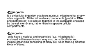
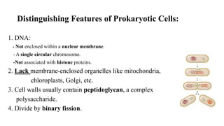
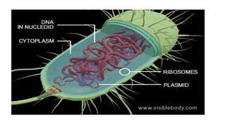
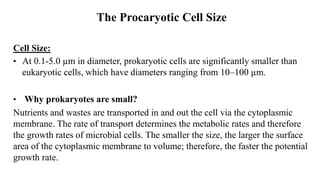
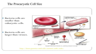
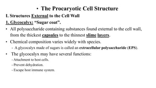
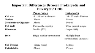
PROKARYOTIC CELL
STRUCTURE
By
SAMIRA FATTAH HAMID
Ph.D. Medical Bacteriology
College of Health Sciences
Hawler Medical University
Microorganisms are either:
• Prokaryotes: name comes from the Greek word, Pros means
“before” and karyon means “nut” referring to the nucleus.
or
• Eukaryotes: Eu means “true”, karyon means “nut” referring to
nucleus.
-Prokaryotes
is a unicellular organism that lacks nucleus, mitochondria, or any
other organelle. All the intracellular components (proteins, DNA
and metabolites) are located together in the cytoplasm enclosed
by the cell membrane, rather than in separate cellular
compartments.
-Eukaryotes
cells have a nucleus and organelles (e.g. mitochondria)
enclosed within membranes, may also be multicellular and
include organisms consisting of many cell types forming different
kinds of tissue.
Distinguishing Features of Prokaryotic Cells:
1. DNA:
- Not enclosed within a nuclear membrane.
- A single circular chromosome.
-Not associated with histone proteins.
2. Lack membrane-enclosed organelles like mitochondria,
chloroplasts, Golgi, etc.
3. Cell walls usually contain peptidoglycan, a complex
polysaccharide.
4. Divide by binary fission.
Distinguishing Features of Eukaryotic Cells:
1. DNA:
- Enclosed within a nuclear membrane.
- Several linear chromosomes.
- Associated with histones and other proteins.
2. Have membrane-enclosed organelles like mitochondria, chloroplasts,
Golgi, endoplasmic reticulum, etc.
3. Divide by mitosis.
The Procaryotic Cell Size
Cell Size:
• At 0.1-5.0 µm in diameter, prokaryotic cells are significantly smaller than
eukaryotic cells, which have diameters ranging from 10–100 µm.
• Why prokaryotes are small?
Nutrients and wastes are transported in and out the cell via the cytoplasmic
membrane. The rate of transport determines the metabolic rates and therefore
the growth rates of microbial cells. The smaller the size, the larger the surface
area of the cytoplasmic membrane to volume; therefore, the faster the potential
growth rate.
The Procaryotic Cell Size
• The Procaryotic Cell Structure
I. Structures External to the Cell Wall
1. Glycocalyx: “Sugar coat”.
• All polysaccharide containing substances found external to the cell wall,
from the thickest capsules to the thinnest slime layers.
• Chemical composition varies widely with species.
- A glycocalyx made of sugars is called an extracellular polysaccharide (EPS).
• The glycocalyx may have several functions:
- Attachment to host cells.
- Prevent dehydration.
- Escape host immune system.
I. Structures External to the Cell Wall
1. Glycocalyx: “Sugar coat”.
A. Capsules: Organized polysaccharide substance that is firmly
attached to the cell wall.
• Not formed by all bacteria.
• Important in virulence.
- Only Streptococcus pneumoniae with capsule cause pneumonia.
• Help bacteria escape the host immune system, by preventing destruction by
phagocytosis.
• When bacteria lose their capsules they become less likely to cause disease and
more susceptible to destruction.
I. Structures External to the Cell Wall
1. Glycocalyx:
B. Slime Layer: Thin polysaccharide substance that is loosely attached
to the cell wall.
• Not formed by all bacteria.
• Important for virulence.
-Oral bacteria stick to teeth due to slime layer and with time produce dental plaque.
• Allow bacteria to adhere to objects in their environment so they can remain
near sources of nutrients or oxygen.
- Rock surfaces
- Plant roots
I. Structures External to the Cell Wall
2. Flagella (Sing. Flagellum):
• The prokaryotic flagellum provide the power of motility
• This allows a cell to swim freely through an aqueous habitat.
• The bacterial flagellum when viewed under high magnification
displays three distinct parts:
- The filament
- the hook (sheath)
- the basal body.
I. Structures External to the Cell Wall
3. Fimbriae (Sing: fimbria):
• Found in Gram-negative bacteria.
• Like glycocalyx, enable bacteria to adhere to surfaces. Important for
colonization of host tissue.
• Neisseria gonorrhoeae: Causes gonorrhea. Attach to sperm cells and mucous
membranes through fimbriae.
• Bacteria can attach to broth surface via fimbriae, forming a film-like layer called
pellicle.
II. The Cell Wall
• Semirigid structure that lies outside the cell membrane in almost all
bacteria.
• Two major functions:
1. Maintains characteristic shape of cell.
2. Prevents the cell from bursting when fluids flow into the cell by osmosis.
• Composition:
Peptidoglycan (Murein): Made up of a repeating disaccharide attached by
polypeptides to form a lattice.
II. The Cell Wall
Peptidoglycan (Murein):
• Disaccharide component: Made up of
two monoscaccharides:
-N-acetylglucosamine (NAG)
- N-acetylmuramic acid (NAM)
• Alternating disaccharides (NAG-NAM)
are linked together in rows of 10 to 65
molecules.
II. The Cell Wall
Peptidoglycan (Murein):
• Adjacent disaccharide rows are linked together by polypeptide chains which vary in
composition, but always contain tetrapeptide side chains.
• Parallel tetrapeptide side chains may be directly linked together or linked by a
polypeptide cross-bridge.
III. Structures Internal to the Cell Wall
1. The Plasma (Cytoplasmic) Membrane:
• Thin structure inside of cell wall that surrounds the cytoplasm.
• Phospholipid bilayer with proteins.
- Integral membrane proteins: Penetrate membrane completely.
- Peripheral membrane proteins: On inner or outer membrane surface.
• Lack sterols and are less rigid than eukaryotic membranes.
- Exception: Mycoplasmas
Structure of Plasma Membrane
III. Structures Internal to the Cell Wall
Functions of the Plasma Membrane:
1. Selective barrier that regulates the passage of materials in and out of the
cell.
- Impermeable to large proteins, ions, and most polar molecules.
- Permeable to water, oxygen, carbon dioxide, some simple sugars, and small nonpolar
substances.
2. Synthesis of cell wall components
3. Site of photosynthesis: Photosynthetic bacteria have membrane
extensions called thylakoids, where photosynthesis occurs.
4. Secretes proteins
5. Contains bases of flagella
III. Structures Internal to the Cell Wall
2. Cytoplasm
• Substance inside the cell membrane.
Contains:
- 80% water
- Proteins
- Carbohydrates
- Lipids
- Inorganic ions
- Low molecular weight compounds
• Lacks a cytoskeleton.
III. Structures Internal to the Cell Wall
3. The Nuclear Area (Nucleoid):
• Contains a single chromosome, a long circular molecule of double
stranded DNA.
• The chromosome is attached to the plasma membrane.
• May occupy up to 20% of the intracellular volume.
Plasmids:
• Small, circular, double stranded DNA molecules. Found in many
bacterial cells in addition to chromosomal DNA.
• May contain from 5 to 100 genes that are usually not essential for
survival.
• Antibiotic resistance genes
• Toxins
III. Structures Internal to the Cell Wall
4. Ribosomes:
• The site of protein synthesis (translation).
• Found in all eukaryotic and prokaryotic cells.
• Prokaryotic ribosomes have two subunits:
- Small subunit: 30S
- Large subunit: 50S
• Several antibiotics work by inhibiting protein synthesis by prokaryotic
ribosomes, without affecting eukaryotic ribosomes.
Important Differences Between Prokaryotic and
Eukaryotic Cells
Prokaryotes Eukaryotes
Cell size 0.1-5.0 um in diameter 10-100 um in diameter
Nucleus Absent Present
Membranous Organelle Absent Present
Cell Wall Chemically complex When present, simple
Ribosomes Smaller (70S) Larger (80S)
DNA Single circular chromosome Multiple linear
chromosomes (histones)
Cell Division Binary fission Mitosis
Cytoskeleton Absent Present
Next lecture
Classification of Microorganisms
HOMEWORK
Write a report about:
Eukaryotic Cell Structure
(not more than 3 papers- to be submitted during lecture next week)

Prokaryotic Cell Structure

  • 1.
    PROKARYOTIC CELL STRUCTURE By SAMIRA FATTAHHAMID Ph.D. Medical Bacteriology College of Health Sciences Hawler Medical University
  • 2.
    Microorganisms are either: •Prokaryotes: name comes from the Greek word, Pros means “before” and karyon means “nut” referring to the nucleus. or • Eukaryotes: Eu means “true”, karyon means “nut” referring to nucleus.
  • 3.
    -Prokaryotes is a unicellularorganism that lacks nucleus, mitochondria, or any other organelle. All the intracellular components (proteins, DNA and metabolites) are located together in the cytoplasm enclosed by the cell membrane, rather than in separate cellular compartments. -Eukaryotes cells have a nucleus and organelles (e.g. mitochondria) enclosed within membranes, may also be multicellular and include organisms consisting of many cell types forming different kinds of tissue.
  • 4.
    Distinguishing Features ofProkaryotic Cells: 1. DNA: - Not enclosed within a nuclear membrane. - A single circular chromosome. -Not associated with histone proteins. 2. Lack membrane-enclosed organelles like mitochondria, chloroplasts, Golgi, etc. 3. Cell walls usually contain peptidoglycan, a complex polysaccharide. 4. Divide by binary fission.
  • 6.
    Distinguishing Features ofEukaryotic Cells: 1. DNA: - Enclosed within a nuclear membrane. - Several linear chromosomes. - Associated with histones and other proteins. 2. Have membrane-enclosed organelles like mitochondria, chloroplasts, Golgi, endoplasmic reticulum, etc. 3. Divide by mitosis.
  • 8.
    The Procaryotic CellSize Cell Size: • At 0.1-5.0 µm in diameter, prokaryotic cells are significantly smaller than eukaryotic cells, which have diameters ranging from 10–100 µm. • Why prokaryotes are small? Nutrients and wastes are transported in and out the cell via the cytoplasmic membrane. The rate of transport determines the metabolic rates and therefore the growth rates of microbial cells. The smaller the size, the larger the surface area of the cytoplasmic membrane to volume; therefore, the faster the potential growth rate.
  • 9.
  • 10.
    • The ProcaryoticCell Structure I. Structures External to the Cell Wall 1. Glycocalyx: “Sugar coat”. • All polysaccharide containing substances found external to the cell wall, from the thickest capsules to the thinnest slime layers. • Chemical composition varies widely with species. - A glycocalyx made of sugars is called an extracellular polysaccharide (EPS). • The glycocalyx may have several functions: - Attachment to host cells. - Prevent dehydration. - Escape host immune system.
  • 11.
    I. Structures Externalto the Cell Wall 1. Glycocalyx: “Sugar coat”. A. Capsules: Organized polysaccharide substance that is firmly attached to the cell wall. • Not formed by all bacteria. • Important in virulence. - Only Streptococcus pneumoniae with capsule cause pneumonia. • Help bacteria escape the host immune system, by preventing destruction by phagocytosis. • When bacteria lose their capsules they become less likely to cause disease and more susceptible to destruction.
  • 12.
    I. Structures Externalto the Cell Wall 1. Glycocalyx: B. Slime Layer: Thin polysaccharide substance that is loosely attached to the cell wall. • Not formed by all bacteria. • Important for virulence. -Oral bacteria stick to teeth due to slime layer and with time produce dental plaque. • Allow bacteria to adhere to objects in their environment so they can remain near sources of nutrients or oxygen. - Rock surfaces - Plant roots
  • 13.
    I. Structures Externalto the Cell Wall 2. Flagella (Sing. Flagellum): • The prokaryotic flagellum provide the power of motility • This allows a cell to swim freely through an aqueous habitat. • The bacterial flagellum when viewed under high magnification displays three distinct parts: - The filament - the hook (sheath) - the basal body.
  • 15.
    I. Structures Externalto the Cell Wall 3. Fimbriae (Sing: fimbria): • Found in Gram-negative bacteria. • Like glycocalyx, enable bacteria to adhere to surfaces. Important for colonization of host tissue. • Neisseria gonorrhoeae: Causes gonorrhea. Attach to sperm cells and mucous membranes through fimbriae. • Bacteria can attach to broth surface via fimbriae, forming a film-like layer called pellicle.
  • 16.
    II. The CellWall • Semirigid structure that lies outside the cell membrane in almost all bacteria. • Two major functions: 1. Maintains characteristic shape of cell. 2. Prevents the cell from bursting when fluids flow into the cell by osmosis. • Composition: Peptidoglycan (Murein): Made up of a repeating disaccharide attached by polypeptides to form a lattice.
  • 17.
    II. The CellWall Peptidoglycan (Murein): • Disaccharide component: Made up of two monoscaccharides: -N-acetylglucosamine (NAG) - N-acetylmuramic acid (NAM) • Alternating disaccharides (NAG-NAM) are linked together in rows of 10 to 65 molecules.
  • 18.
    II. The CellWall Peptidoglycan (Murein): • Adjacent disaccharide rows are linked together by polypeptide chains which vary in composition, but always contain tetrapeptide side chains. • Parallel tetrapeptide side chains may be directly linked together or linked by a polypeptide cross-bridge.
  • 19.
    III. Structures Internalto the Cell Wall 1. The Plasma (Cytoplasmic) Membrane: • Thin structure inside of cell wall that surrounds the cytoplasm. • Phospholipid bilayer with proteins. - Integral membrane proteins: Penetrate membrane completely. - Peripheral membrane proteins: On inner or outer membrane surface. • Lack sterols and are less rigid than eukaryotic membranes. - Exception: Mycoplasmas
  • 20.
  • 21.
    III. Structures Internalto the Cell Wall Functions of the Plasma Membrane: 1. Selective barrier that regulates the passage of materials in and out of the cell. - Impermeable to large proteins, ions, and most polar molecules. - Permeable to water, oxygen, carbon dioxide, some simple sugars, and small nonpolar substances. 2. Synthesis of cell wall components 3. Site of photosynthesis: Photosynthetic bacteria have membrane extensions called thylakoids, where photosynthesis occurs. 4. Secretes proteins 5. Contains bases of flagella
  • 22.
    III. Structures Internalto the Cell Wall 2. Cytoplasm • Substance inside the cell membrane. Contains: - 80% water - Proteins - Carbohydrates - Lipids - Inorganic ions - Low molecular weight compounds • Lacks a cytoskeleton.
  • 23.
    III. Structures Internalto the Cell Wall 3. The Nuclear Area (Nucleoid): • Contains a single chromosome, a long circular molecule of double stranded DNA. • The chromosome is attached to the plasma membrane. • May occupy up to 20% of the intracellular volume. Plasmids: • Small, circular, double stranded DNA molecules. Found in many bacterial cells in addition to chromosomal DNA. • May contain from 5 to 100 genes that are usually not essential for survival. • Antibiotic resistance genes • Toxins
  • 25.
    III. Structures Internalto the Cell Wall 4. Ribosomes: • The site of protein synthesis (translation). • Found in all eukaryotic and prokaryotic cells. • Prokaryotic ribosomes have two subunits: - Small subunit: 30S - Large subunit: 50S • Several antibiotics work by inhibiting protein synthesis by prokaryotic ribosomes, without affecting eukaryotic ribosomes.
  • 26.
    Important Differences BetweenProkaryotic and Eukaryotic Cells Prokaryotes Eukaryotes Cell size 0.1-5.0 um in diameter 10-100 um in diameter Nucleus Absent Present Membranous Organelle Absent Present Cell Wall Chemically complex When present, simple Ribosomes Smaller (70S) Larger (80S) DNA Single circular chromosome Multiple linear chromosomes (histones) Cell Division Binary fission Mitosis Cytoskeleton Absent Present
  • 27.
  • 28.
    HOMEWORK Write a reportabout: Eukaryotic Cell Structure (not more than 3 papers- to be submitted during lecture next week)