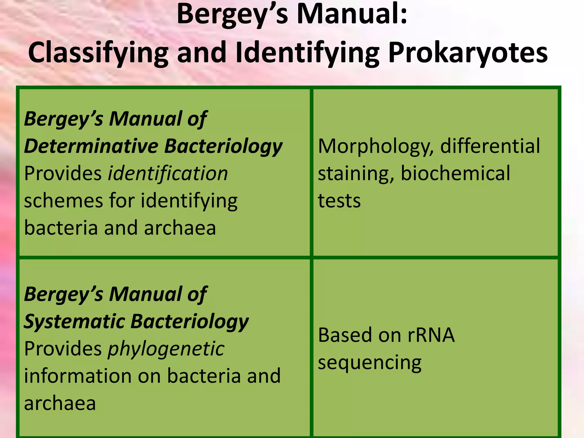
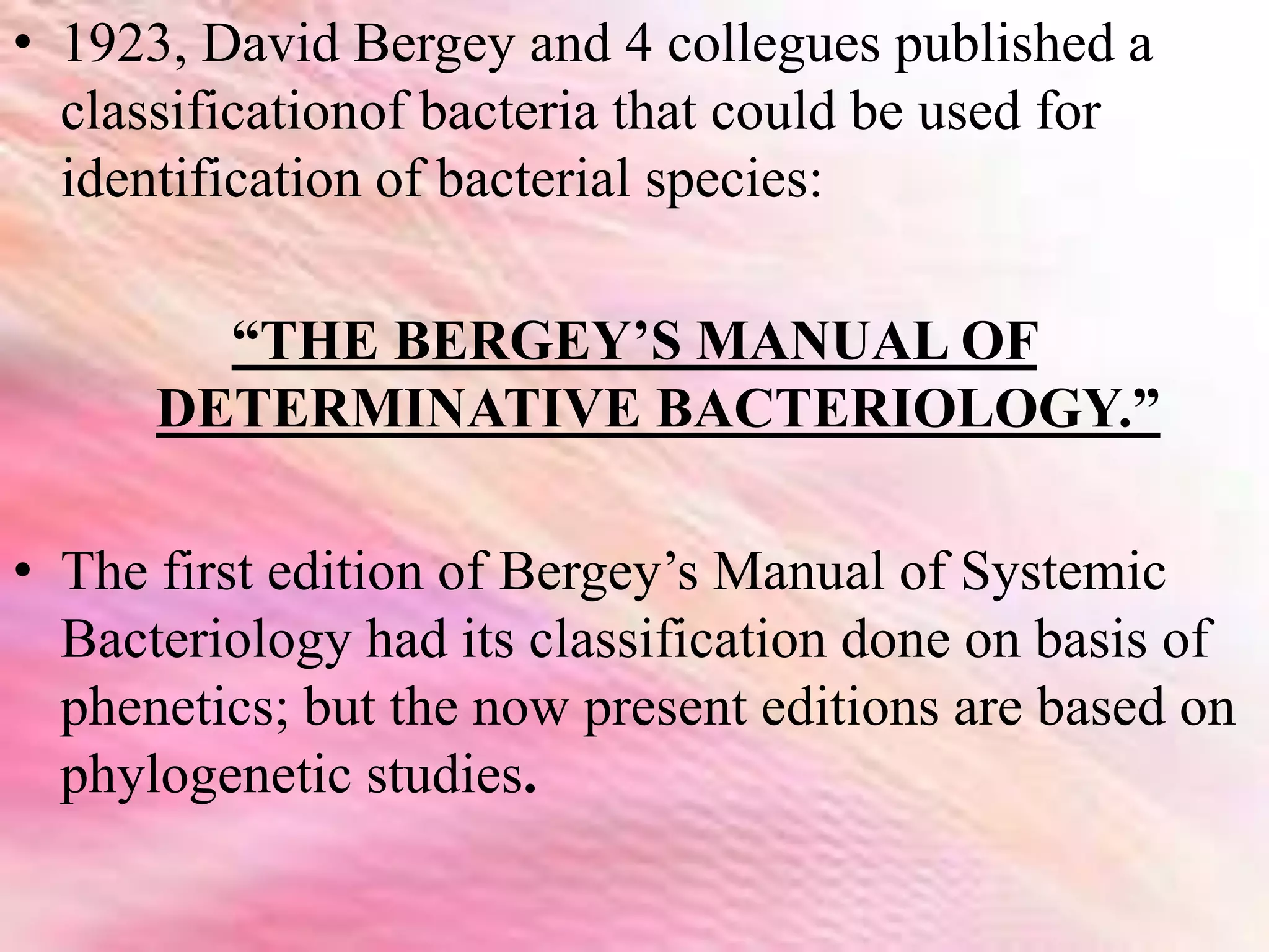
Bergey's Manual and it's classification. A brief concised presentation prepared for taking seminar and classes.
Volume II (Edition 2) described more in detail.
Bergey's Manual and it's classification. A brief concised presentation prepared for taking seminar and classes.
Volume II (Edition 2) described more in detail.
The archaebacteria
group members
Rameen nadeem
Syeda iqra hussain
Hina zamir
Mahnoor khan
Maleeha inayat
Background
Biologists have long organized living things into large groups called kingdoms.
There are six of them:
Archaebacteria
Eubacteria
Protista
Fungi
Plantae
Animalia
Some recent findings…
In 1996, scientists decided to split Monera into two groups of bacteria:
Archaebacteria and Eubacteria
Because these two groups of bacteria were different in many ways scientists created a new level of classification called a DOMAIN.
Now we have 3 domains
Bacteria
Archaea
Eukarya
KingdomArchaebacteria
Any of a large group of primitive bacteria having unusual cell walls, membrane lipids, ribosomes, and RNA sequences, and having the ability to produce methane and to live in anaerobic, extremely hot, salty, or acidic conditions
The Domain Archaea
“ancient” bacteria
Some of the first archaebacteria were discovered in Yellowstone National Park’s hot springs
Prokaryotes are structurally simple, but biochemically complex
Basic Facts
They live in extreme environments (like hot springs or salty lakes) and normal environments (like soil and ocean water).
All are unicellular (each individual is only one cell).
No peptidoglycan in their cell wall.
Some have a flagella that aids in their locomotion.
Most don’t need oxygen to survive
They can produce ATP (energy) from sunlight
They can survive enormous temperature extremes
They can survive under rocks and in ocean floor vents deep below the ocean’s surface
They can tolerate huge pressure differences
STRUCTURE
Size
Archaea are slightly less than 1 micron long.
A micron is 1/1,000 of a millimeter.
In order to see their cellular features, scientists use powerful electron microscopes.
Shape
Shapes can be spherical or ball shaped and are called coccus.
Others are rod shaped, long and thin, and labeled bacillus.
Variations of cells have been discovered in square and triangular shapes.
STRUCTURE
Locomotion
Some archaea have flagella, hair-like structures that assist in movement.
There can be one or many attached to the cell's outer membrane. Protein networks can also be found on the cell membrane, which allow cells to attach themselves in groups.
Cell Features
Within the cell membrane, the archaea cell contains cytoplasm and DNA, which are in single-looped forms called plasmids.
Most archaeal cells also have a semi-rigid cell wall that helps it to maintain its shape and chemical balance.
This protects the cytoplasm, which is the semi-liquid gel that fills the cell and enables the various parts to function.
STRUCTURE
Phospholipids
The molecules that make up cell membranes are called phospholipids, which act as building blocks for the cell.
In archaea, these molecules are made of glycerol-ether lipids.
Ether Bonding
The ether bonding makes it possible for archaea to survive in environments that are extremely acidic or al
Direct methods of measurement of microbial growth includes various methods of enumeration of both viable and non viable cell also includes growth curve. Helpful for UG and PG programs of microbiology
Microbial Taxonomy - Dr. R Subashkumar, Associate Professor in Biotechnology, Sri Ramakrishna College of Arts and Science (Autonomous), Coimbatore-641006
Taxonomy of Prokaryotes that include Bacteria and Archea, Brief description of taxonomy
history
types of classification
numerical taxonomy
different classification system
basic characters used for classification
International code of Nomenclature of Bacteria
Bergey's manual
pathovar concept
This presentation has been designed to give the foundation of taxonomy in general and Plant Taxonomy in particular as a matter of pleasure to explore the diversity of the plant world.
Similar to Bergey’s manual of bacterial classification (20)
(May 29th, 2024) Advancements in Intravital Microscopy- Insights for Preclini...Scintica Instrumentation
Intravital microscopy (IVM) is a powerful tool utilized to study cellular behavior over time and space in vivo. Much of our understanding of cell biology has been accomplished using various in vitro and ex vivo methods; however, these studies do not necessarily reflect the natural dynamics of biological processes. Unlike traditional cell culture or fixed tissue imaging, IVM allows for the ultra-fast high-resolution imaging of cellular processes over time and space and were studied in its natural environment. Real-time visualization of biological processes in the context of an intact organism helps maintain physiological relevance and provide insights into the progression of disease, response to treatments or developmental processes.
In this webinar we give an overview of advanced applications of the IVM system in preclinical research. IVIM technology is a provider of all-in-one intravital microscopy systems and solutions optimized for in vivo imaging of live animal models at sub-micron resolution. The system’s unique features and user-friendly software enables researchers to probe fast dynamic biological processes such as immune cell tracking, cell-cell interaction as well as vascularization and tumor metastasis with exceptional detail. This webinar will also give an overview of IVM being utilized in drug development, offering a view into the intricate interaction between drugs/nanoparticles and tissues in vivo and allows for the evaluation of therapeutic intervention in a variety of tissues and organs. This interdisciplinary collaboration continues to drive the advancements of novel therapeutic strategies.
Nutraceutical market, scope and growth: Herbal drug technologyLokesh Patil
As consumer awareness of health and wellness rises, the nutraceutical market—which includes goods like functional meals, drinks, and dietary supplements that provide health advantages beyond basic nutrition—is growing significantly. As healthcare expenses rise, the population ages, and people want natural and preventative health solutions more and more, this industry is increasing quickly. Further driving market expansion are product formulation innovations and the use of cutting-edge technology for customized nutrition. With its worldwide reach, the nutraceutical industry is expected to keep growing and provide significant chances for research and investment in a number of categories, including vitamins, minerals, probiotics, and herbal supplements.
What is greenhouse gasses and how many gasses are there to affect the Earth.moosaasad1975
What are greenhouse gasses how they affect the earth and its environment what is the future of the environment and earth how the weather and the climate effects.
Observation of Io’s Resurfacing via Plume Deposition Using Ground-based Adapt...Sérgio Sacani
Since volcanic activity was first discovered on Io from Voyager images in 1979, changes
on Io’s surface have been monitored from both spacecraft and ground-based telescopes.
Here, we present the highest spatial resolution images of Io ever obtained from a groundbased telescope. These images, acquired by the SHARK-VIS instrument on the Large
Binocular Telescope, show evidence of a major resurfacing event on Io’s trailing hemisphere. When compared to the most recent spacecraft images, the SHARK-VIS images
show that a plume deposit from a powerful eruption at Pillan Patera has covered part
of the long-lived Pele plume deposit. Although this type of resurfacing event may be common on Io, few have been detected due to the rarity of spacecraft visits and the previously low spatial resolution available from Earth-based telescopes. The SHARK-VIS instrument ushers in a new era of high resolution imaging of Io’s surface using adaptive
optics at visible wavelengths.
Slide 1: Title Slide
Extrachromosomal Inheritance
Slide 2: Introduction to Extrachromosomal Inheritance
Definition: Extrachromosomal inheritance refers to the transmission of genetic material that is not found within the nucleus.
Key Components: Involves genes located in mitochondria, chloroplasts, and plasmids.
Slide 3: Mitochondrial Inheritance
Mitochondria: Organelles responsible for energy production.
Mitochondrial DNA (mtDNA): Circular DNA molecule found in mitochondria.
Inheritance Pattern: Maternally inherited, meaning it is passed from mothers to all their offspring.
Diseases: Examples include Leber’s hereditary optic neuropathy (LHON) and mitochondrial myopathy.
Slide 4: Chloroplast Inheritance
Chloroplasts: Organelles responsible for photosynthesis in plants.
Chloroplast DNA (cpDNA): Circular DNA molecule found in chloroplasts.
Inheritance Pattern: Often maternally inherited in most plants, but can vary in some species.
Examples: Variegation in plants, where leaf color patterns are determined by chloroplast DNA.
Slide 5: Plasmid Inheritance
Plasmids: Small, circular DNA molecules found in bacteria and some eukaryotes.
Features: Can carry antibiotic resistance genes and can be transferred between cells through processes like conjugation.
Significance: Important in biotechnology for gene cloning and genetic engineering.
Slide 6: Mechanisms of Extrachromosomal Inheritance
Non-Mendelian Patterns: Do not follow Mendel’s laws of inheritance.
Cytoplasmic Segregation: During cell division, organelles like mitochondria and chloroplasts are randomly distributed to daughter cells.
Heteroplasmy: Presence of more than one type of organellar genome within a cell, leading to variation in expression.
Slide 7: Examples of Extrachromosomal Inheritance
Four O’clock Plant (Mirabilis jalapa): Shows variegated leaves due to different cpDNA in leaf cells.
Petite Mutants in Yeast: Result from mutations in mitochondrial DNA affecting respiration.
Slide 8: Importance of Extrachromosomal Inheritance
Evolution: Provides insight into the evolution of eukaryotic cells.
Medicine: Understanding mitochondrial inheritance helps in diagnosing and treating mitochondrial diseases.
Agriculture: Chloroplast inheritance can be used in plant breeding and genetic modification.
Slide 9: Recent Research and Advances
Gene Editing: Techniques like CRISPR-Cas9 are being used to edit mitochondrial and chloroplast DNA.
Therapies: Development of mitochondrial replacement therapy (MRT) for preventing mitochondrial diseases.
Slide 10: Conclusion
Summary: Extrachromosomal inheritance involves the transmission of genetic material outside the nucleus and plays a crucial role in genetics, medicine, and biotechnology.
Future Directions: Continued research and technological advancements hold promise for new treatments and applications.
Slide 11: Questions and Discussion
Invite Audience: Open the floor for any questions or further discussion on the topic.
Multi-source connectivity as the driver of solar wind variability in the heli...Sérgio Sacani
The ambient solar wind that flls the heliosphere originates from multiple
sources in the solar corona and is highly structured. It is often described
as high-speed, relatively homogeneous, plasma streams from coronal
holes and slow-speed, highly variable, streams whose source regions are
under debate. A key goal of ESA/NASA’s Solar Orbiter mission is to identify
solar wind sources and understand what drives the complexity seen in the
heliosphere. By combining magnetic feld modelling and spectroscopic
techniques with high-resolution observations and measurements, we show
that the solar wind variability detected in situ by Solar Orbiter in March
2022 is driven by spatio-temporal changes in the magnetic connectivity to
multiple sources in the solar atmosphere. The magnetic feld footpoints
connected to the spacecraft moved from the boundaries of a coronal hole
to one active region (12961) and then across to another region (12957). This
is refected in the in situ measurements, which show the transition from fast
to highly Alfvénic then to slow solar wind that is disrupted by the arrival of
a coronal mass ejection. Our results describe solar wind variability at 0.5 au
but are applicable to near-Earth observatories.
Richard's entangled aventures in wonderlandRichard Gill
Since the loophole-free Bell experiments of 2020 and the Nobel prizes in physics of 2022, critics of Bell's work have retreated to the fortress of super-determinism. Now, super-determinism is a derogatory word - it just means "determinism". Palmer, Hance and Hossenfelder argue that quantum mechanics and determinism are not incompatible, using a sophisticated mathematical construction based on a subtle thinning of allowed states and measurements in quantum mechanics, such that what is left appears to make Bell's argument fail, without altering the empirical predictions of quantum mechanics. I think however that it is a smoke screen, and the slogan "lost in math" comes to my mind. I will discuss some other recent disproofs of Bell's theorem using the language of causality based on causal graphs. Causal thinking is also central to law and justice. I will mention surprising connections to my work on serial killer nurse cases, in particular the Dutch case of Lucia de Berk and the current UK case of Lucy Letby.
Introduction:
RNA interference (RNAi) or Post-Transcriptional Gene Silencing (PTGS) is an important biological process for modulating eukaryotic gene expression.
It is highly conserved process of posttranscriptional gene silencing by which double stranded RNA (dsRNA) causes sequence-specific degradation of mRNA sequences.
dsRNA-induced gene silencing (RNAi) is reported in a wide range of eukaryotes ranging from worms, insects, mammals and plants.
This process mediates resistance to both endogenous parasitic and exogenous pathogenic nucleic acids, and regulates the expression of protein-coding genes.
What are small ncRNAs?
micro RNA (miRNA)
short interfering RNA (siRNA)
Properties of small non-coding RNA:
Involved in silencing mRNA transcripts.
Called “small” because they are usually only about 21-24 nucleotides long.
Synthesized by first cutting up longer precursor sequences (like the 61nt one that Lee discovered).
Silence an mRNA by base pairing with some sequence on the mRNA.
Discovery of siRNA?
The first small RNA:
In 1993 Rosalind Lee (Victor Ambros lab) was studying a non- coding gene in C. elegans, lin-4, that was involved in silencing of another gene, lin-14, at the appropriate time in the
development of the worm C. elegans.
Two small transcripts of lin-4 (22nt and 61nt) were found to be complementary to a sequence in the 3' UTR of lin-14.
Because lin-4 encoded no protein, she deduced that it must be these transcripts that are causing the silencing by RNA-RNA interactions.
Types of RNAi ( non coding RNA)
MiRNA
Length (23-25 nt)
Trans acting
Binds with target MRNA in mismatch
Translation inhibition
Si RNA
Length 21 nt.
Cis acting
Bind with target Mrna in perfect complementary sequence
Piwi-RNA
Length ; 25 to 36 nt.
Expressed in Germ Cells
Regulates trnasposomes activity
MECHANISM OF RNAI:
First the double-stranded RNA teams up with a protein complex named Dicer, which cuts the long RNA into short pieces.
Then another protein complex called RISC (RNA-induced silencing complex) discards one of the two RNA strands.
The RISC-docked, single-stranded RNA then pairs with the homologous mRNA and destroys it.
THE RISC COMPLEX:
RISC is large(>500kD) RNA multi- protein Binding complex which triggers MRNA degradation in response to MRNA
Unwinding of double stranded Si RNA by ATP independent Helicase
Active component of RISC is Ago proteins( ENDONUCLEASE) which cleave target MRNA.
DICER: endonuclease (RNase Family III)
Argonaute: Central Component of the RNA-Induced Silencing Complex (RISC)
One strand of the dsRNA produced by Dicer is retained in the RISC complex in association with Argonaute
ARGONAUTE PROTEIN :
1.PAZ(PIWI/Argonaute/ Zwille)- Recognition of target MRNA
2.PIWI (p-element induced wimpy Testis)- breaks Phosphodiester bond of mRNA.)RNAse H activity.
MiRNA:
The Double-stranded RNAs are naturally produced in eukaryotic cells during development, and they have a key role in regulating gene expression .
This presentation explores a brief idea about the structural and functional attributes of nucleotides, the structure and function of genetic materials along with the impact of UV rays and pH upon them.
3. TAXONOMY
• Is defined as the science of biological classification.
•It consists of 3 separate but interrelated parts:
•Classification
•Nomenclature
•Identification
•Once a classification scheme is selected, it is used to group
organisms into groups called “taxa” based on mutual
similarity.
4. NOMENCLATURE
• It is the branch of taxonomy concerned with
the assignment of names to taxonomic groups
in agreement with published rules.
5. IDENTIFICATION
It is the practical side of taxonomy,
the process of determining if a
particular isolate belongs to a
recognized taxon.
6. SYSTEMATICS
• This term is often used for taxonomy.
• Many taxonomists define systematics as “ The
scientific studies of organisms with the
ultimate object of characterizing and
arranging them in an orderly manner.”
• Thus it ecompasses disciplines such as
‘morphology’, ‘ecology’, ‘epidemiology’,
‘biochemistry’, ‘molecular biology’ and
‘physiology’.
7. • One of the oldest classification systems was the
“ the natural classification”.
• It included arrangement of organisms into
groups whose members share many characteristc
and reflect as much as possible the biological
nature of the organism.
• CAROLUS LINNAEUS developed the first
natural classification, based largely on
anatomical characteristics, in the mid 18thC.
8. • In practice, the determination of the genusand
the species of a newly discovered prokaryote is
based on polyphasic taxonomy.
• It includes the following approaches:
• Phenetic classification
• Phylogenetic classification
• Genotypic classification
9. PHYLOGENETIC CLASSIFICATION
• It groups organisms together based on the
mutual similarity of their phenotypic
characteristics.
• The term “phylogeny” is a Greek word where
phylon=tribe or race; and genesis= generation
or origin.
• It sought to compare organisms based on their
evolutionary relationships.
10. GENOTYPIC CLASSIFICATION
• It seeks to compare thee genetic similarity
between organisms by comparing either
individual genes or whole genomes.
12. Bergey’s Manual:
Classifying and Identifying Prokaryotes
Bergey’s Manual of
Determinative Bacteriology
Provides identification
schemes for identifying
bacteria and archaea
Morphology, differential
staining, biochemical
tests
Bergey’s Manual of
Systematic Bacteriology
Provides phylogenetic
information on bacteria and
archaea
Based on rRNA
sequencing
13. • 1923, David Bergey and 4 collegues published a
classificationof bacteria that could be used for
identification of bacterial species:
“THE BERGEY’S MANUAL OF
DETERMINATIVE BACTERIOLOGY.”
• The first edition of Bergey’s Manual of Systemic
Bacteriology had its classification done on basis of
phenetics; but the now present editions are based on
phylogenetic studies.
14. THE SECOND EDITION OF THE BERGEY’S MANUAL has
the following volumes where it doesn’t group all
the clinically important prokaryotes together as the
1stdeition does, here the pathogenic bacteria are
placed phylogenetically, and so are distributed
throughtout in all the volumes:
15. • VOL.1: The Archae, and the Deeply Branching and
the Phototropic Bacteria.
• VOL.2: The Proteobacteria.
• VOL.3: The Low G+C Gram positive Bacteria
• VOL.4: The High G+C Gram Positive Bacteria
• VOL.5: The Planctomycetes, Spyrochaetes,
Fibrobacters, Bacteriodetes, Fusobacteria,
Chlamydiae, Acidobacteria, Verrumicrobia and
Dictoglomus.