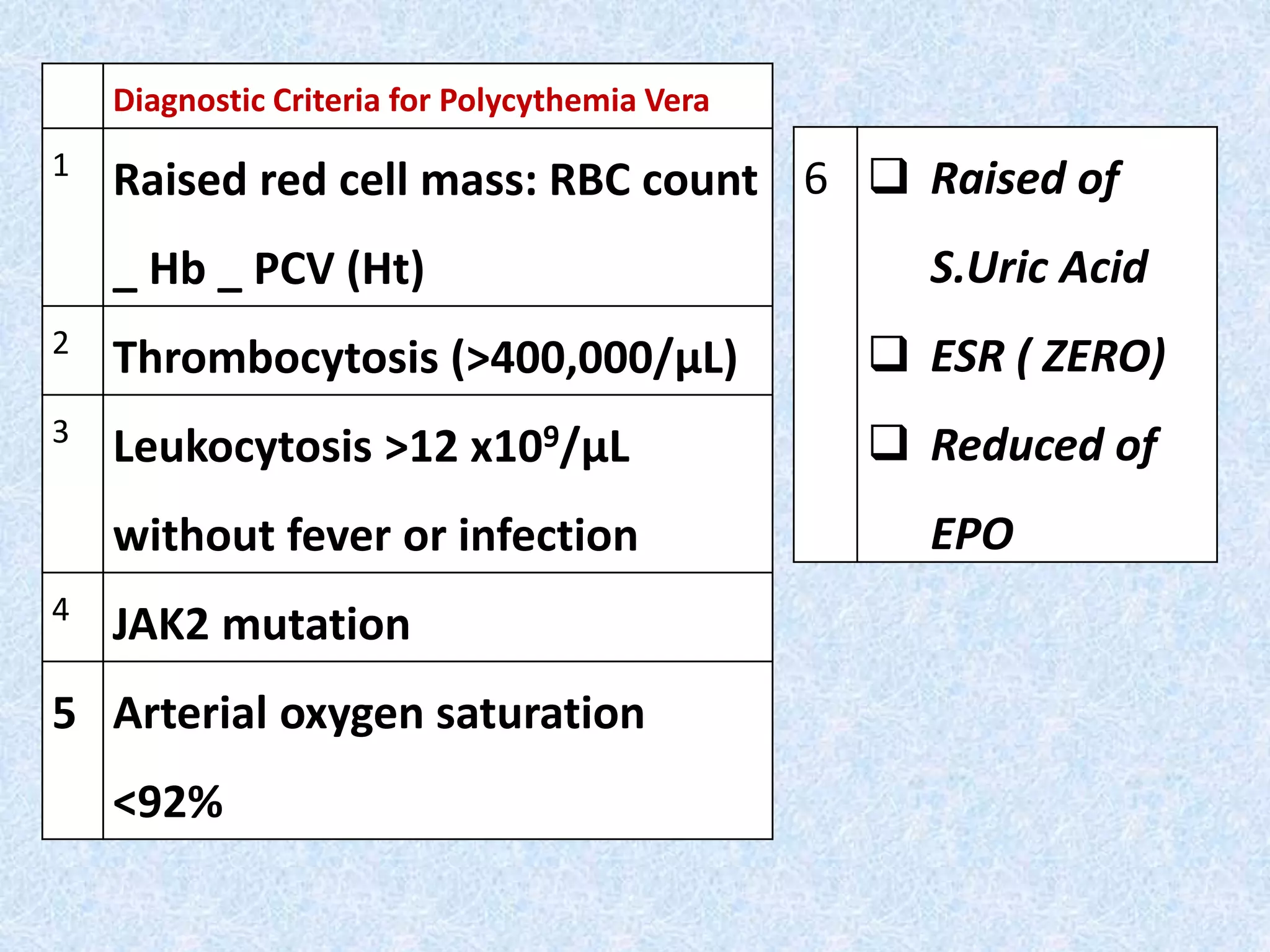
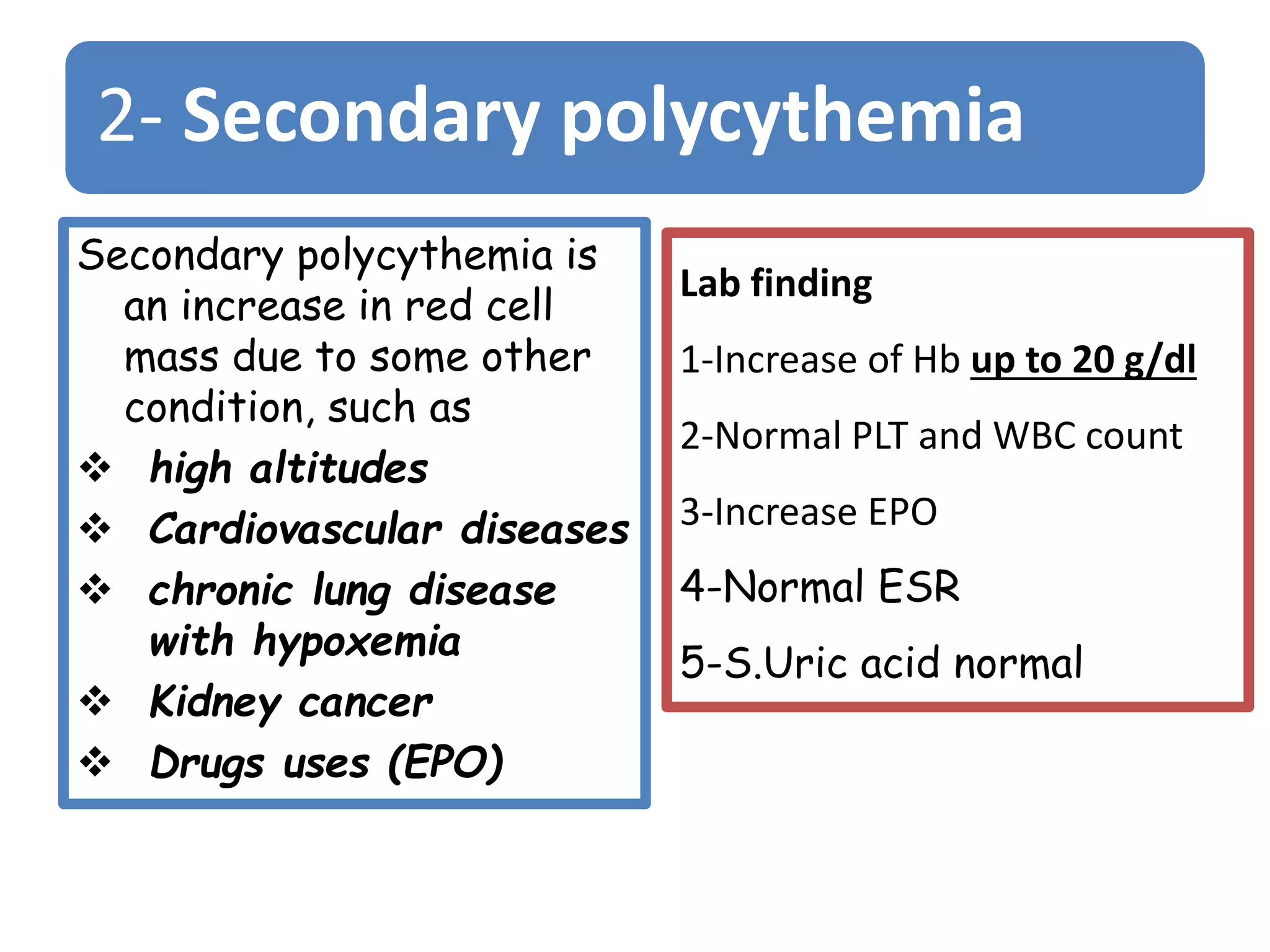
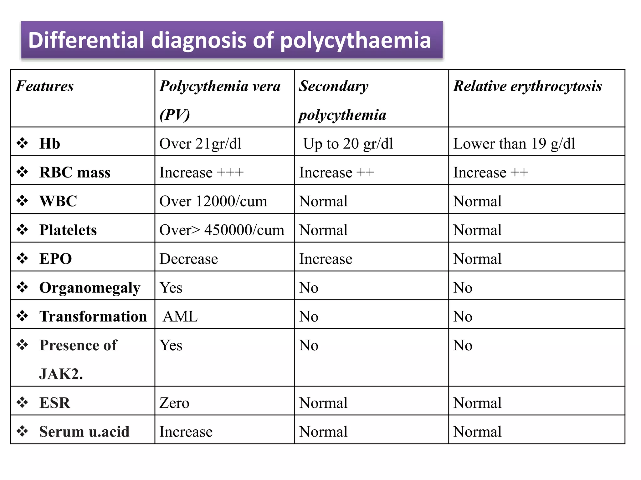
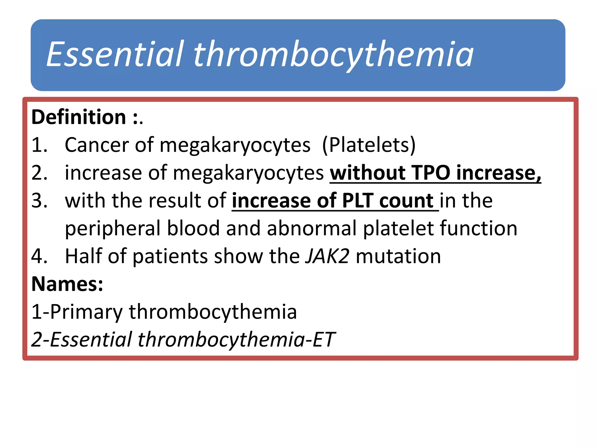
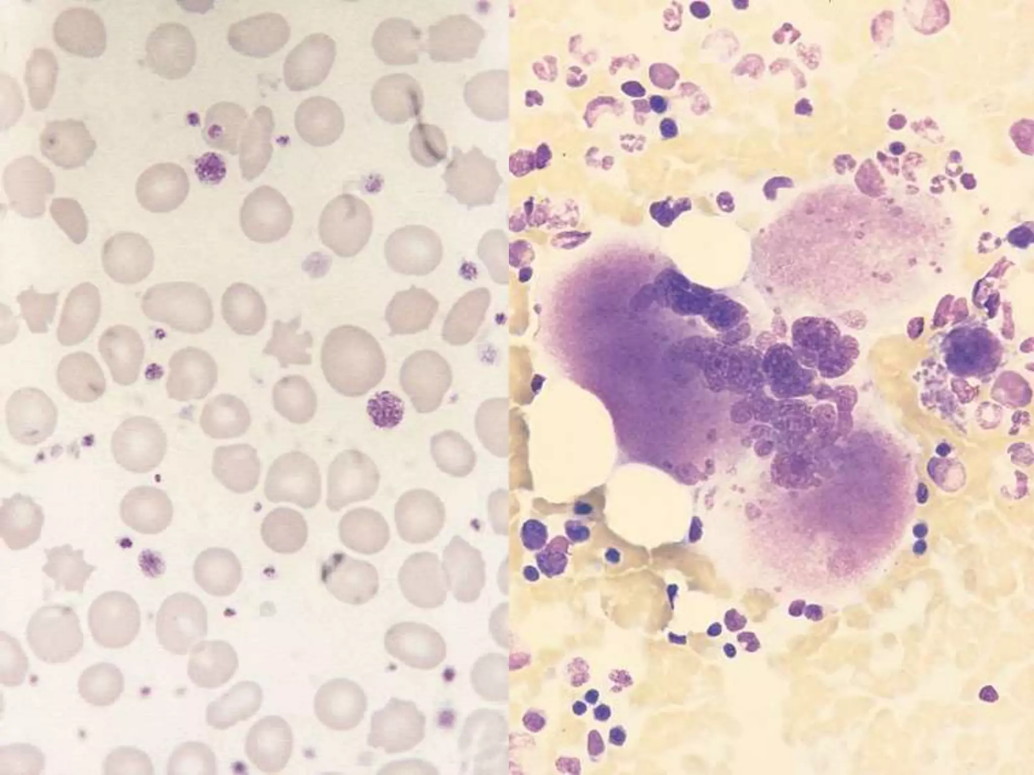
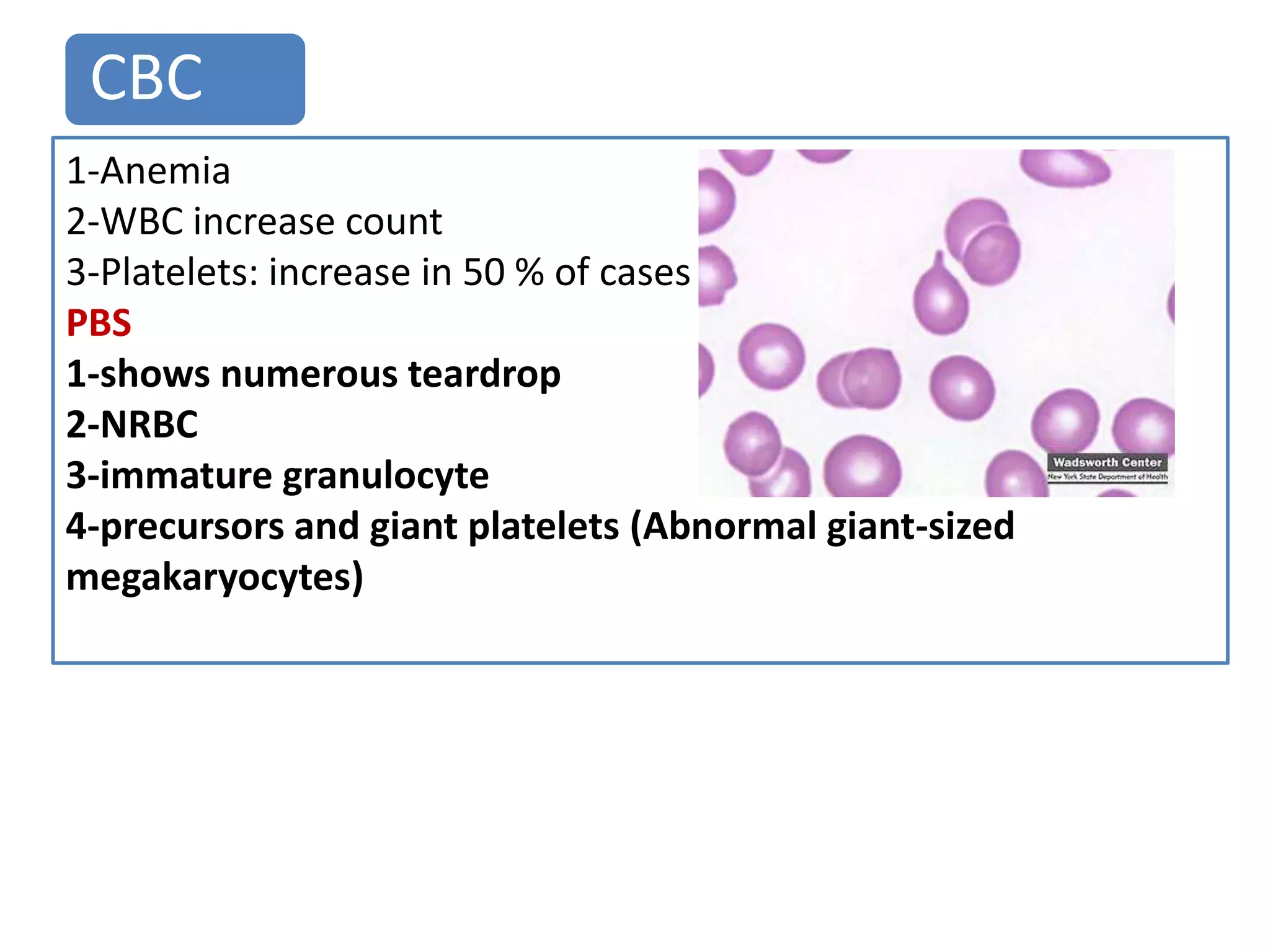
Lesson 5-6
1 Polycythaemia rubra vera (PRV).
2 Essential thrombocythaemia (ET).
3 Myelofibrosis
Polycythaemia (erythrocytosis) is defined as an increase in the
haemoglobin concentration above the upper limit of normal for
the patient's age and sex.
Classification of polycythaemia
primary
polycythaemia
(PRV)
Relative polycythaemia
Absolute polycythaemia
Secondary
polycythaemia
Polycythaemia (rubra) vera (PRV)
Definition:
•Polycythemia.vera is a cancer of the erythrocytes
characterized by
•excessive production of all blood cells lines
1-RBC, PCV, Hb Male > 0.51 (50%)-Female > 0.48 (48%)
2-WBC
3-PLTs
•independent of erythropoietin
Names:
1-Polycythemia vera (P. Vera)
2-Polycythemia rubra vera (PRVera)
Classified as negative Philadelphia chromosome
Janus-associated kinase 2 JAK2)
Genetic Causes of PRV
 The disease results from somatic mutation of
a single stem cell (MSC) this mutation called
“The JAK2 mutation” is present in
haemopoietic cells in almost 100% of patients.
 the increase of “JAK2” in red cells is the
diagnostic finding, in many patients
Diagnostic Criteria for Polycythemia Vera
1
Raised red cell mass: RBC count
_ Hb _ PCV (Ht)
2
Thrombocytosis (>400,000/µL)
3
Leukocytosis >12 x109/µL
without fever or infection
4
JAK2 mutation
5 Arterial oxygen saturation
<92%
6  Raised of
S.Uric Acid
 ESR ( ZERO)
 Reduced of
EPO
Peripheral blood findings for polycythemia vera include:
1. Normal red blood cell morphology,
2. Normoblasts may be present
3. Mild to moderate leukocytosis
4. Mild neutrophilia and/or basophilia
5. Thrombocytosis
Complication of PV:
1. -Thrombosis, - Bleeding
2. -Bone marrow fibrosis “ Burnt out”
3. -Transforming to AML
2- Secondary polycythemia
Secondary polycythemia is
an increase in red cell
mass due to some other
condition, such as
 high altitudes
 Cardiovascular diseases
 chronic lung disease
with hypoxemia
 Kidney cancer
 Drugs uses (EPO)
Lab finding
1-Increase of Hb up to 20 g/dl
2-Normal PLT and WBC count
3-Increase EPO
4-Normal ESR
5-S.Uric acid normal
3- Relative erythrocytosis
• It is a case of an increase
of
• RBC mass. Hb .PCV
• It is not malignant case;
the term is synonymous
with ‘polycythaemia
Causes
• 1-Heavy smoking
• 2-dehydration and
hemoconcentration.
Lab finding:
 Increase Hb not over
19g/dl
 Normal PLT and WBC
count,
 Normal EPO,
 Normal ESR
 S.Uric acid normal
Features Polycythemia vera
(PV)
Secondary
polycythemia
Relative erythrocytosis
 Hb Over 21gr/dl Up to 20 gr/dl Lower than 19 g/dl
 RBC mass Increase +++ Increase ++ Increase ++
 WBC Over 12000/cum Normal Normal
 Platelets Over> 450000/cum Normal Normal
 EPO Decrease Increase Normal
 Organomegaly Yes No No
 Transformation AML No No
 Presence of
JAK2.
Yes No No
 ESR Zero Normal Normal
 Serum u.acid Increase Normal Normal
Differential diagnosis of polycythaemia
Primary or Essential
Thrombocythemia
ET
Essential thrombocythemia
Definition :.
1. Cancer of megakaryocytes (Platelets)
2. increase of megakaryocytes without TPO increase,
3. with the result of increase of PLT count in the
peripheral blood and abnormal platelet function
4. Half of patients show the JAK2 mutation
Names:
1-Primary thrombocythemia
2-Essential thrombocythemia-ET
Points on ET
 Disease of middle aged persons
 sometimes occurs in children
 disease is inactive for 10-20 years or more.
 Transform to myelofibrosis
 May be transform to AML 10-20% of patients
 discovered only by routine PB examination
 neither splenomegaly nor hepatomegaly
 recurrent abortions
 Cause of death: thromboembolic events
1. Bleeding: (at GTI, urinary
tract , joint, and brain )
2. Neurologic events:
Headaches,
paresthesias,visual
disturbances etc )
3. Microvascular occlusions:
(pain, acrocyanosis gangrene
of the fingertips or toes)
4. Large vessel thromboses
Complication:
1-Thrombosis
2-Bleeding.
3-Transformation to AML
(rare)
4-myelofbrosis
Symptoms of ET
Diagnostic criteria
1. platelet count > 600 x 10 9/L
2. Hgb < 13.0 g/dl or normal red cell mass
3. No Ph1 or evidence of bcr/abl hybrid
1. Leukocytes: <20.000/ cum
2. mild eosinophilia/basophilia
1. RBC normal unless concurrent
iron deficiency
1. PLTs count: over 600.000/cum
could be reach > 1.000.000
/cum
2. thrombocytosis with giant
platelets ; bizarre forms
3. increased MPV
4. circulating megakaryocytes and
megakaryocytic fragments
Peripheral blood findings
PLT
WBC
RBC
Bone marrow:
1. Marked clustering of megakaryocytes
2. Half of patients show the JAK2 (Va1617Phe) mutation
Reactive
Thrombocytosis:
 Benign case of thrombocytosis
 PLT`s count over 600,000/Cum
Cases has thrombocytosis over million/cum
1. infection 22%
2. Tissue damage 20%
3. Chronic inflammation 13%
4. Malignancy 6%
Lesson: 7
Myelofibrosis
Myeloid Metaplasia (MMM)
1-Leukoerythroblastic reaction:
which means presence in the peripheral blood of
•Nucleated RBC
•Immature granulocytes
2-Extramedullary hematopoiesis
3-Fibrosis of the bone marrow/reticulin silver stain
4-Teardrop RBCs
5-Absence of the Philadelphia chromosome
6-Hepatosplenomegaly
Definition: it is a leukoerythroblastic reaction in the
blood or it is a clonal stem cell defect characterized by
Points to remember
•Hematopoietic stem cells grow out of control
•Age: middle age50-70yrs
•Incidence: male and female are equal
•Risk factor: benzene and also to ionizing
radiation breast cancer, prostate cancer, Hodgkin's
disease
•Chronic: many years the patient is healthy
•In late stage, anemia, fever weight loss etc
CBC
1-Anemia
2-WBC increase count
3-Platelets: increase in 50 % of cases
PBS
1-shows numerous teardrop
2-NRBC
3-immature granulocyte
4-precursors and giant platelets (Abnormal giant-sized
megakaryocytes)
Chemistry:
Increased of Serum
• 1-uric acid,
• 2-LDH,
• 3-alkaline
phosphatase,
• 4-and vitamin B12
Bone Marrow
•1-The bone marrow usually
cannot be aspirated (“dry
tap”).
•2-The biopsy can give the
diagnosis
•3-Philadelphia chromosome
negative
Summary and points to remember about MMM
MMM is characterized by
marrow fibrosis,
extramedullary hematopoiesis
leukoerythroblastic blood smear.
MMM, the accelerating fibrosis –with leukopenia and
thrombocytopenia
Bone marrow biopsy- dry tap.
MMM has the worst prognosis of all of the
myeloproliferative disorders.
Can transform to acute leukaemia in 10-20% of cases
Dr.Abdulrazzaq Alagbary 2009-
2010
Tear drops NRBC and tear drops
Dr.Abdulrazzaq Alagbary 2009-
2010
Myelofibrosis
Relationship between the three myeloproliferative diseases

Polycythemia VERA.ppt

  • 1.
    Lesson 5-6 1 Polycythaemiarubra vera (PRV). 2 Essential thrombocythaemia (ET). 3 Myelofibrosis
  • 2.
    Polycythaemia (erythrocytosis) isdefined as an increase in the haemoglobin concentration above the upper limit of normal for the patient's age and sex. Classification of polycythaemia primary polycythaemia (PRV) Relative polycythaemia Absolute polycythaemia Secondary polycythaemia
  • 4.
    Polycythaemia (rubra) vera(PRV) Definition: •Polycythemia.vera is a cancer of the erythrocytes characterized by •excessive production of all blood cells lines 1-RBC, PCV, Hb Male > 0.51 (50%)-Female > 0.48 (48%) 2-WBC 3-PLTs •independent of erythropoietin Names: 1-Polycythemia vera (P. Vera) 2-Polycythemia rubra vera (PRVera) Classified as negative Philadelphia chromosome
  • 5.
    Janus-associated kinase 2JAK2) Genetic Causes of PRV  The disease results from somatic mutation of a single stem cell (MSC) this mutation called “The JAK2 mutation” is present in haemopoietic cells in almost 100% of patients.  the increase of “JAK2” in red cells is the diagnostic finding, in many patients
  • 6.
    Diagnostic Criteria forPolycythemia Vera 1 Raised red cell mass: RBC count _ Hb _ PCV (Ht) 2 Thrombocytosis (>400,000/µL) 3 Leukocytosis >12 x109/µL without fever or infection 4 JAK2 mutation 5 Arterial oxygen saturation <92% 6  Raised of S.Uric Acid  ESR ( ZERO)  Reduced of EPO
  • 7.
    Peripheral blood findingsfor polycythemia vera include: 1. Normal red blood cell morphology, 2. Normoblasts may be present 3. Mild to moderate leukocytosis 4. Mild neutrophilia and/or basophilia 5. Thrombocytosis Complication of PV: 1. -Thrombosis, - Bleeding 2. -Bone marrow fibrosis “ Burnt out” 3. -Transforming to AML
  • 8.
    2- Secondary polycythemia Secondarypolycythemia is an increase in red cell mass due to some other condition, such as  high altitudes  Cardiovascular diseases  chronic lung disease with hypoxemia  Kidney cancer  Drugs uses (EPO) Lab finding 1-Increase of Hb up to 20 g/dl 2-Normal PLT and WBC count 3-Increase EPO 4-Normal ESR 5-S.Uric acid normal
  • 9.
    3- Relative erythrocytosis •It is a case of an increase of • RBC mass. Hb .PCV • It is not malignant case; the term is synonymous with ‘polycythaemia Causes • 1-Heavy smoking • 2-dehydration and hemoconcentration. Lab finding:  Increase Hb not over 19g/dl  Normal PLT and WBC count,  Normal EPO,  Normal ESR  S.Uric acid normal
  • 10.
    Features Polycythemia vera (PV) Secondary polycythemia Relativeerythrocytosis  Hb Over 21gr/dl Up to 20 gr/dl Lower than 19 g/dl  RBC mass Increase +++ Increase ++ Increase ++  WBC Over 12000/cum Normal Normal  Platelets Over> 450000/cum Normal Normal  EPO Decrease Increase Normal  Organomegaly Yes No No  Transformation AML No No  Presence of JAK2. Yes No No  ESR Zero Normal Normal  Serum u.acid Increase Normal Normal Differential diagnosis of polycythaemia
  • 11.
  • 12.
    Essential thrombocythemia Definition :. 1.Cancer of megakaryocytes (Platelets) 2. increase of megakaryocytes without TPO increase, 3. with the result of increase of PLT count in the peripheral blood and abnormal platelet function 4. Half of patients show the JAK2 mutation Names: 1-Primary thrombocythemia 2-Essential thrombocythemia-ET
  • 13.
    Points on ET Disease of middle aged persons  sometimes occurs in children  disease is inactive for 10-20 years or more.  Transform to myelofibrosis  May be transform to AML 10-20% of patients  discovered only by routine PB examination  neither splenomegaly nor hepatomegaly  recurrent abortions  Cause of death: thromboembolic events
  • 14.
    1. Bleeding: (atGTI, urinary tract , joint, and brain ) 2. Neurologic events: Headaches, paresthesias,visual disturbances etc ) 3. Microvascular occlusions: (pain, acrocyanosis gangrene of the fingertips or toes) 4. Large vessel thromboses Complication: 1-Thrombosis 2-Bleeding. 3-Transformation to AML (rare) 4-myelofbrosis Symptoms of ET
  • 15.
    Diagnostic criteria 1. plateletcount > 600 x 10 9/L 2. Hgb < 13.0 g/dl or normal red cell mass 3. No Ph1 or evidence of bcr/abl hybrid 1. Leukocytes: <20.000/ cum 2. mild eosinophilia/basophilia 1. RBC normal unless concurrent iron deficiency 1. PLTs count: over 600.000/cum could be reach > 1.000.000 /cum 2. thrombocytosis with giant platelets ; bizarre forms 3. increased MPV 4. circulating megakaryocytes and megakaryocytic fragments Peripheral blood findings PLT WBC RBC Bone marrow: 1. Marked clustering of megakaryocytes 2. Half of patients show the JAK2 (Va1617Phe) mutation
  • 16.
    Reactive Thrombocytosis:  Benign caseof thrombocytosis  PLT`s count over 600,000/Cum Cases has thrombocytosis over million/cum 1. infection 22% 2. Tissue damage 20% 3. Chronic inflammation 13% 4. Malignancy 6%
  • 19.
  • 20.
    1-Leukoerythroblastic reaction: which meanspresence in the peripheral blood of •Nucleated RBC •Immature granulocytes 2-Extramedullary hematopoiesis 3-Fibrosis of the bone marrow/reticulin silver stain 4-Teardrop RBCs 5-Absence of the Philadelphia chromosome 6-Hepatosplenomegaly Definition: it is a leukoerythroblastic reaction in the blood or it is a clonal stem cell defect characterized by
  • 21.
    Points to remember •Hematopoieticstem cells grow out of control •Age: middle age50-70yrs •Incidence: male and female are equal •Risk factor: benzene and also to ionizing radiation breast cancer, prostate cancer, Hodgkin's disease •Chronic: many years the patient is healthy •In late stage, anemia, fever weight loss etc
  • 22.
    CBC 1-Anemia 2-WBC increase count 3-Platelets:increase in 50 % of cases PBS 1-shows numerous teardrop 2-NRBC 3-immature granulocyte 4-precursors and giant platelets (Abnormal giant-sized megakaryocytes)
  • 23.
    Chemistry: Increased of Serum •1-uric acid, • 2-LDH, • 3-alkaline phosphatase, • 4-and vitamin B12 Bone Marrow •1-The bone marrow usually cannot be aspirated (“dry tap”). •2-The biopsy can give the diagnosis •3-Philadelphia chromosome negative
  • 24.
    Summary and pointsto remember about MMM MMM is characterized by marrow fibrosis, extramedullary hematopoiesis leukoerythroblastic blood smear. MMM, the accelerating fibrosis –with leukopenia and thrombocytopenia Bone marrow biopsy- dry tap. MMM has the worst prognosis of all of the myeloproliferative disorders. Can transform to acute leukaemia in 10-20% of cases
  • 25.
    Dr.Abdulrazzaq Alagbary 2009- 2010 Teardrops NRBC and tear drops
  • 26.
  • 27.
    Relationship between thethree myeloproliferative diseases