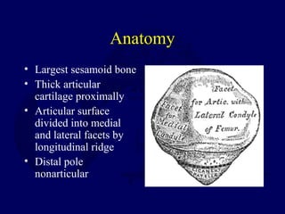
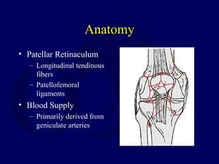
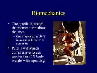
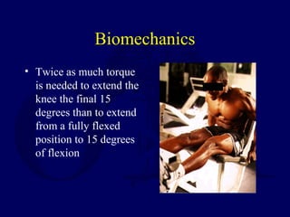
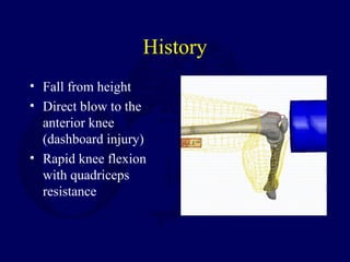
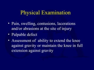
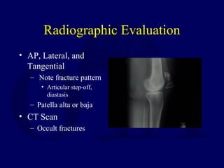
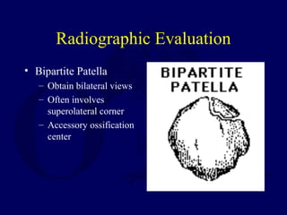
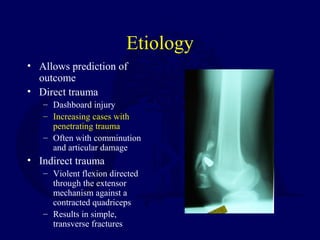
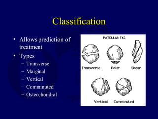
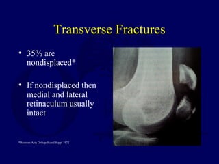
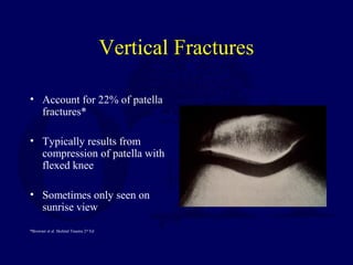
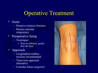
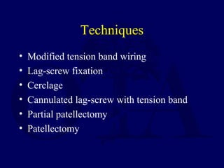
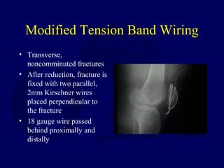
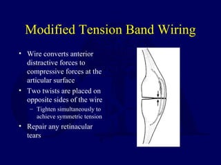
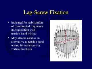
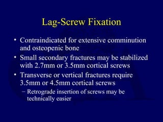
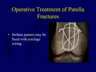
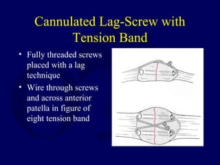
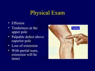
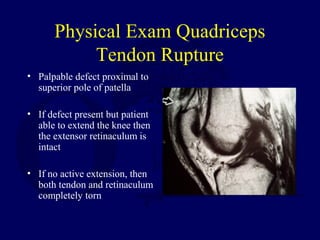
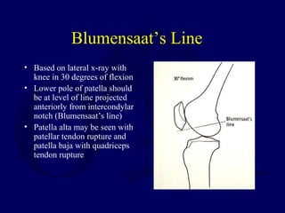
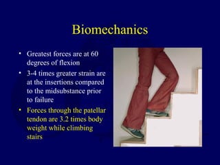
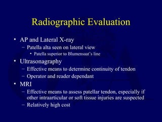
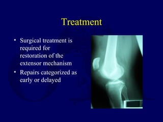
Injuries to the Patella and
Extensor Mechanism
Charles G. Haddad, Jr., MD
Lisa K. Cannada, MD
Emory University
Robert Cantu, MD
Anatomy
• Largest sesamoid bone
• Thick articular
cartilage proximally
• Articular surface
divided into medial
and lateral facets by
longitudinal ridge
• Distal pole
nonarticular
Anatomy
• Patellar Retinaculum
– Longitudinal tendinous
fibers
– Patellofemoral
ligaments
• Blood Supply
– Primarily derived from
geniculate arteries
Biomechanics
• The patella undergoes approximately 7 cm
of translation from full flexion to extension
• Only 13-38% of the patellar surface is in
contact with the femur throughout its range
of motion
Biomechanics
• The patella increases
the moment arm about
the knee
– Contributes up to 30%
increase in force with
extension
• Patella withstands
compressive forces
greater than 7X body
weight with squatting
Biomechanics
• Twice as much torque
is needed to extend the
knee the final 15
degrees than to extend
from a fully flexed
position to 15 degrees
of flexion
History
• Fall from height
• Direct blow to the
anterior knee
(dashboard injury)
• Rapid knee flexion
with quadriceps
resistance
Physical Examination
• Pain, swelling, contusions, lacerations
and/or abrasions at the site of injury
• Palpable defect
• Assessment of ability to extend the knee
against gravity or maintain the knee in full
extension against gravity
Radiographic Evaluation
• AP, Lateral, and
Tangential
– Note fracture pattern
• Articular step-off,
diastasis
– Patella alta or baja
• CT Scan
– Occult fractures
Radiographic Evaluation
• Bipartite Patella
– Obtain bilateral views
– Often involves
superolateral corner
– Accessory ossification
center
Etiology
• Allows prediction of
outcome
• Direct trauma
– Dashboard injury
– Increasing cases with
penetrating trauma
– Often with comminution
and articular damage
• Indirect trauma
– Violent flexion directed
through the extensor
mechanism against a
contracted quadriceps
– Results in simple,
transverse fractures
Classification
• Allows prediction of
treatment
• Types
– Transverse
– Marginal
– Vertical
– Comminuted
– Osteochondral
Transverse Fractures
• 35% are
nondisplaced*
• If nondisplaced then
medial and lateral
retinaculum usually
intact
*Bostrom Acta Orthop Scand Suppl 1972
Vertical Fractures
• Account for 22% of patella
fractures*
• Typically results from
compression of patella with
flexed knee
• Sometimes only seen on
sunrise view
*Browner et al. Skeletal Trauma 2nd
Ed
Stellate Fractures
• Result from direct
blow
• 65% of stellate
fractures are displaced
• May have associated
articular damage to
femoral condyles
Nonoperative Treatment
• Indicated for nondisplaced fractures
– <2mm of articular stepoff and <3mm of
diastasis with an intact extensor mechanism
• May also be considered for minimally
displaced fractures in the elderly
• Patients with a extensive medical
comorbidities
Nonoperative Treatment
• Long leg cylinder cast for 4-6 weeks
– May consider a knee immobilizer for the
elderly
• Immediate weightbearing as tolerated
• Rehabilitation includes range of motion
exercises with gradual quadriceps
strengthening
Operative Treatment
• Goals
– Preserve extensor function
– Restore articular
congruency
• Preoperative Setup
– Tourniquet
• Prior to inflation, gently
flex the knee
• Approach
– Longitudinal midline
incision recommended
– Transverse approach
alternative
– Consider future surgeries!
Techniques
• Modified tension band wiring
• Lag-screw fixation
• Cerclage
• Cannulated lag-screw with tension band
• Partial patellectomy
• Patellectomy
Modified Tension Band Wiring
• Transverse,
noncomminuted fractures
• After reduction, fracture is
fixed with two parallel,
2mm Kirschner wires
placed perpendicular to
the fracture
• 18 gauge wire passed
behind proximally and
distally
Modified Tension Band Wiring
• Wire converts anterior
distractive forces to
compressive forces at the
articular surface
• Two twists are placed on
opposite sides of the wire
– Tighten simultaneously to
achieve symmetric tension
• Repair any retinacular
tears
Lag-Screw Fixation
• Indicated for stabilization
of comminuted fragments
in conjunction with
tension band wiring
• May also be used as an
alternative to tension band
wiring for transverse or
vertical fractures
Lag-Screw Fixation
• Contraindicated for extensive comminution
and osteopenic bone
• Small secondary fractures may be stabilized
with 2.7mm or 3.5mm cortical screws
• Transverse or vertical fractures require
3.5mm or 4.5mm cortical screws
– Retrograde insertion of screws may be
technically easier
Operative Treatment of Patella
Fractures
• Stellate pattern may be
fixed with cerclage
wiring
Cannulated Lag-Screw with
Tension Band
• Fully threaded screws
placed with a lag
technique
• Wire through screws
and across anterior
patella in figure of
eight tension band
Cannulated Lag-Screw with
Tension Band
• Most stable construct
– Screws and tension band wire combination
eliminates both possible separation seen at the
fracture site with modified tension band and
screw failure due to excessive three point
bending
Partial Patellectomy
• Indicated for fractures
involving extensive
comminution not
amenable to fixation
• Larger fragments repaired
with screws to preserve
maximum cartilage
• Smaller fragments excised
– Usually involving the distal
pole
Partial Patellectomy
• Tendon is attached to fragment with
nonabsorbable suture passed through drill holes in
the fragment
– Drill holes should be near the articular surface to
prevent tilting of the tendon and minimize articular
step-off
• Load sharing wire passed through drill holes in the
tibial tubercle and patella may be used to protect
the repair and facilitate early range of motion
• Watch for patellar tilt!
Total Patellectomy
• Indicated for displaced, comminuted
fractures not amenable to reconstruction
• Bone fragments sharply dissected
• Defect may be repaired through a variety of
techniques
• Usually results in extensor lag and loss of
strength
Postoperative Management
• Immobilization with knee brace
• Immediate WBAT
• Early range of motion
– Based on intraoperative assessment of repair
– Active flexion with passive extension
• Quadriceps strengthening
– Begun when there is radiographic evidence of
healing, usually around 6 weeks
Complications
• Knee Stiffness
– Most common
complication
• Infection
– Rare, depends on soft
tissue compromise
• Loss of Fixation
– Hardware failure in up
to 20% of cases
• Osteoarthritis
– May result from
articular damage or
incongruity
• Nonunion < 1% with
surgical repair
• Painful hardware
– Removal required in
approximately 15%
Quadriceps Tendon Rupture
• Typically occurs in patients > 40 years old
• Usually 0-2 cm above the superior pole
• Level often associated with age
– Rupture occurs at the bone-tendon junction in
majority of patients > 40 years old
– Rupture occurs at midsubstance in majority of
patients < 40 years old
Quadriceps Tendon Rupture
• Risk Factors
– Chronic tendonitis
– Anabolic steroid use
– Local steroid injection
– Diabetes mellitus
– Inflammatory
arthropathy
– Chronic renal failure
History
• Sensation of a sudden pop while stressing
the extensor mechanism
• Pain at the site of injury
• Inability/difficulty weightbearing
Physical Exam
• Effusion
• Tenderness at the
upper pole
• Palpable defect above
superior pole
• Loss of extension
• With partial tears,
extension will be
intact
Physical Exam Quadriceps
Tendon Rupture
• Palpable defect proximal to
superior pole of patella
• If defect present but patient
able to extend the knee then
the extensor retinaculum is
intact
• If no active extension, then
both tendon and retinaculum
completely torn
Quadriceps Tendon Rupture
Radiographic
Evaluation
• X-ray- AP, Lateral,
and Tangential
(Sunrise, Merchant)
– Distal displacement of
the patella
• MRI
– Useful when diagnosis
is unclear
Treatment
• Nonoperative
– Partial tears and strains
• Operative
– For complete ruptures
Blumensaat’s Line
• Based on lateral x-ray with
knee in 30 degrees of flexion
• Lower pole of patella should
be at level of line projected
anteriorly from intercondylar
notch (Blumensaat’s line)
• Patella alta may be seen with
patellar tendon rupture and
patella baja with quadriceps
tendon rupture
Nonoperative Treatment
Quadriceps Tendon Rupture
• Reserved for incomplete tears in which active, full
knee extension is preserved
• Immobilize leg in extension for approximately 4-6
weeks
• Progressive physical therapy required to regain
strength and motion
Operative Treatment
• Reapproximation of tendon to bone using
nonabsorbable sutures
– Locking stitch (Bunnel, Krakow) with No. 5
ethibond passed through transverse bone
tunnels
– Repair tendon close to articular surface to avoid
patellar tilting
Operative Treatment
• Midsubstance tears may
undergo end-to-end repair
after edges are freshened
and slightly overlapped
– May benefit from
reinforcement from distally
based partial thickness
quadriceps tendon turned
down across the repair site
(Scuderi Technique)
Treatment
• Chronic tears may
require a V-Y
advancement of a
retracted quadriceps
tendon (Codivilla V-
Y-plasty Technique)
Postoperative Management
• Knee immobilizer or cylinder cast for 5-6
weeks
• Immediate vs. delayed (3 weeks)
weightbearing as tolerated
• At 2-3 weeks, hinged knee brace starting
with 45 degrees active range of motion with
10-15 degrees of progression each week
Complications
• Rerupture
• Persistent quadriceps
atrophy/weakness
• Loss of motion
• Infection
Patellar Tendon Rupture
• Less common than quadriceps tendon
rupture
• Most often occurs in patients < 40 years old
• Associated with degenerative changes of
the tendon
• Rupture often occurs at inferior pole
insertion site
Patellar Tendon Rupture
• Risk Factors
– Rheumatoid
– Systemic Lupus
Erythematosus
– Diabetes
– Chronic Renal Failure
– Systemic Corticosteroid
Therapy
– Local Steroid Injection
– Chronic patellar tendonitis
Anatomy
• Patellar tendon
– Averages 4 mm thick but widens to 5-6 mm at
the tibial tubercle insertion
– Merges with the medial and lateral retinaculum
– 90% type I collagen
Blood Supply
• Fat pad vessels supply posterior aspect of tendon
via inferior medial and lateral geniculate arteries
• Retinacular vessels supply anterior portion of
tendon via the inferior medial geniculate and
recurrent tibial arteries
• Proximal and distal insertion areas are relatively
avascular and subsequently are a common site of
rupture
Biomechanics
• Greatest forces are at 60
degrees of flexion
• 3-4 times greater strain are
at the insertions compared
to the midsubstance prior
to failure
• Forces through the patellar
tendon are 3.2 times body
weight while climbing
stairs
History
• Often a report of
forceful quadriceps
contraction against a
flexed knee
• May experience and
audible “pop”
• Inability to weightbear
or extend the knee
Physical Examination
• Palpable defect
• Hemarthrosis
• Painful passive knee flexion
• Partial or complete loss of active extension
• Quadriceps atrophy with chronic injury
Radiographic Evaluation
• AP and Lateral X-ray
– Patella alta seen on lateral view
• Patella superior to Blumensaat’s line
• Ultrasonagraphy
– Effective means to determine continuity of tendon
– Operator and reader dependant
• MRI
– Effective means to assess patellar tendon, especially if
other intraarticular or soft tissue injuries are suspected
– Relatively high cost
Classification
• No widely accepted means of classification
• Can be categorized by:
– Location of tear
• Proximal insertion most common
– Timing between injury and surgery
• Most important factor for prognosis
• Acute- within two weeks
• Chronic- greater than two weeks
Treatment
• Surgical treatment is
required for
restoration of the
extensor mechanism
• Repairs categorized as
early or delayed
Nonoperative Treatment of
Patellar Tedon Rupture
• Nonoperative treatment reserved for partial
tears in which patient able to fully extend
knee
• Treatment is immobilization in full
extension for 3-6 weeks
Early Repair
• Better overall outcome
• Primary repair of the tendon
• Surgical approach is through a midline incision
– Incise just lateral to tibial tubercle as skin thicker with
better blood supply to decrease wound complications
• Patellar tendon rupture and retinacular tears are
exposed
Early Repair
• Frayed edges and
hematoma are debrided
• With a Bunnell or Krakow
stitch, two ethibond
sutures or their equivalent
are used to repair the
tendon to the patella
• Sutures passed through
three parallel, longitudinal
bone tunnels and tied
proximally
• Repair retinacular
tears
• May reinforce with
wire, cable or
umbilical tape
• Assess repair
intraoperatively with
flexion
Operative Treatment of Patellar
Tendon Rupture
• For rupture at osteotendinous
junction, tendon reattached to
patella with heavy,
nonabsorbable sutures passed
through drill holes in patella
• Medial and lateral retinacula
repaired with heavy
absorbable suture
• Knee immobilized in
extension for 6 weeks
Operative Treatment of Patellar
Tendon Ruptures
• 18 gauge wire can be used to reinforce
repair
• Repair should be tested with flexion of knee
in OR and any loose sutures replaced
Postoperative Management
• Hinged knee brace locked at 20 degrees
• Immediate isometric quadriceps exercises
• Active flexion with passive extension at two
weeks; start with 0-45 degrees and advance
30 degrees each week
• Active extension at three weeks
Postoperative Management
• Initial toe-touch weightbearing is gradually
advanced to full weightbearing by six weeks
• Maintain hinged knee brace which is gradually
increased as motion increases
• All restrictions are lifted after full range of motion
and 90% of the contralateral quadriceps strength
are obtained; usually at 4-6 months
Delayed Repair
• > 6 weeks from initial injury
• Often results in poorer outcome
• Quadriceps contraction and patellar migration are
encountered
• Adhesions between the patella and femur may be
present
• Options include hamstring and fascia lata
autograft augmentation of primary repair or
Achilles tendon allograft
Postoperative Management
• More conservative when compared to early
repair
• Bivalved cylinder cast for 6 weeks; may
start passive range of motion
• Active range of motion is started at 6 weeks
Complications
• Knee stiffness
• Persistent quadriceps weakness
• Rerupture
• Infection
• Patella baja
Return to
Lower Extremity
Index

L07 extensor mechnsm injury

  • 1.
    Injuries to thePatella and Extensor Mechanism Charles G. Haddad, Jr., MD Lisa K. Cannada, MD Emory University Robert Cantu, MD
  • 2.
    Anatomy • Largest sesamoidbone • Thick articular cartilage proximally • Articular surface divided into medial and lateral facets by longitudinal ridge • Distal pole nonarticular
  • 3.
    Anatomy • Patellar Retinaculum –Longitudinal tendinous fibers – Patellofemoral ligaments • Blood Supply – Primarily derived from geniculate arteries
  • 4.
    Biomechanics • The patellaundergoes approximately 7 cm of translation from full flexion to extension • Only 13-38% of the patellar surface is in contact with the femur throughout its range of motion
  • 5.
    Biomechanics • The patellaincreases the moment arm about the knee – Contributes up to 30% increase in force with extension • Patella withstands compressive forces greater than 7X body weight with squatting
  • 6.
    Biomechanics • Twice asmuch torque is needed to extend the knee the final 15 degrees than to extend from a fully flexed position to 15 degrees of flexion
  • 7.
    History • Fall fromheight • Direct blow to the anterior knee (dashboard injury) • Rapid knee flexion with quadriceps resistance
  • 8.
    Physical Examination • Pain,swelling, contusions, lacerations and/or abrasions at the site of injury • Palpable defect • Assessment of ability to extend the knee against gravity or maintain the knee in full extension against gravity
  • 9.
    Radiographic Evaluation • AP,Lateral, and Tangential – Note fracture pattern • Articular step-off, diastasis – Patella alta or baja • CT Scan – Occult fractures
  • 10.
    Radiographic Evaluation • BipartitePatella – Obtain bilateral views – Often involves superolateral corner – Accessory ossification center
  • 11.
    Etiology • Allows predictionof outcome • Direct trauma – Dashboard injury – Increasing cases with penetrating trauma – Often with comminution and articular damage • Indirect trauma – Violent flexion directed through the extensor mechanism against a contracted quadriceps – Results in simple, transverse fractures
  • 12.
    Classification • Allows predictionof treatment • Types – Transverse – Marginal – Vertical – Comminuted – Osteochondral
  • 13.
    Transverse Fractures • 35%are nondisplaced* • If nondisplaced then medial and lateral retinaculum usually intact *Bostrom Acta Orthop Scand Suppl 1972
  • 14.
    Vertical Fractures • Accountfor 22% of patella fractures* • Typically results from compression of patella with flexed knee • Sometimes only seen on sunrise view *Browner et al. Skeletal Trauma 2nd Ed
  • 15.
    Stellate Fractures • Resultfrom direct blow • 65% of stellate fractures are displaced • May have associated articular damage to femoral condyles
  • 16.
    Nonoperative Treatment • Indicatedfor nondisplaced fractures – <2mm of articular stepoff and <3mm of diastasis with an intact extensor mechanism • May also be considered for minimally displaced fractures in the elderly • Patients with a extensive medical comorbidities
  • 17.
    Nonoperative Treatment • Longleg cylinder cast for 4-6 weeks – May consider a knee immobilizer for the elderly • Immediate weightbearing as tolerated • Rehabilitation includes range of motion exercises with gradual quadriceps strengthening
  • 18.
    Operative Treatment • Goals –Preserve extensor function – Restore articular congruency • Preoperative Setup – Tourniquet • Prior to inflation, gently flex the knee • Approach – Longitudinal midline incision recommended – Transverse approach alternative – Consider future surgeries!
  • 19.
    Techniques • Modified tensionband wiring • Lag-screw fixation • Cerclage • Cannulated lag-screw with tension band • Partial patellectomy • Patellectomy
  • 20.
    Modified Tension BandWiring • Transverse, noncomminuted fractures • After reduction, fracture is fixed with two parallel, 2mm Kirschner wires placed perpendicular to the fracture • 18 gauge wire passed behind proximally and distally
  • 21.
    Modified Tension BandWiring • Wire converts anterior distractive forces to compressive forces at the articular surface • Two twists are placed on opposite sides of the wire – Tighten simultaneously to achieve symmetric tension • Repair any retinacular tears
  • 22.
    Lag-Screw Fixation • Indicatedfor stabilization of comminuted fragments in conjunction with tension band wiring • May also be used as an alternative to tension band wiring for transverse or vertical fractures
  • 23.
    Lag-Screw Fixation • Contraindicatedfor extensive comminution and osteopenic bone • Small secondary fractures may be stabilized with 2.7mm or 3.5mm cortical screws • Transverse or vertical fractures require 3.5mm or 4.5mm cortical screws – Retrograde insertion of screws may be technically easier
  • 24.
    Operative Treatment ofPatella Fractures • Stellate pattern may be fixed with cerclage wiring
  • 25.
    Cannulated Lag-Screw with TensionBand • Fully threaded screws placed with a lag technique • Wire through screws and across anterior patella in figure of eight tension band
  • 26.
    Cannulated Lag-Screw with TensionBand • Most stable construct – Screws and tension band wire combination eliminates both possible separation seen at the fracture site with modified tension band and screw failure due to excessive three point bending
  • 27.
    Partial Patellectomy • Indicatedfor fractures involving extensive comminution not amenable to fixation • Larger fragments repaired with screws to preserve maximum cartilage • Smaller fragments excised – Usually involving the distal pole
  • 28.
    Partial Patellectomy • Tendonis attached to fragment with nonabsorbable suture passed through drill holes in the fragment – Drill holes should be near the articular surface to prevent tilting of the tendon and minimize articular step-off • Load sharing wire passed through drill holes in the tibial tubercle and patella may be used to protect the repair and facilitate early range of motion • Watch for patellar tilt!
  • 29.
    Total Patellectomy • Indicatedfor displaced, comminuted fractures not amenable to reconstruction • Bone fragments sharply dissected • Defect may be repaired through a variety of techniques • Usually results in extensor lag and loss of strength
  • 30.
    Postoperative Management • Immobilizationwith knee brace • Immediate WBAT • Early range of motion – Based on intraoperative assessment of repair – Active flexion with passive extension • Quadriceps strengthening – Begun when there is radiographic evidence of healing, usually around 6 weeks
  • 31.
    Complications • Knee Stiffness –Most common complication • Infection – Rare, depends on soft tissue compromise • Loss of Fixation – Hardware failure in up to 20% of cases • Osteoarthritis – May result from articular damage or incongruity • Nonunion < 1% with surgical repair • Painful hardware – Removal required in approximately 15%
  • 32.
    Quadriceps Tendon Rupture •Typically occurs in patients > 40 years old • Usually 0-2 cm above the superior pole • Level often associated with age – Rupture occurs at the bone-tendon junction in majority of patients > 40 years old – Rupture occurs at midsubstance in majority of patients < 40 years old
  • 33.
    Quadriceps Tendon Rupture •Risk Factors – Chronic tendonitis – Anabolic steroid use – Local steroid injection – Diabetes mellitus – Inflammatory arthropathy – Chronic renal failure
  • 34.
    History • Sensation ofa sudden pop while stressing the extensor mechanism • Pain at the site of injury • Inability/difficulty weightbearing
  • 35.
    Physical Exam • Effusion •Tenderness at the upper pole • Palpable defect above superior pole • Loss of extension • With partial tears, extension will be intact
  • 36.
    Physical Exam Quadriceps TendonRupture • Palpable defect proximal to superior pole of patella • If defect present but patient able to extend the knee then the extensor retinaculum is intact • If no active extension, then both tendon and retinaculum completely torn
  • 37.
    Quadriceps Tendon Rupture Radiographic Evaluation •X-ray- AP, Lateral, and Tangential (Sunrise, Merchant) – Distal displacement of the patella • MRI – Useful when diagnosis is unclear Treatment • Nonoperative – Partial tears and strains • Operative – For complete ruptures
  • 38.
    Blumensaat’s Line • Basedon lateral x-ray with knee in 30 degrees of flexion • Lower pole of patella should be at level of line projected anteriorly from intercondylar notch (Blumensaat’s line) • Patella alta may be seen with patellar tendon rupture and patella baja with quadriceps tendon rupture
  • 39.
    Nonoperative Treatment Quadriceps TendonRupture • Reserved for incomplete tears in which active, full knee extension is preserved • Immobilize leg in extension for approximately 4-6 weeks • Progressive physical therapy required to regain strength and motion
  • 40.
    Operative Treatment • Reapproximationof tendon to bone using nonabsorbable sutures – Locking stitch (Bunnel, Krakow) with No. 5 ethibond passed through transverse bone tunnels – Repair tendon close to articular surface to avoid patellar tilting
  • 41.
    Operative Treatment • Midsubstancetears may undergo end-to-end repair after edges are freshened and slightly overlapped – May benefit from reinforcement from distally based partial thickness quadriceps tendon turned down across the repair site (Scuderi Technique)
  • 42.
    Treatment • Chronic tearsmay require a V-Y advancement of a retracted quadriceps tendon (Codivilla V- Y-plasty Technique)
  • 43.
    Postoperative Management • Kneeimmobilizer or cylinder cast for 5-6 weeks • Immediate vs. delayed (3 weeks) weightbearing as tolerated • At 2-3 weeks, hinged knee brace starting with 45 degrees active range of motion with 10-15 degrees of progression each week
  • 44.
    Complications • Rerupture • Persistentquadriceps atrophy/weakness • Loss of motion • Infection
  • 45.
    Patellar Tendon Rupture •Less common than quadriceps tendon rupture • Most often occurs in patients < 40 years old • Associated with degenerative changes of the tendon • Rupture often occurs at inferior pole insertion site
  • 46.
    Patellar Tendon Rupture •Risk Factors – Rheumatoid – Systemic Lupus Erythematosus – Diabetes – Chronic Renal Failure – Systemic Corticosteroid Therapy – Local Steroid Injection – Chronic patellar tendonitis
  • 47.
    Anatomy • Patellar tendon –Averages 4 mm thick but widens to 5-6 mm at the tibial tubercle insertion – Merges with the medial and lateral retinaculum – 90% type I collagen
  • 48.
    Blood Supply • Fatpad vessels supply posterior aspect of tendon via inferior medial and lateral geniculate arteries • Retinacular vessels supply anterior portion of tendon via the inferior medial geniculate and recurrent tibial arteries • Proximal and distal insertion areas are relatively avascular and subsequently are a common site of rupture
  • 49.
    Biomechanics • Greatest forcesare at 60 degrees of flexion • 3-4 times greater strain are at the insertions compared to the midsubstance prior to failure • Forces through the patellar tendon are 3.2 times body weight while climbing stairs
  • 50.
    History • Often areport of forceful quadriceps contraction against a flexed knee • May experience and audible “pop” • Inability to weightbear or extend the knee
  • 51.
    Physical Examination • Palpabledefect • Hemarthrosis • Painful passive knee flexion • Partial or complete loss of active extension • Quadriceps atrophy with chronic injury
  • 52.
    Radiographic Evaluation • APand Lateral X-ray – Patella alta seen on lateral view • Patella superior to Blumensaat’s line • Ultrasonagraphy – Effective means to determine continuity of tendon – Operator and reader dependant • MRI – Effective means to assess patellar tendon, especially if other intraarticular or soft tissue injuries are suspected – Relatively high cost
  • 53.
    Classification • No widelyaccepted means of classification • Can be categorized by: – Location of tear • Proximal insertion most common – Timing between injury and surgery • Most important factor for prognosis • Acute- within two weeks • Chronic- greater than two weeks
  • 54.
    Treatment • Surgical treatmentis required for restoration of the extensor mechanism • Repairs categorized as early or delayed
  • 55.
    Nonoperative Treatment of PatellarTedon Rupture • Nonoperative treatment reserved for partial tears in which patient able to fully extend knee • Treatment is immobilization in full extension for 3-6 weeks
  • 56.
    Early Repair • Betteroverall outcome • Primary repair of the tendon • Surgical approach is through a midline incision – Incise just lateral to tibial tubercle as skin thicker with better blood supply to decrease wound complications • Patellar tendon rupture and retinacular tears are exposed
  • 57.
    Early Repair • Frayededges and hematoma are debrided • With a Bunnell or Krakow stitch, two ethibond sutures or their equivalent are used to repair the tendon to the patella • Sutures passed through three parallel, longitudinal bone tunnels and tied proximally • Repair retinacular tears • May reinforce with wire, cable or umbilical tape • Assess repair intraoperatively with flexion
  • 58.
    Operative Treatment ofPatellar Tendon Rupture • For rupture at osteotendinous junction, tendon reattached to patella with heavy, nonabsorbable sutures passed through drill holes in patella • Medial and lateral retinacula repaired with heavy absorbable suture • Knee immobilized in extension for 6 weeks
  • 59.
    Operative Treatment ofPatellar Tendon Ruptures • 18 gauge wire can be used to reinforce repair • Repair should be tested with flexion of knee in OR and any loose sutures replaced
  • 60.
    Postoperative Management • Hingedknee brace locked at 20 degrees • Immediate isometric quadriceps exercises • Active flexion with passive extension at two weeks; start with 0-45 degrees and advance 30 degrees each week • Active extension at three weeks
  • 61.
    Postoperative Management • Initialtoe-touch weightbearing is gradually advanced to full weightbearing by six weeks • Maintain hinged knee brace which is gradually increased as motion increases • All restrictions are lifted after full range of motion and 90% of the contralateral quadriceps strength are obtained; usually at 4-6 months
  • 62.
    Delayed Repair • >6 weeks from initial injury • Often results in poorer outcome • Quadriceps contraction and patellar migration are encountered • Adhesions between the patella and femur may be present • Options include hamstring and fascia lata autograft augmentation of primary repair or Achilles tendon allograft
  • 63.
    Postoperative Management • Moreconservative when compared to early repair • Bivalved cylinder cast for 6 weeks; may start passive range of motion • Active range of motion is started at 6 weeks
  • 64.
    Complications • Knee stiffness •Persistent quadriceps weakness • Rerupture • Infection • Patella baja Return to Lower Extremity Index

Editor's Notes

  • #13 Based on mechanism-OTA-predicts outcome Erase types and pull back each bullet? Picture of classification
  • #18 May add slide referencing JAAOS ref #1, 90% good-excellent results, 1% poor results OTA manual –protected ROM 0-45 X 4 weeks???
  • #20 OK to change title??