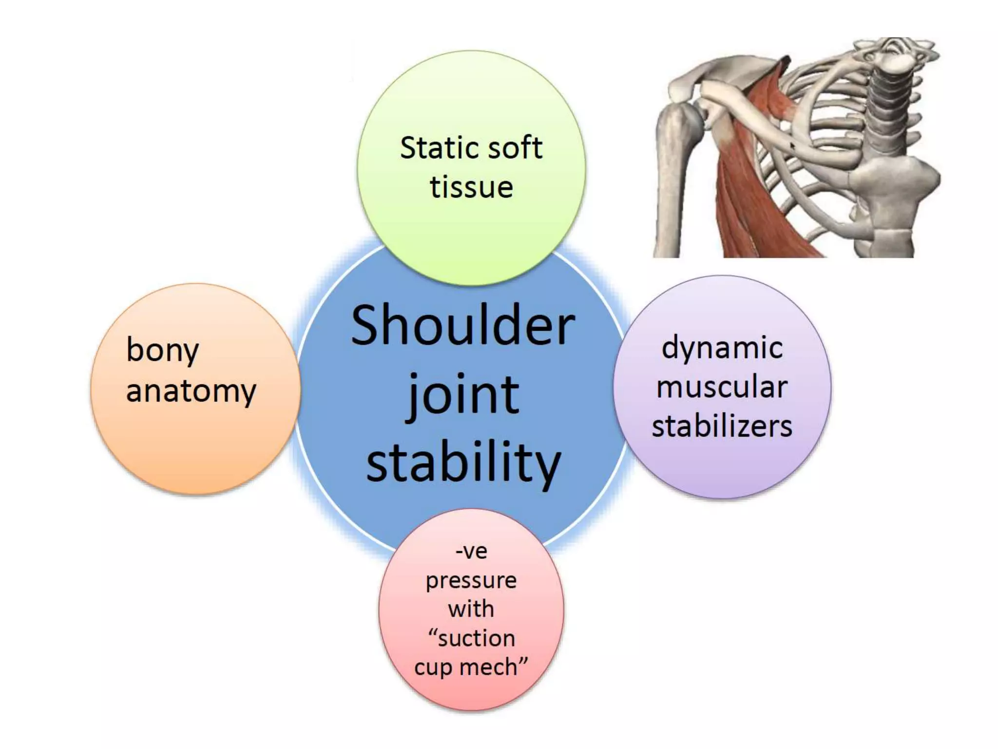
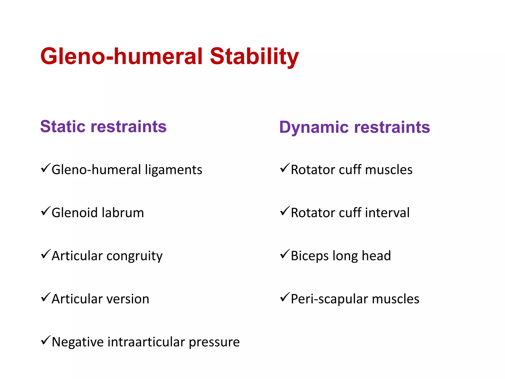
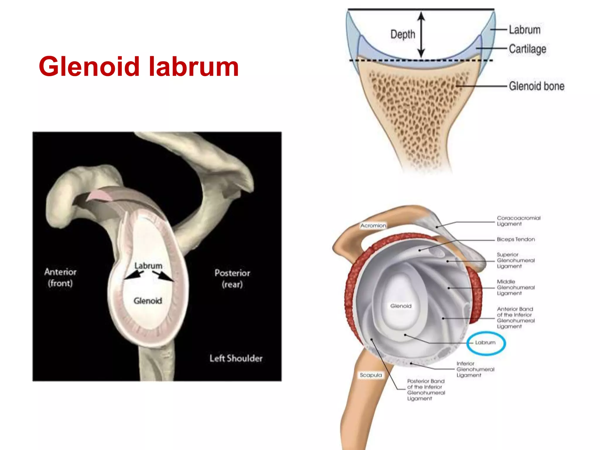
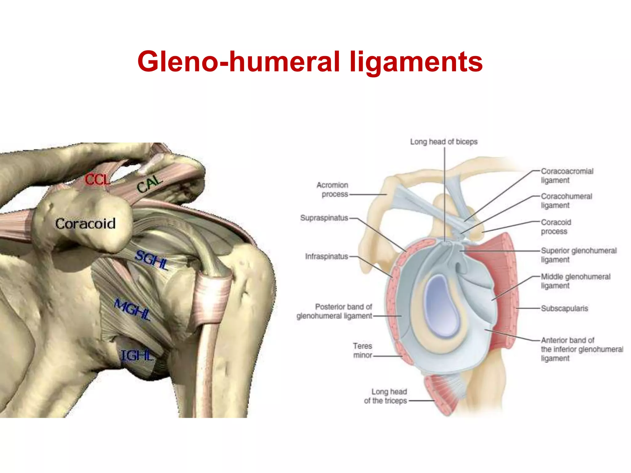
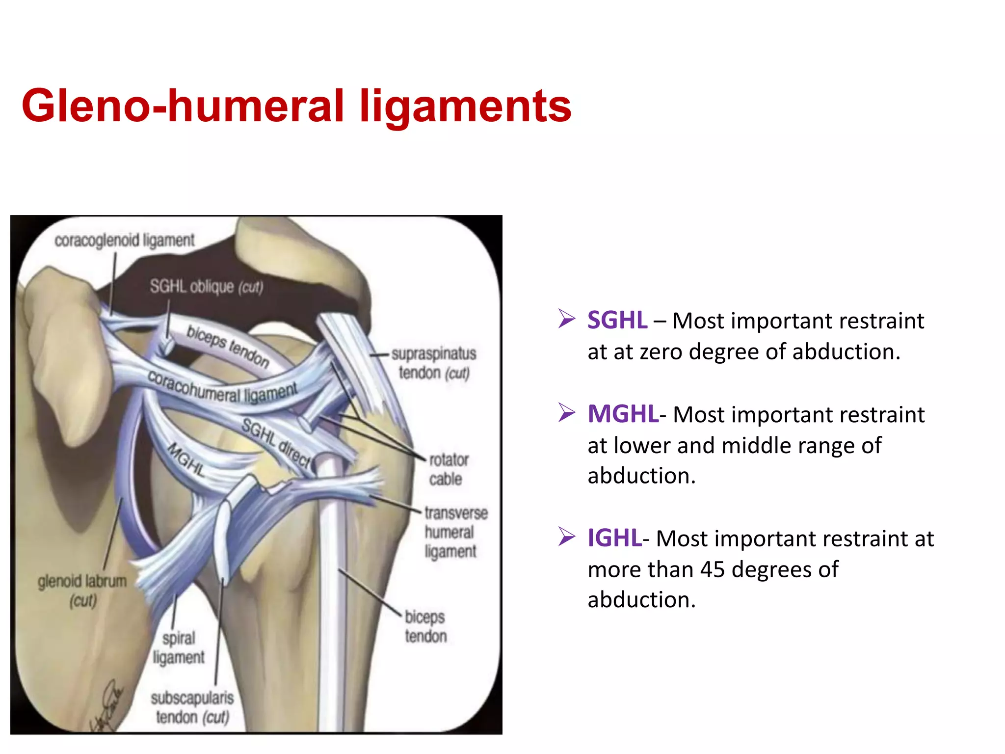
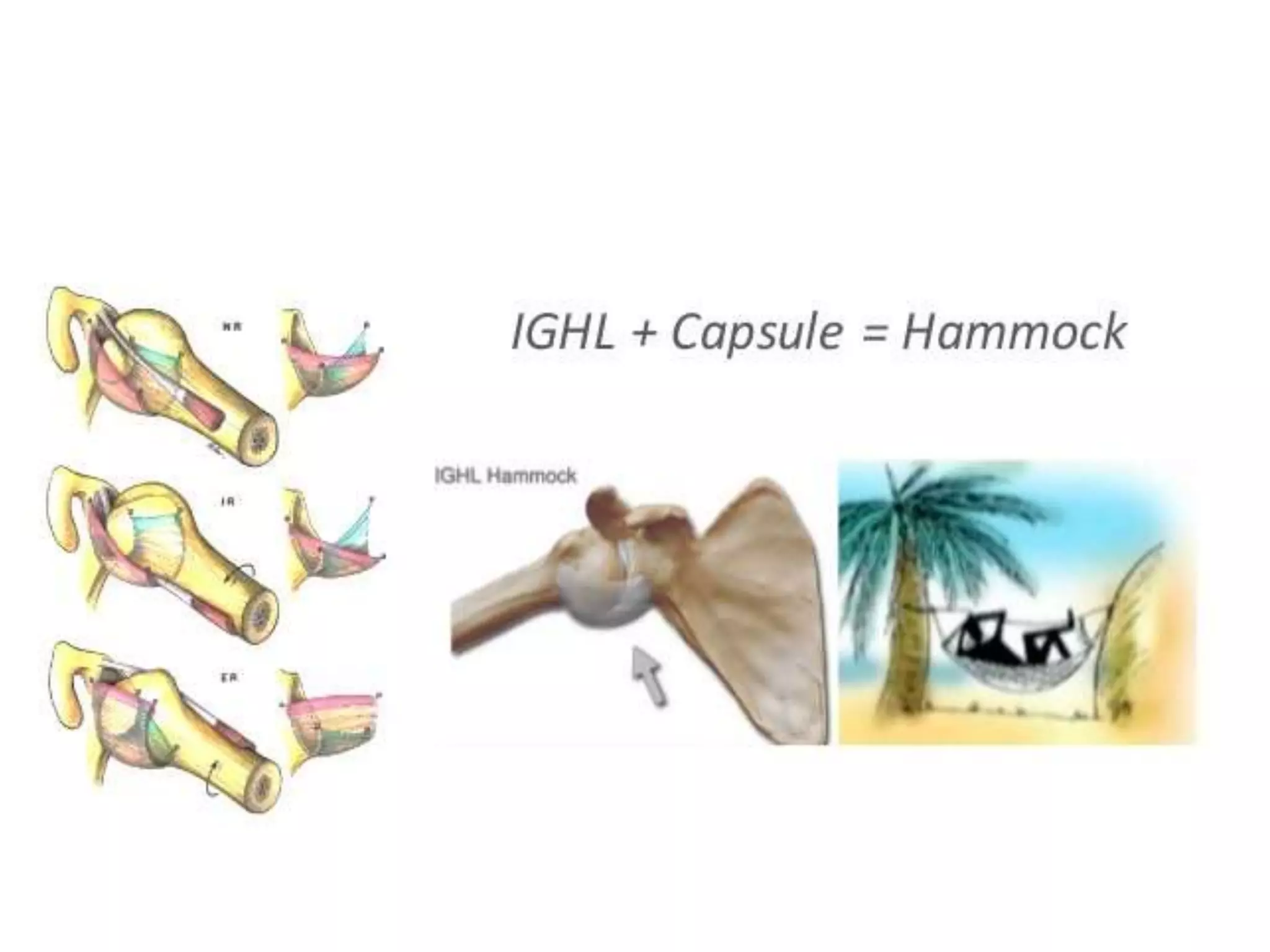
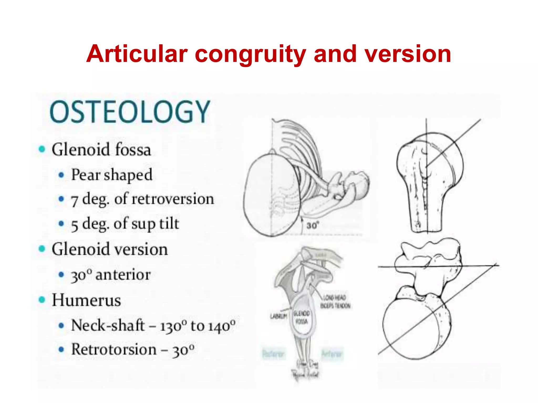
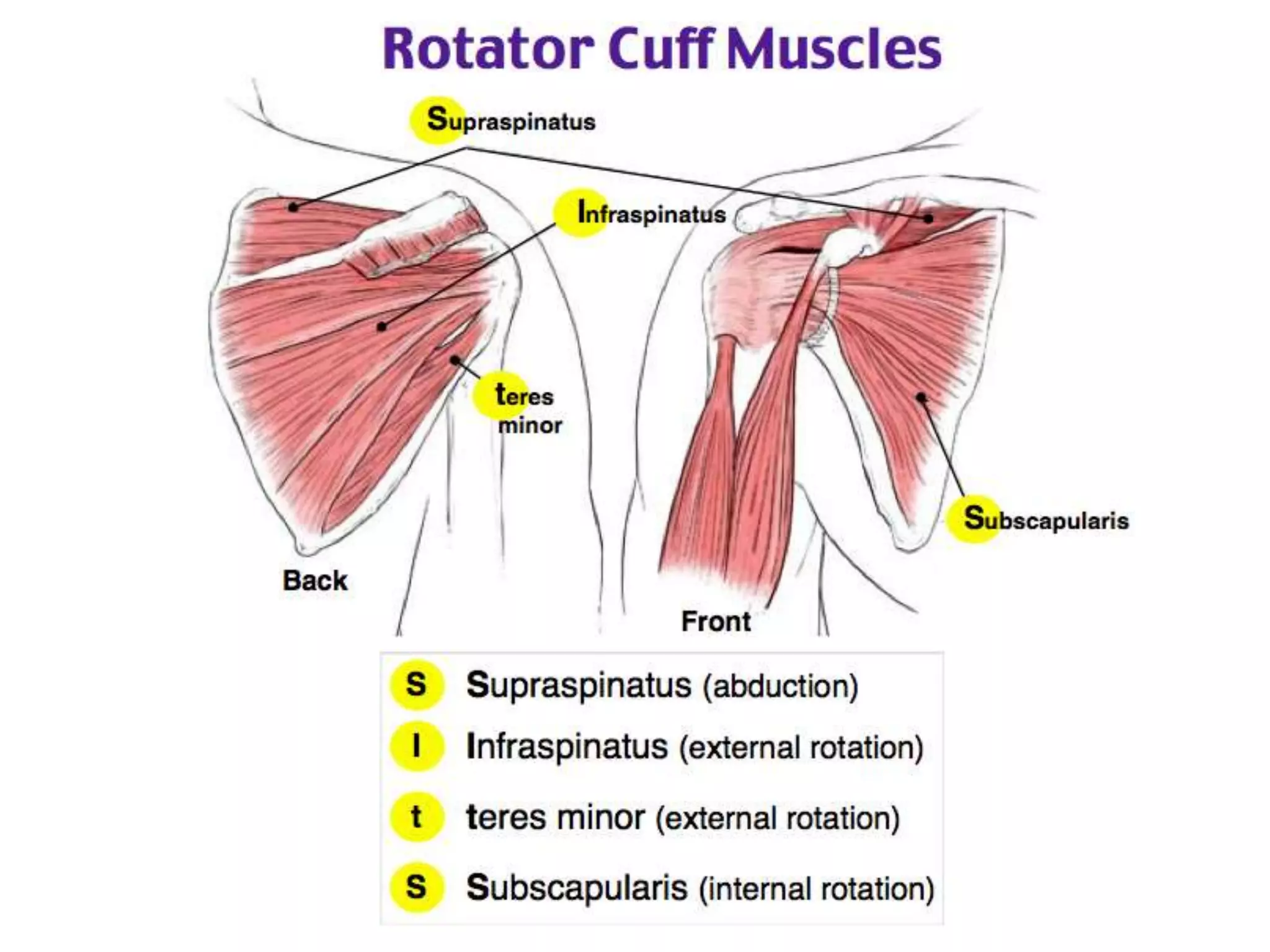
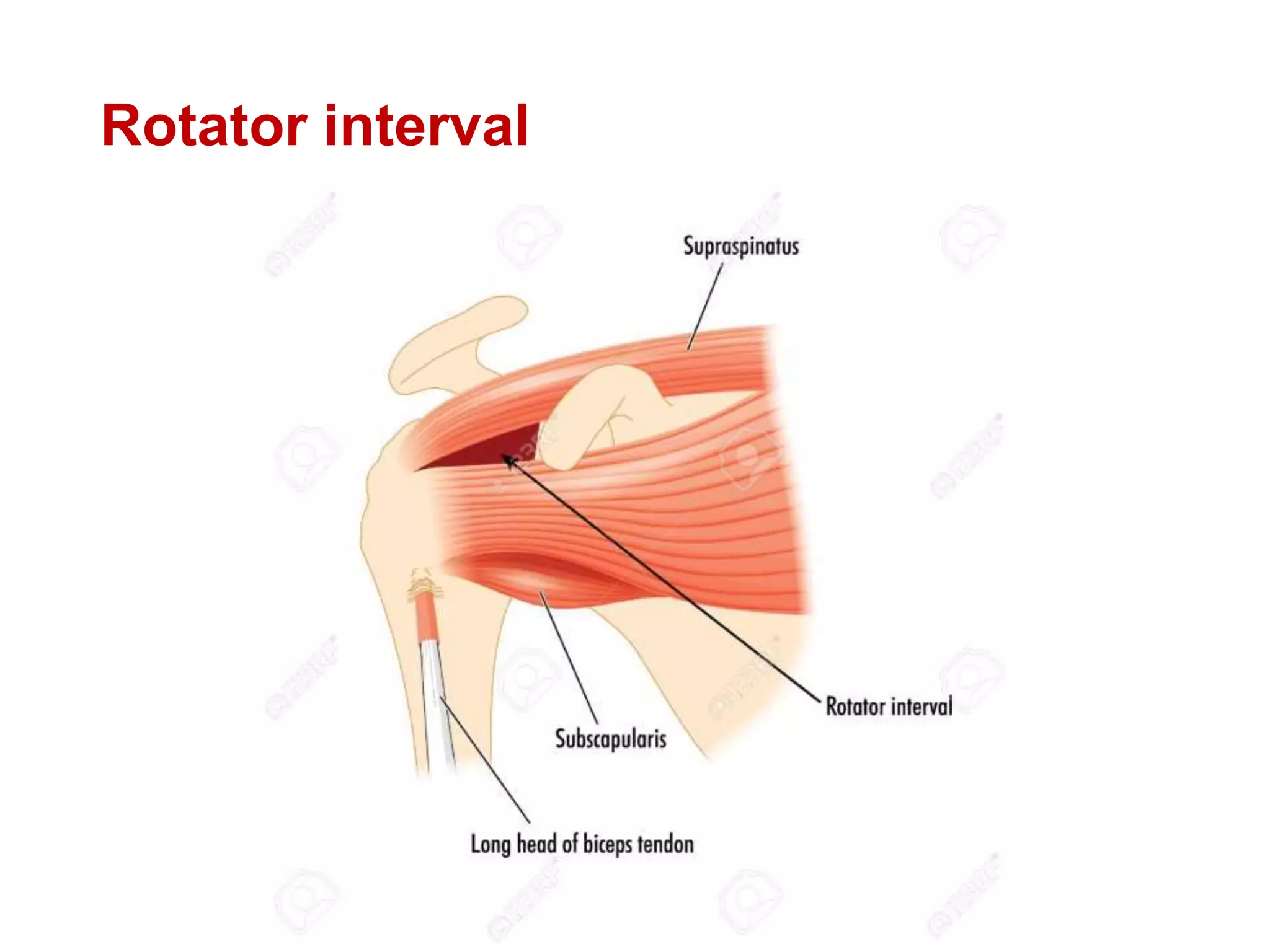
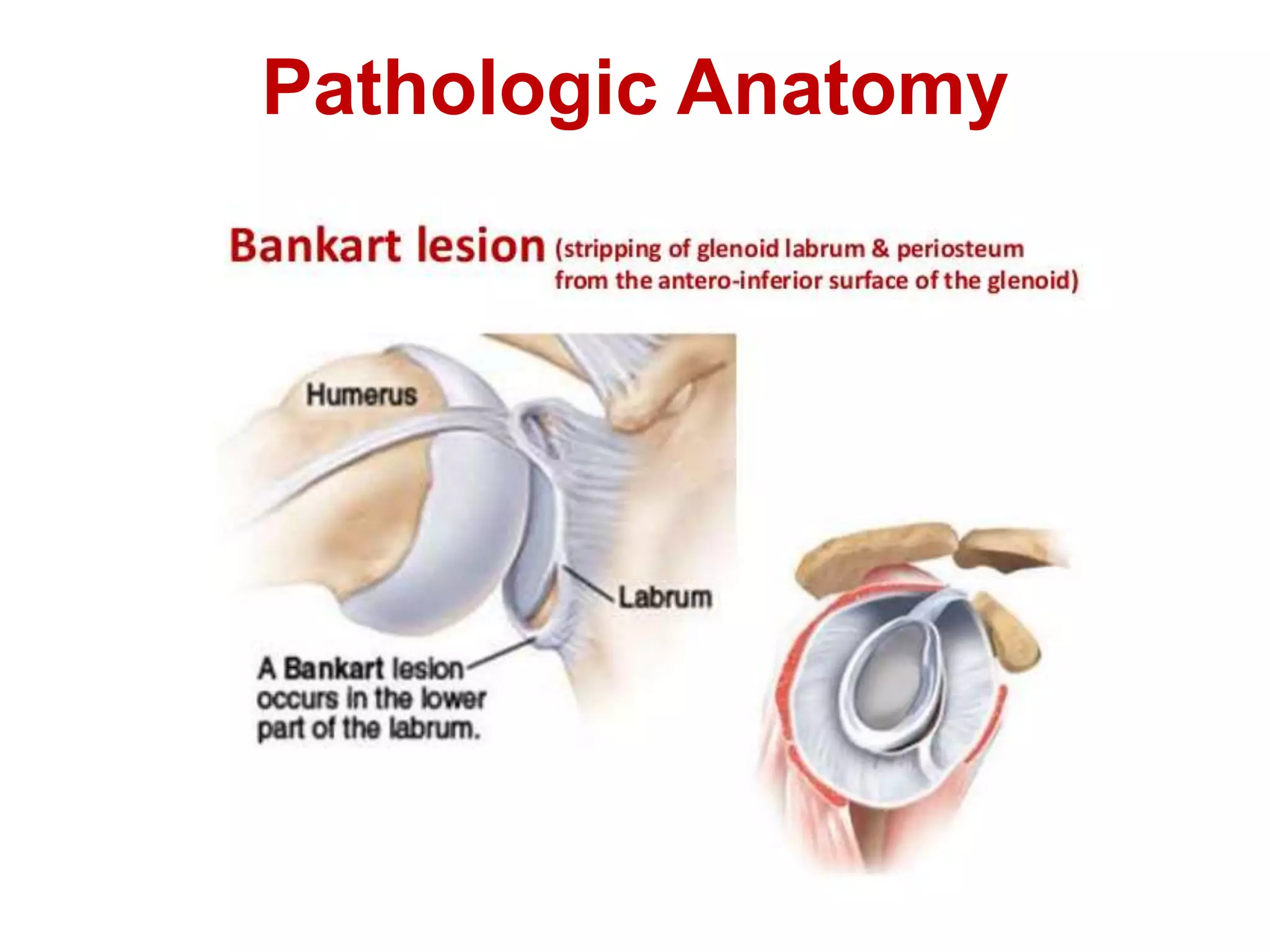
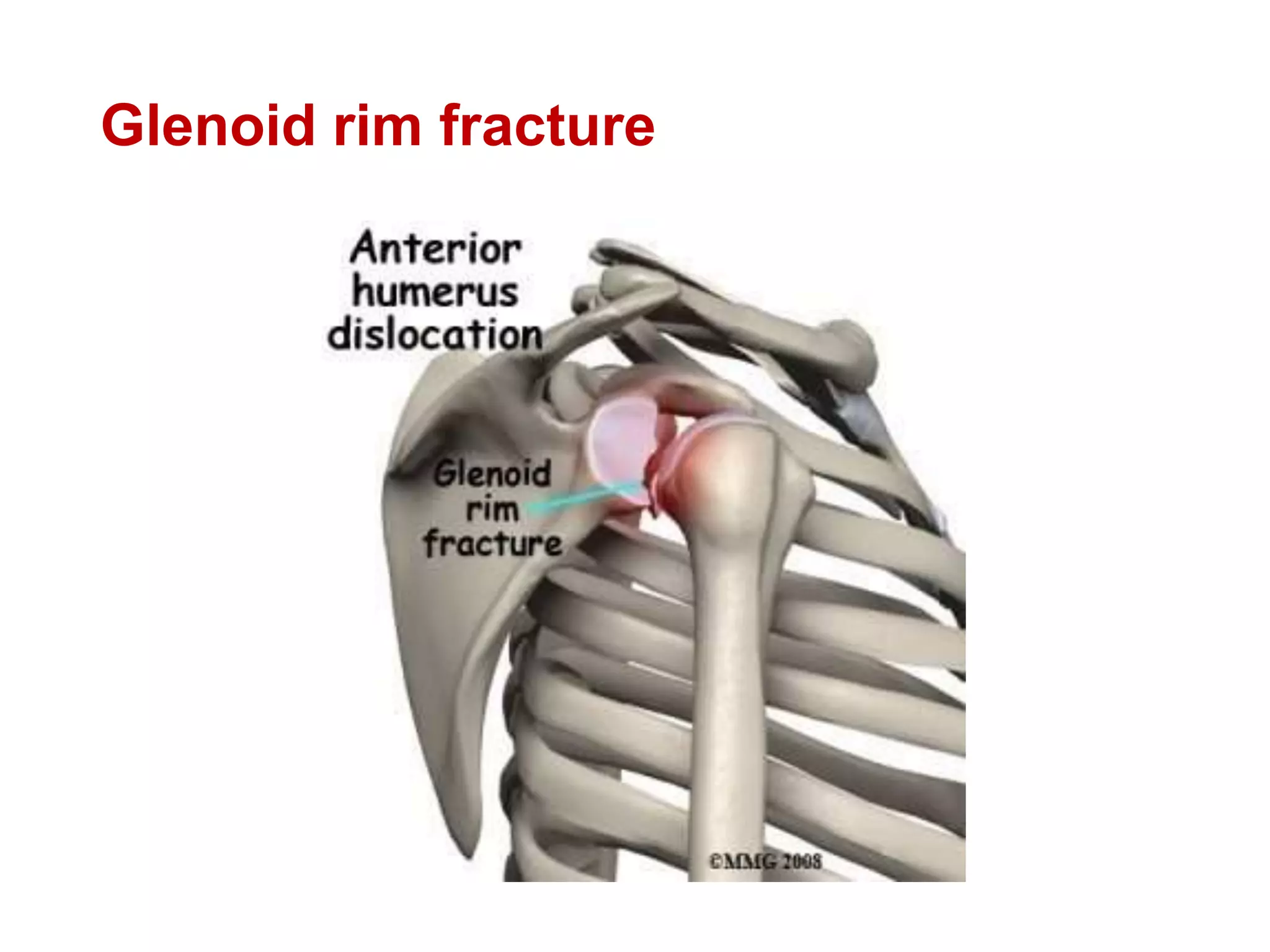
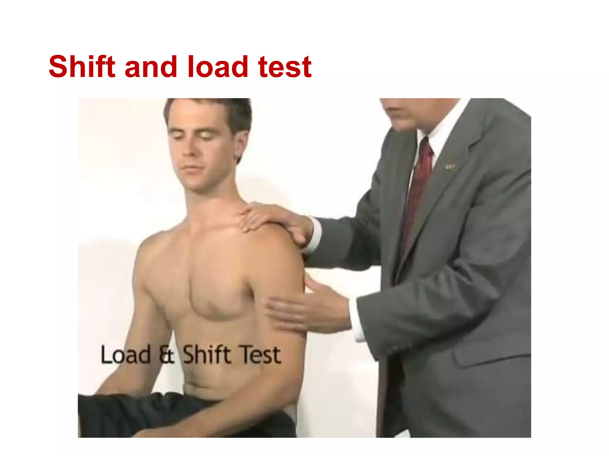
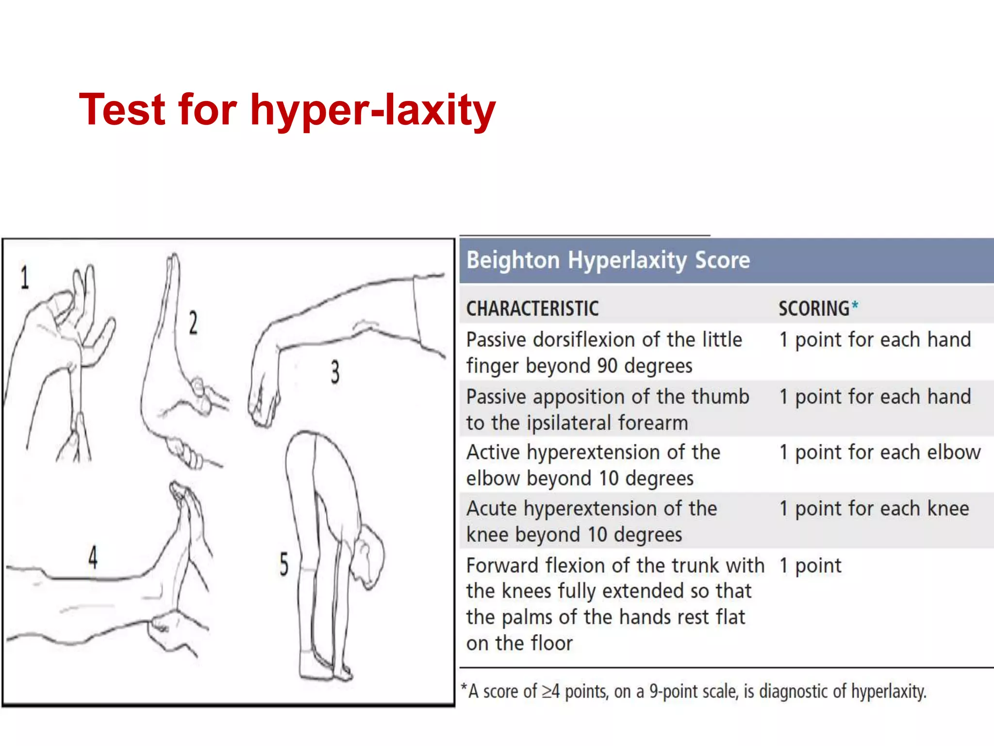
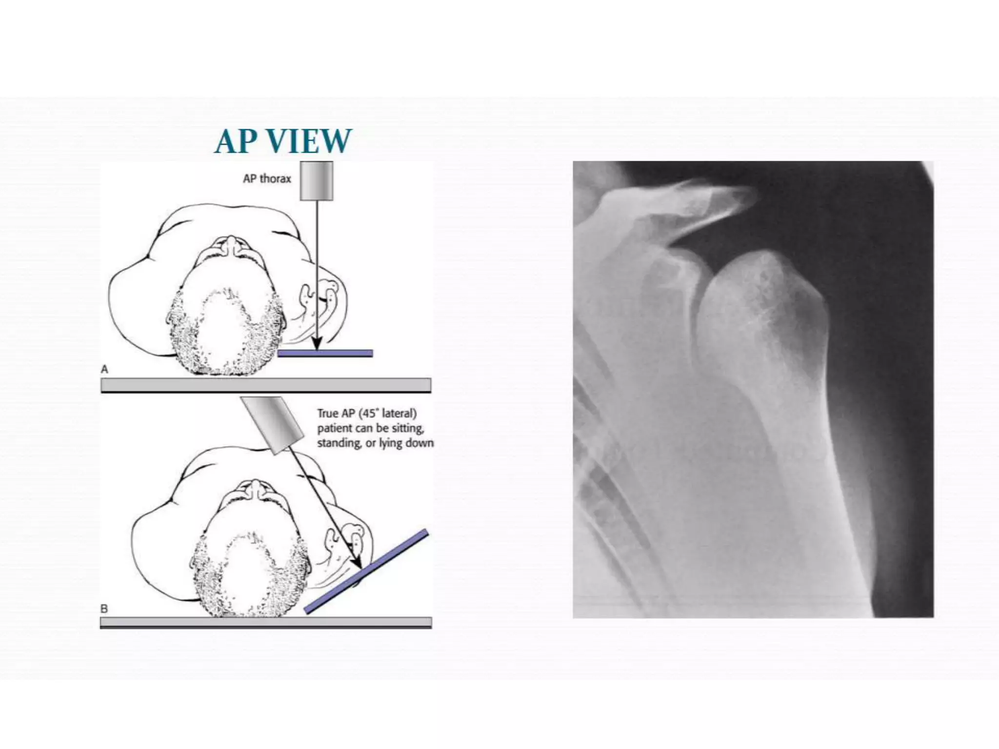
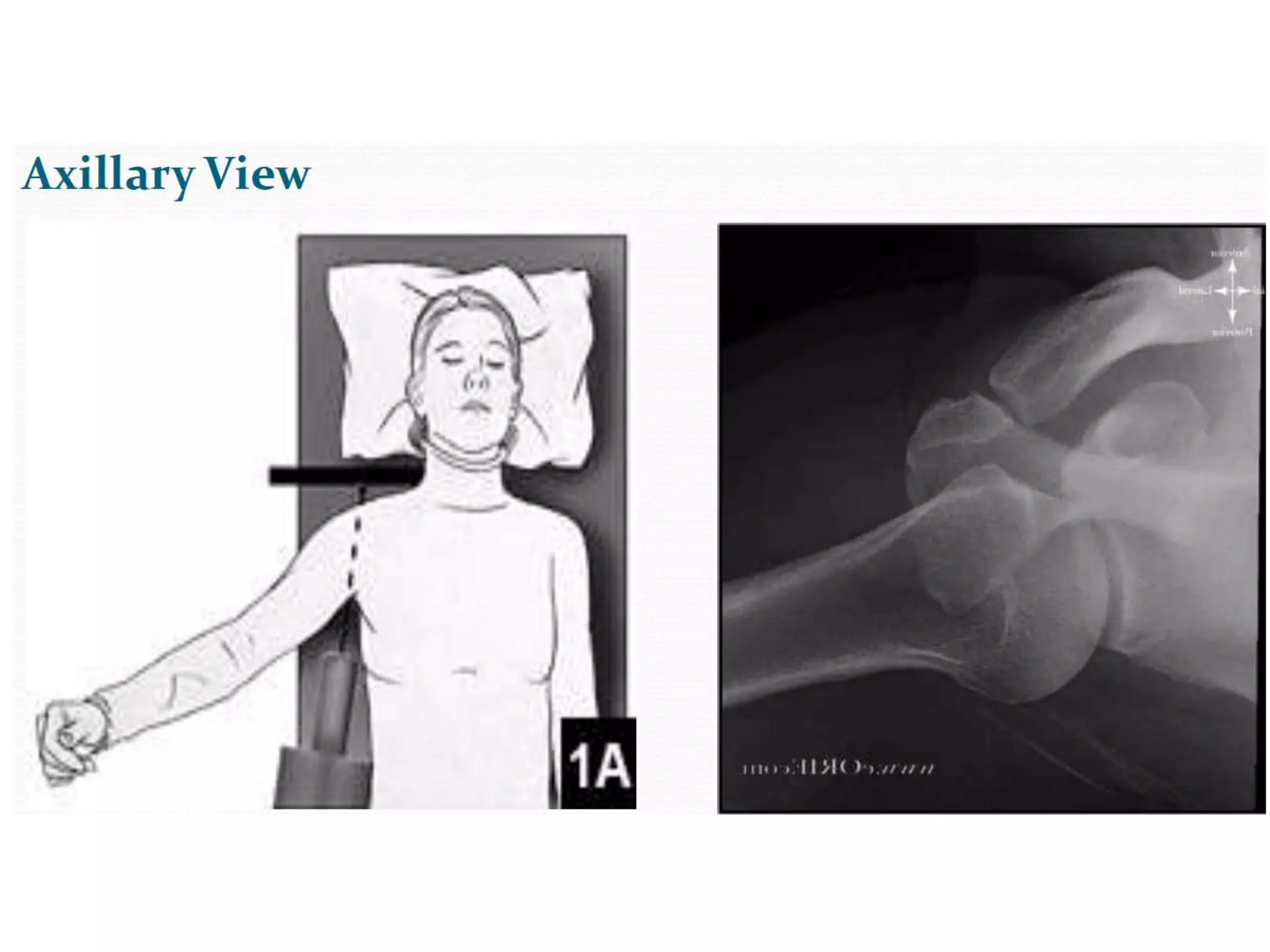
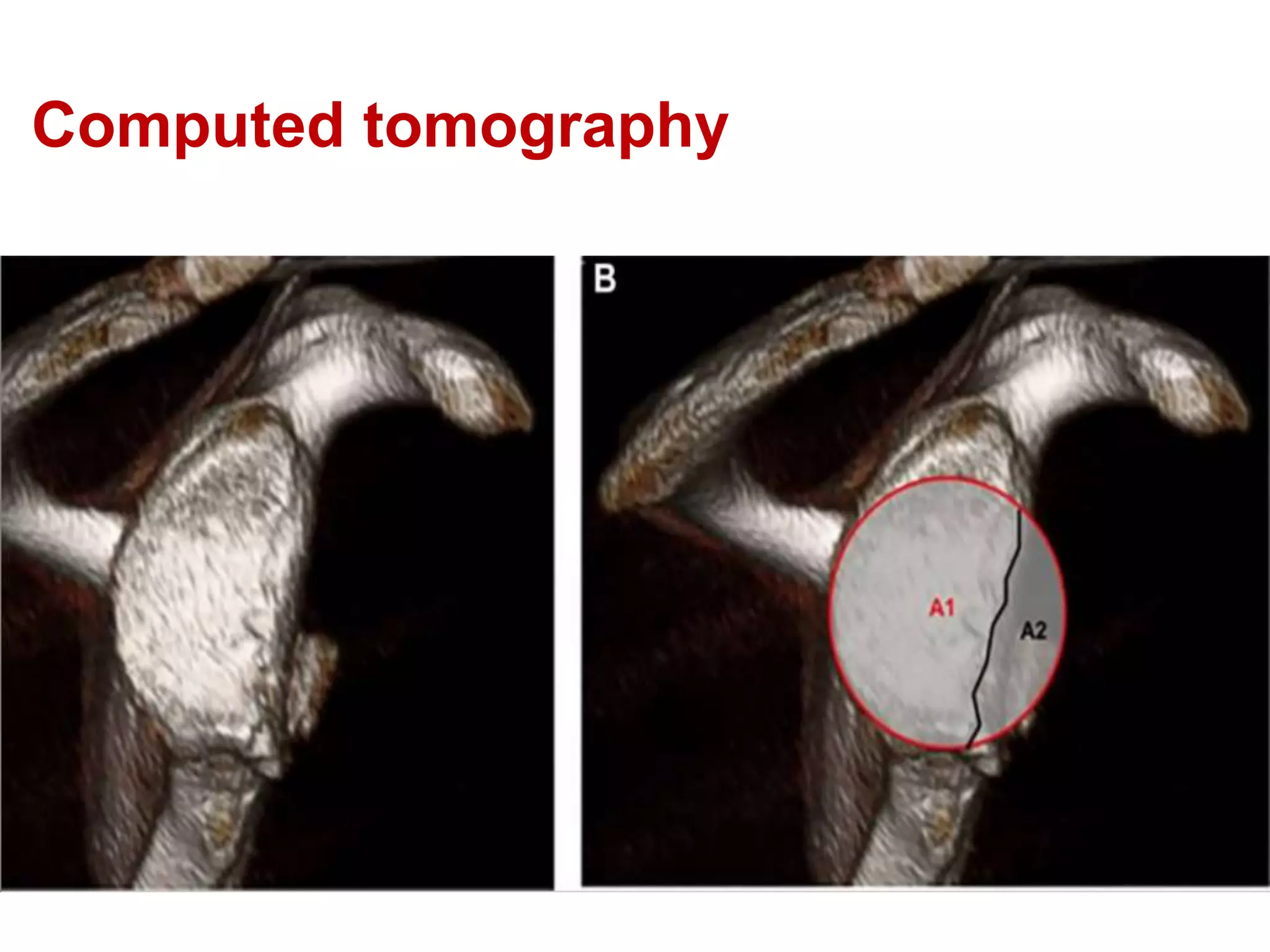
This document discusses shoulder instability, including the normal anatomy, causes of instability, classifications, clinical evaluation, radiographic evaluation, and treatment options. The glenohumeral joint has the highest mobility of any joint but lacks stability. Instability can be caused by excessive ligament laxity, bone defects, or trauma. Clinical exams include special tests like the apprehension and relocation tests. Treatment may involve arthroscopic or open stabilization surgery like Bankart repair, with post-op rehabilitation progressing from immobilization to strengthening and return to activity.
Deformity: It’s the position of a limb/Joint, from which it cannot be brought back to its normal anatomical position.
Described as abnormalities of :
Length
Angulation
Rotation
Translation
Combination
Deformity: It’s the position of a limb/Joint, from which it cannot be brought back to its normal anatomical position.
Described as abnormalities of :
Length
Angulation
Rotation
Translation
Combination
Basics of patellofemoral instability for postgraduates. Gives brief introduction about patellofemoral joint anatomy, causes, examintaion and treatment for patellofemoral instability
Assessment and Management of Frozen ShoulderThe Arm Clinic
The Arm Clinic's Mr Mike Walton presents his thoughts on assessment and management of Frozen Shoulder. Presentation for The Arm Clinic educational event #stiffshoulder at The Wilmslow Hospital, 29th April 2016.
micro teaching on communication m.sc nursing.pdfAnurag Sharma
Microteaching is a unique model of practice teaching. It is a viable instrument for the. desired change in the teaching behavior or the behavior potential which, in specified types of real. classroom situations, tends to facilitate the achievement of specified types of objectives.
These lecture slides, by Dr Sidra Arshad, offer a quick overview of physiological basis of a normal electrocardiogram.
Learning objectives:
1. Define an electrocardiogram (ECG) and electrocardiography
2. Describe how dipoles generated by the heart produce the waveforms of the ECG
3. Describe the components of a normal electrocardiogram of a typical bipolar leads (limb II)
4. Differentiate between intervals and segments
5. Enlist some common indications for obtaining an ECG
Study Resources:
1. Chapter 11, Guyton and Hall Textbook of Medical Physiology, 14th edition
2. Chapter 9, Human Physiology - From Cells to Systems, Lauralee Sherwood, 9th edition
3. Chapter 29, Ganong’s Review of Medical Physiology, 26th edition
4. Electrocardiogram, StatPearls - https://www.ncbi.nlm.nih.gov/books/NBK549803/
5. ECG in Medical Practice by ABM Abdullah, 4th edition
6. ECG Basics, http://www.nataliescasebook.com/tag/e-c-g-basics
Ethanol (CH3CH2OH), or beverage alcohol, is a two-carbon alcohol
that is rapidly distributed in the body and brain. Ethanol alters many
neurochemical systems and has rewarding and addictive properties. It
is the oldest recreational drug and likely contributes to more morbidity,
mortality, and public health costs than all illicit drugs combined. The
5th edition of the Diagnostic and Statistical Manual of Mental Disorders
(DSM-5) integrates alcohol abuse and alcohol dependence into a single
disorder called alcohol use disorder (AUD), with mild, moderate,
and severe subclassifications (American Psychiatric Association, 2013).
In the DSM-5, all types of substance abuse and dependence have been
combined into a single substance use disorder (SUD) on a continuum
from mild to severe. A diagnosis of AUD requires that at least two of
the 11 DSM-5 behaviors be present within a 12-month period (mild
AUD: 2–3 criteria; moderate AUD: 4–5 criteria; severe AUD: 6–11 criteria).
The four main behavioral effects of AUD are impaired control over
drinking, negative social consequences, risky use, and altered physiological
effects (tolerance, withdrawal). This chapter presents an overview
of the prevalence and harmful consequences of AUD in the U.S.,
the systemic nature of the disease, neurocircuitry and stages of AUD,
comorbidities, fetal alcohol spectrum disorders, genetic risk factors, and
pharmacotherapies for AUD.
TEST BANK for Operations Management, 14th Edition by William J. Stevenson, Ve...kevinkariuki227
TEST BANK for Operations Management, 14th Edition by William J. Stevenson, Verified Chapters 1 - 19, Complete Newest Version.pdf
Couples presenting to the infertility clinic- Do they really have infertility...Sujoy Dasgupta
Dr Sujoy Dasgupta presented the study on "Couples presenting to the infertility clinic- Do they really have infertility? – The unexplored stories of non-consummation" in the 13th Congress of the Asia Pacific Initiative on Reproduction (ASPIRE 2024) at Manila on 24 May, 2024.
These simplified slides by Dr. Sidra Arshad present an overview of the non-respiratory functions of the respiratory tract.
Learning objectives:
1. Enlist the non-respiratory functions of the respiratory tract
2. Briefly explain how these functions are carried out
3. Discuss the significance of dead space
4. Differentiate between minute ventilation and alveolar ventilation
5. Describe the cough and sneeze reflexes
Study Resources:
1. Chapter 39, Guyton and Hall Textbook of Medical Physiology, 14th edition
2. Chapter 34, Ganong’s Review of Medical Physiology, 26th edition
3. Chapter 17, Human Physiology by Lauralee Sherwood, 9th edition
4. Non-respiratory functions of the lungs https://academic.oup.com/bjaed/article/13/3/98/278874
Explore natural remedies for syphilis treatment in Singapore. Discover alternative therapies, herbal remedies, and lifestyle changes that may complement conventional treatments. Learn about holistic approaches to managing syphilis symptoms and supporting overall health.
Flu Vaccine Alert in Bangalore Karnatakaaddon Scans
As flu season approaches, health officials in Bangalore, Karnataka, are urging residents to get their flu vaccinations. The seasonal flu, while common, can lead to severe health complications, particularly for vulnerable populations such as young children, the elderly, and those with underlying health conditions.
Dr. Vidisha Kumari, a leading epidemiologist in Bangalore, emphasizes the importance of getting vaccinated. "The flu vaccine is our best defense against the influenza virus. It not only protects individuals but also helps prevent the spread of the virus in our communities," he says.
This year, the flu season is expected to coincide with a potential increase in other respiratory illnesses. The Karnataka Health Department has launched an awareness campaign highlighting the significance of flu vaccinations. They have set up multiple vaccination centers across Bangalore, making it convenient for residents to receive their shots.
To encourage widespread vaccination, the government is also collaborating with local schools, workplaces, and community centers to facilitate vaccination drives. Special attention is being given to ensuring that the vaccine is accessible to all, including marginalized communities who may have limited access to healthcare.
Residents are reminded that the flu vaccine is safe and effective. Common side effects are mild and may include soreness at the injection site, mild fever, or muscle aches. These side effects are generally short-lived and far less severe than the flu itself.
Healthcare providers are also stressing the importance of continuing COVID-19 precautions. Wearing masks, practicing good hand hygiene, and maintaining social distancing are still crucial, especially in crowded places.
Protect yourself and your loved ones by getting vaccinated. Together, we can help keep Bangalore healthy and safe this flu season. For more information on vaccination centers and schedules, residents can visit the Karnataka Health Department’s official website or follow their social media pages.
Stay informed, stay safe, and get your flu shot today!
Report Back from SGO 2024: What’s the Latest in Cervical Cancer?bkling
Are you curious about what’s new in cervical cancer research or unsure what the findings mean? Join Dr. Emily Ko, a gynecologic oncologist at Penn Medicine, to learn about the latest updates from the Society of Gynecologic Oncology (SGO) 2024 Annual Meeting on Women’s Cancer. Dr. Ko will discuss what the research presented at the conference means for you and answer your questions about the new developments.
ARTIFICIAL INTELLIGENCE IN HEALTHCARE.pdfAnujkumaranit
Artificial intelligence (AI) refers to the simulation of human intelligence processes by machines, especially computer systems. It encompasses tasks such as learning, reasoning, problem-solving, perception, and language understanding. AI technologies are revolutionizing various fields, from healthcare to finance, by enabling machines to perform tasks that typically require human intelligence.
3. Introduction
• Highest mobility, at the expense of stability
• Most frequently dislocated
50 % of all dislocations
2 % incidence in general population
• Factors predisposing to recurrent dislocation
Age
Return to contact sports
Hyper-laxity
Significant Bony defect in glenoid or humeral head
4. What is instability?
• Inability to maintain the humeral head centered in the glenoid
fossa and coracoacromial arch throughout ROM.
• Instability ≠ Joint laxity
• Laxity = Incomplete loss of glenohumeral articulation
unassociated with pain
• Subluxation = Partial loss of glenohumeral articulation with
symptoms
• Dislocation = Complete loss of glenohumeral articulation
12. Gleno-humeral ligaments
SGHL – Most important restraint
at at zero degree of abduction.
MGHL- Most important restraint
at lower and middle range of
abduction.
IGHL- Most important restraint at
more than 45 degrees of
abduction.
16. Negative intra-articular pressure
• - 42 cm H2O in cadavers
• Secondary to high osmotic pressure in interstitial tissues
• Only clinically important in the arm at rest in adduction
• Lost with lax capsule and defect
28. “Circle stability concept”
• For a full dislocation to occur, both sides of the capsule and
ligaments must be damaged.
• The capsule preventing the direction of location would be
considered the primary restraint and the opposite side would be
considered the secondary restraint.
33. Questions to be answered while evaluating
a patient!
• Is the problem in the gleno-humeral joint ?
• Is the problem one of failure to maintain the humeral head in the
centered position?
• What mechanical factors are contributing to instability ?
• Are these factors amenable to surgical repair or reconstruction?
34. History
• Age
• Trauma- duration
• Associated pain
• Sports, throwing or overhead activities
• Voluntary subluxation
• ”Clunk” or knock
• Fear- limitation of movements
• Hx dislocations and energy associated
• Hx 1st dislocation or injury
• Subsequent dislocations/ subluxations
35. Physical examination
• Atrophy or asymmetry around shoulder
• Tenderness around anterior or posterior capsule
• Active and passive ROM
• Strengths of the deltoid, rotator cuff and scapular stabilizers.
• Assessment of nerve injury and impingement (primary vs secondary)
• Special tests:
Shift and load test
Sulcus test
Apprehension test
Drawer’s test
Jobe’s relocation test
70. Phases of rehabilitation
• Phase I (Post op to 3 weeks)
Rest and immobilization. Pain control with nonsteroidal anti-inflammatory
drugs and ice applied to the shoulder.
• Phase II (3 to 6 weeks)
Isometric strengthening, Isotonic strengthening. Begin exercises with shoulder
in adducted, forward- flexed position, progressing to abducted position.
• Phase III ( 6 weeks to 3 months)
Endurance building along with strengthening exercises. Goal: the patient
reaches 90% strength in the injured shoulder compared with the uninjured
shoulder.
• Phase IV (3 months to 6 months)
Increase activity to sport- or job-specific activities
Editor's Notes
Shoulder joint is a complex anatomical and biomechanical structure which functions in a manner that several stabilizers play role in a special harmony in different stages of motion.
Contact surface of the humeral head with the glenoid is about 30%, which means that the joint has a limited osseous constraint so that the primary stability is due to other soft tissue components rather than the osseous contact.
The glenoid is deepened by 50% by the presence of the glenoid labrum and helps create cavity-compression. The labrum increases the humeral contact to 75%. The labrum is composed of fibro-cartilaginous tissue
Integral to the glenoid labrum is the insertion of the tendon of the long head of the biceps, which inserts on the superior aspect of the joint and blends to become indistinguishable from the posterior glenoid labrum. Matsen et al. suggested that the labrum may serve as a “chock block” to prevent excessive humeral head rollback.
Normal variant - the labrum attached to the glenoid rim and a flat/broad middle glenohumeral ligament is the most common “normal” variation. A cord-like middle glenohumeral ligament is often present in 86% of population.
Sub labral foramen- seen in 12 % population
Buford complex- absent antero superior labrum with cord like MGHL
seen in ~1.5% of population
- Cordlike middle glenohumeral ligament with attachment to base of biceps anchor and complete absence of the anterosuperior labrum
- Attaching a Buford complex will lead to painful and restricted external rotation and elevation.
The shoulder joint capsule is lax and thin and, by itself, offers little resistance or stability. Anteriorly, the capsule is reinforced by three capsular thickenings
or ligaments that are intimately fused with the labral attachment to the glenoid rim.
Superior glenohumeral ligament attaches to the glenoid rim near the apex of the labrum conjoined with the long head of the biceps. On the humerus, it is attached to the anterior aspect of the anatomic neck of the humerus.
The middle glenohumeral ligament has a wide attachment extending from the superior glenohumeral ligament along the anterior margin of the glenoid down as far as the junction of the middle and inferior thirds of the glenoid rim. On the humerus, it also is attached to the anterior aspect of the anatomic neck.
The inferior glenohumeral ligament attaches to the glenoid margin from the 2- to 3-o’clock positions anteriorly to the 8- to 9-o’clock positions posteriorly. The humeral attachment is below the level of the horizontally oriented physis into the inferior aspect of the anatomic and surgical neck of the humerus.
The inferior glenohumeral ligament is the strongest and most important of the glenohumeral ligaments.
O'Brien and Warren looked in detail at the gross and microscopic nature of this ligament. They likened the ligament to a hammock, strung between the glenoid and the humeral head. The two ropes holding the hammock are the anterior superior band and, at the back, the posterior superior band.
With external rotation, the hammock slides anteriorly and superiorly. The anterior band tightens, and the posterior band fans out. With internal rotation, the opposite occurs.
Laxity of the rotator interval results in a visible sulcus sign with inferior laxity with the shoulder in adduction.
o includes the capsule, SGHL, coracohumeral ligament and long head biceps tendon that bridge the gap between the supraspinatus and the subscapularis.
o boundaries
medially by lateral coracoid base
superiorly by anterior edge of supraspinatus
inferiorly by superior border of subscapularis
lateral apex formed by transverse humeral ligament
Biceps Long Head (dynamic)
o long head of biceps acts as humeral head depressor.
o variable origin from superior labrum
o forms weak links that predisposes to SLAP tear
o SGHL and subscapularis thought to play role in stabilizing long head of biceps
Scapular dyskinesis is an alteration of the normal position or motion of the scapula during coupled scapulo humeral movements and can occur after overuse of and repeated injuries to the shoulder joint. A particular overuse muscle fatigue syndrome has been designated the SICK scapula: scapular malposition, inferior medial border prominence, coracoid pain and malposition, and dyskinesis of scapular movement.
Perthes- a labro-ligamentous avulsion like Bankart but with a medially stripped intact periosteum.
ALPSA- Anterior labral periosteal sleeve avulsion
GLAD- Glenoid labrum articular disruption
A humeral head impaction fracture produced as the shoulder is dislocated, and the humeral head is impacted against the rim of the glenoid at the time of dislocation. This Hill-Sachs lesion is a defect in the posterolateral aspect of the humeral head. Instability results when the defect engages the glenoid rim in the functional arc of motion at 90 degrees abduction and external rotation.
Excessive laxity of the shoulder capsule also causes instability of the shoulder joint. Excessive laxity can be caused by a congenital collagen deficiency, shown by hyperlaxity of other joints, or by plastic deformation of the capsulo-ligamentous complex from a single macrotraumatic event or repetitive microtraumatic events. Hyperlaxity has been implicated as a cause of failure in surgical correction of chronic shoulder instability.
Glenoid rim fractures also can occur with an anterior or posterior dislocation. If these lesions involve more than 20% to 25% of the glenoid, they can result in
recurrent instability despite having an excellent soft-tissue repair.
These lesions are difficult to see on plain radiographs; three-dimensional CT is the best method for evaluating the extent of the defect
Chronic or recurrent is more than 6 weeks.
From a clinical and therapeutic point of view, three polar types of disorder can be identified:
Type I Traumatic structural instability.
Type II Atraumatic (or minimally traumatic)
structural instability.
Type III Atraumatic non-structural instability
(muscular dyskinesia).
The triangular relationship between these conditions allows for the fact there are intermediate types that lie between the ‘poles’; the balance of abnormalities
can shift and patients may ‘move’ from one group to another over time or present with a combination of pathologies: for example, a purely structural disorder
which, if allowed to persist, becomes associated with abnormal muscle patterning to the extent that both conditions need to be treated and the problems grow
in complexity. The system also recognizes that there is a gradation in the opposite direction, from dyskinetic muscle patterning to structural abnormality.
Sulcus test is done with the arm in 0 degrees and 45 degrees of abduction. This test is done by pulling distally on the extremity and observing for a sulcus or dimple between the humeral head and the acromion that does not reduce with 45 degrees of external rotation. The distance between the humeral head and acromion should be graded from 0 to 3 with the arm in 0 degrees and 45 degrees of abduction, with 1+ indicating subluxation of less than 1 cm, 2+ indicating 1 to 2 cm of subluxation, and 3+ indicating more than 2 cm of inferior subluxation that does not reduce with external rotation. Subluxation at 0 degrees of abduction is more
indicative of laxity at the rotator interval, and subluxation at 45 degrees indicates laxity of the inferior gleno-humeral ligament complex.
Shoulder anterior drawer test should be performed with the patient supine and the extremity in various degrees of abduction and external rotation in the plane of the scapula. When examining the patient’s right shoulder, the examiner’s left hand is used to grasp the proximal humerus while the right hand is used to hold the elbow lightly. Anterior stress is applied to the proximal humerus using the left hand, and the
amount of translation and the end point are evaluated.
Anterior stress is applied with the shoulder in various degrees of abduction and external rotation, and posterior stress is applied to evaluate for posterior instability with the arm in 90 degrees of abduction and various degrees of flexion. Apprehension is evaluated with anterior and posterior stress during these procedures.
Bony deformity of the glenoid or humerus is indicated by apprehension or instability at low ranges of motion (< 90 degrees of abduction) and when inferior instability is prominent.
The Jobe relocation test can be used for evaluating instability in athletes involved in sports requiring overhead motion. This test is done with the patient supine and the shoulder in 90 degrees of abduction and external rotation. Various degrees of abduction are evaluated while anterior stress is applied by the examiner’s hand to the posterior part of the humerus. If this produces pain or apprehension, posteriorly directed force is applied to the humerus to relocate the humeral head in the glenohumeral joint while the shoulder is placed in abduction and external rotation. The posteriorly directed stress used to relocate the humerus is released.
A feeling of apprehension or subluxation on the part of the patient indicates anterior instability.
Hyperlaxity is indicated by a positive sulcus test, a positive Gagey hyperabduction test, and by the Beighton hyperlaxity scale. The hyperabduction test is done by stabilizing the scapula with one hand placed superiorly while passively abducting the shoulder with the other hand. A side-to-side difference of more than 20 degrees is suggestive of inferior capsular laxity. External rotation of more than 85 degrees at 0 degrees of abduction is indicative of hyperlaxity, which may need to be corrected with rotator interval closure.
The initial radiographic examination should include anteroposterior and axillary lateral views of the shoulder. If the initial radiographic evaluation
is inconclusive, special views, gadolinium-enhanced MRI, or CT arthrography can be used to show posttraumatic changes not otherwise detected.
The most common special views that can be obtained in the office are the anteroposterior view of the shoulder in internal rotation, the West Point or
Rokous view, and the Stryker notch view. An anteroposterior radiograph of the shoulder in internal rotation often shows a Hill-Sachs lesion that may not be apparent on routine views.
Helps to evaluate the relationship of humeral head with glenoid. Humeral head should be at the bifurcation of the Y.
The Stryker notch view is obtained with the patient supine and the elbow elevated over the head. The x-ray beam is directed 10 degrees cephalad
Garth et al. also described an apical oblique radiograph that frequently shows posterior humeral head defects that might not be seen on routine films.
The West Point view is used to show calcification or small fractures at the anteroinferior glenoid rim. This is a modified, prone, axillary lateral view of the shoulder obtained with the shoulder abducted 90 degrees and the elbow bent with the arm hanging over the side of the table. The x-ray beam is directed 25 degrees medially and 25 degrees cephalad with the cassette placed above the shoulder perpendicular to the table.
CT, particularly three-dimensional CT, is the most sensitive test for detecting and measuring bone deficiency or retroversion of the glenoid or humerus. CT is indicated when there is blunting of the glenoid cortical outline or an obvious bone defect on plain radiographs. CT also is indicated for evaluating recurrences that occur with trivial trauma, low angle instability, and failed surgical procedures.
MRI or MRA is useful in evaluating soft-tissue lesions associated with instability.
Posterior capsule and infraspinatus tendon sutured into the Hill-Sachs lesion. May be performed with concomitant Bankart repair
Latarjet procedure is an open bony procedure which involves a coracoid transfer to the anterior inferior glenoid bone defect done in cases of glenoid bony defects that cannot be repaired.
The Latarjet procedure involves the removal and transfer of a section of the coracoid process and its attached muscles to the front of the glenoid. This placement of the coracoid acts as a bone block which, combined with the transferred muscles acting as a strut, prevents further dislocation of the joint.[3] In layman's terms, this procedure involves removing a piece of bone from another part of the shoulder, and attaching it to the front of the shoulder socket. The bone will then act as a barrier which will physically block the shoulder from slipping out of the socket, while the muscles which are transferred with the bone will give additional stability to the joint.
The mechanism of action has been described as a triple blocking effect:
-conjoint tendon acting as a sling on the subscapularis and capsule with the arm abducted and externally rotated;
-increasing or restoring the glenoid bone; and
-repair of the capsule to the stump of coracoacromial ligament.
Magnuson-Stack is performed with lateral advancement of subscapularis (lateral to bicipital groove and at times to greater tuberosity)
The initial treatment of posterior shoulder instability should be nonoperative. The regimen includes having the patient avoid provocative activities and educating the patient to avoid specific voluntary maneuvers that would cause the posterior subluxation. A strengthening exercise program aimed at the external rotators and posterior deltoid is carried out.
If at least 4 to 6 months of an appropriate rehabilitation program has failed, if habitual dislocation has been ruled out, and if the patient is emotionally stable, surgery may be indicated if the pain and instability preclude adequate function of the involved shoulder.
For recurrent posterior dislocation associated with a large anterior medial Hill-Sachs lesion, McLaughlin described transfer of the subscapularis tendon into the defect. Neer and Foster subsequently described transfer of the subscapularis with the lesser tuberosity into the defect and securing it with a bone screw.