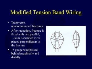
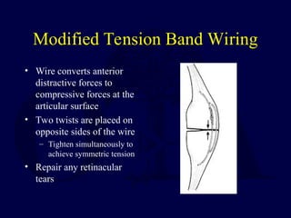
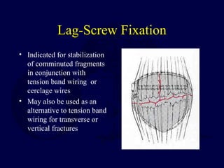
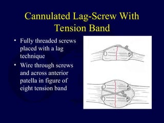
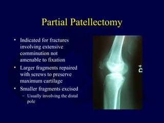
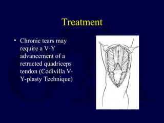
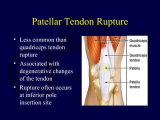
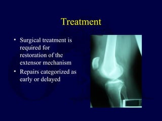
Patella Fractures & Extensor
Mechanism Injuries
Lisa K. Cannada, MD
Original Authors: Charles G. Haddad, Jr., MD, Lisa K. Cannada, MD,
and Robert Cantu, MD; March 2004
New Author: Lisa K. Cannada, MD; Revised January 2006
OTA
Resident’s
Course 2005
Anatomy
• Largest sesamoid bone
• Thick articular
cartilage proximally
• Articular surface
divided into medial
and lateral facets by
longitudinal ridge
• Distal pole
nonarticular
Anatomy
• Patellar Retinaculum
– Longitudinal tendinous
fibers
– Patellofemoral
ligaments
• Blood Supply
– Primarily derived from
geniculate arteries
Biomechanics
• The patella undergoes approximately 7 cm
of translation from full flexion to extension
• Only 13-38% of the patellar surface is in
contact with the femur throughout its range
of motion
Biomechanics
• The patella increases
the moment arm about
the knee
– Contributes up to 30%
increase in force with
extension
• Patella withstands
compressive forces
greater than 7X body
weight with squatting
Biomechanics
• Twice as much torque
is needed to extend the
knee the final 15
degrees than to extend
from a fully flexed
position to 15 degrees
of flexion
History
• Direct blow to the
anterior knee
(dashboard injury)
• Fall from height
• Rapid knee flexion
with quadriceps
resistance
Physical Examination
• Pain, swelling, contusions, lacerations
and/or abrasions at the site of injury
• Palpable defect
• Assessment of ability to extend the knee
against gravity or maintain the knee in full
extension against gravity
Radiographic Evaluation
• AP & Lateral
– Patella alta or baja
– Note fracture pattern
• Articular step-off,
diastasis
• Special views
– Axial or sunrise
• CT Scan
-Occult fractures
Radiographic Evaluation
• Bipartite Patella
– Obtain bilateral views
– Often involves
superolateral corner
– Accessory ossification
center
Etiology
• Allows prediction of outcome
• Direct trauma
– Dashboard injury
– Increasing cases with penetrating
trauma
– Often with comminution and
articular damage
• Indirect trauma
– Violent flexion directed through
the extensor mechanism against a
contracted quadriceps
– Results in simple, transverse
fractures
Classification
• Allows prediction of
treatment
• Types
– Transverse
– Marginal
– Vertical
– Comminuted
– Osteochondral
Nonoperative Treatment
• Indicated for nondisplaced fractures
– <2mm of articular stepoff and <3mm of
diastasis with an intact extensor mechanism
• May also be considered for minimally
displaced fractures in the elderly
• Patients with a extensive medical
comorbidities
Nonoperative Treatment
• Long leg cylinder cast for 4-6 weeks
– May consider a knee immobilizer for the
elderly
• Immediate weightbearing as tolerated
• Rehabilitation includes range of motion
exercises with gradual quadriceps
strengthening
Operative Treatment
• Goals
– Preserve extensor function
– Restore articular congruency
• Preoperative Setup
– Tourniquet
• Prior to inflation, gently flex
the knee
• Approach
– Longitudinal midline incision
recommended
– Transverse approach
alternative
– Consider future surgeries!
Operative Techniques
• Modified tension band wiring
• Lag-screw fixation
• Cannulated lag-screw with tension band
• Partial patellectomy
• Patellectomy
Modified Tension Band Wiring
• Transverse,
noncomminuted fractures
• After reduction, fracture is
fixed with two parallel,
1.6mm Kirschner wires
placed perpendicular to
the fracture
• 18 gauge wire passed
behind proximally and
distally
Modified Tension Band Wiring
• Wire converts anterior
distractive forces to
compressive forces at the
articular surface
• Two twists are placed on
opposite sides of the wire
– Tighten simultaneously to
achieve symmetric tension
• Repair any retinacular
tears
Lag-Screw Fixation
• Indicated for stabilization
of comminuted fragments
in conjunction with
tension band wiring or
cerclage wires
• May also be used as an
alternative to tension band
wiring for transverse or
vertical fractures
Lag-Screw Fixation
• Contraindicated for extensive comminution
and osteopenic bone
• Small secondary fractures may be stabilized
with 2.7mm or 3.5mm cortical screws
• Transverse or vertical fractures require
3.5mm or 4.5mm cortical screws
– Retrograde insertion of screws may be
technically easier
Cannulated Lag-Screw With
Tension Band
• Fully threaded screws
placed with a lag
technique
• Wire through screws
and across anterior
patella in figure of
eight tension band
Cannulated Lag-Screw With
Tension Band
• Most stable construct
– Screws and tension band wire combination
eliminates both possible separation seen at the
fracture site with modified tension band and
screw failure due to excessive three point
bending
Suture vs. Wire Tension Band
Gosal et al Injury 2001
• Wire v. #5 Ethibond
• 37 patients
• Reoperation 38% wire
group vs. 6%
• Infection 3 pts wire
group vs. 0
Patel et al, Injury 2000
McGreal et al, J Med
Eng Tech, 1999
• Cadaveric models
• Quality and stability
of fixation comparable
to wire
• Conclude suture an
acceptable alternative
Partial Patellectomy
• Indicated for fractures
involving extensive
comminution not
amenable to fixation
• Larger fragments repaired
with screws to preserve
maximum cartilage
• Smaller fragments excised
– Usually involving the distal
pole
Partial Patellectomy
• Tendon is attached to fragment with
nonabsorbable suture passed through drill holes in
the fragment
– Drill holes should be near the articular surface to
prevent tilting of the tendon and minimize articular
step-off
• Watch for patellar tilt!
• Load sharing wire passed through drill holes in the
tibial tubercle and patella may be used to protect
the repair and facilitate early range of motion
Total Patellectomy
• Indicated for displaced, comminuted
fractures not amenable to reconstruction
• Bone fragments sharply dissected
• Defect may be repaired through a variety of
techniques
• Usually results in extensor lag and loss of
strength
Postoperative Management
• Immobilization with knee brace
• Immediate WBAT
• Early range of motion
– Based on intraoperative assessment of repair
– Active flexion with passive extension
• Quadriceps strengthening
– Begun when there is radiographic evidence of
healing, usually around 6 weeks
Complications
• Knee Stiffness
– Most common
complication
• Infection
– Rare, depends on soft
tissue compromise
• Loss of Fixation
– Hardware failure in up
to 20% of cases
• Osteoarthritis
– May result from
articular damage or
incongruity
• Nonunion < 1% with
surgical repair
• Painful hardware
– Removal required in
approximately 15%
Extensor Tendon Ruptures
• Patellar and quadriceps
tendon ruptures are
uncommon injuries
• Patients are typically males
in their 30’s or 40’s
– Patellar < 40 yo
– Quadriceps > 40 yo
• Fall, sports, MVA
Quadriceps Tendon Rupture
• Typically occurs in patients > 40 years old
• Usually 0-2 cm above the superior pole
• Level often associated with age
– Rupture occurs at the bone-tendon junction in
majority of patients > 40 years old
– Rupture occurs at midsubstance in majority of
patients < 40 years old
Quadriceps Tendon Ruptures
• Risk Factors
– Chronic tendonitis
– Anabolic steroid use
– Local steroid injection
– Inflammatory
arthropathy
– Chronic renal failure
– Systemic disease
History
• Sensation of a sudden pop while stressing
the extensor mechanism
• Pain at the site of injury
• Inability/difficulty weightbearing
Physical Exam
• Effusion
• Tenderness at the
upper pole
• Palpable defect above
superior pole
• Loss of extension
• With partial tears,
extension will be
intact
Quadriceps Tendon Rupture
Radiographic
Evaluation
• X-ray- AP, Lateral,
and Tangential
(Sunrise, Merchant)
– Distal displacement of
the patella
• MRI
– Useful when diagnosis
is unclear
Treatment
• Nonoperative
– Partial tears and strains
• Operative
– For complete ruptures
Operative Treatment
• Reapproximation of tendon to bone using
nonabsorbable sutures with tears at the
muscultendonous junction
– Locking stitch (Bunnel, Krakow) with No. 5
ethibond passed through vertical bone tunnels
– Repair tendon close to articular surface to avoid
patellar tilting
Operative Treatment
• Midsubstance tears may
undergo end-to-end repair
after edges are freshened
and slightly overlapped
– May benefit from
reinforcement from distally
based partial thickness
quadriceps tendon turned
down across the repair site
(Scuderi Technique)
Treatment
• Chronic tears may
require a V-Y
advancement of a
retracted quadriceps
tendon (Codivilla V-
Y-plasty Technique)
Postoperative Management
• Knee immobilizer or cylinder cast for 5-6
weeks
• Immediate vs. delayed (3 weeks)
weightbearing as tolerated
• At 2-3 weeks, hinged knee brace starting
with 45 degrees active range of motion with
10-15 degrees of progression each week
Complications
• Rerupture
• Persistent quadriceps
atrophy/weakness
• Loss of motion
• Infection
Patellar Tendon Rupture
• Less common than
quadriceps tendon
rupture
• Associated with
degenerative changes
of the tendon
• Rupture often occurs
at inferior pole
insertion site
Patellar Tendon Rupture
• Risk Factors
– Rheumatoid
– Systemic Lupus
Erythematosus
– Diabetes
– Chronic Renal Failure
– Systemic Corticosteroid
Therapy
– Local Steroid Injection
– Chronic patellar tendonitis
Anatomy
• Patellar tendon
– Averages 4 mm thick but widens to 5-6 mm at
the tibial tubercle insertion
– Merges with the medial and lateral retinaculum
– 90% type I collagen
Blood Supply
• Fat pad vessels supply posterior aspect of tendon
via inferior medial and lateral geniculate arteries
• Retinacular vessels supply anterior portion of
tendon via the inferior medial geniculate and
recurrent tibial arteries
• Proximal and distal insertion areas are relatively
avascular and subsequently are a common site of
rupture
Biomechanics
• Greatest forces are at 60
degrees of flexion
• 3-4 times greater strain are
at the insertions compared
to the midsubstance prior
to failure
• Forces through the patellar
tendon are 3.2 times body
weight while climbing
stairs
History
• Often a report of
forceful quadriceps
contraction against a
flexed knee
• May experience and
audible “pop”
• Inability to weightbear
or extend the knee
Physical Examination
• Palpable defect
• Hemarthrosis
• Painful passive knee
flexion
• Partial or complete
loss of active
extension
• High riding patella on
radiographs
Radiographic Evaluation
• AP and Lateral X-ray
– Patella alta seen on lateral view
• Patella superior to Blumensaat’s line
• Ultrasonagraphy
– Effective means to determine continuity of tendon
– Operator and reader dependant
• MRI
– Effective means to assess patellar tendon, especially if
other intraarticular or soft tissue injuries are suspected
– Relatively high cost
Classification
• No widely accepted means of classification
• Can be categorized by:
– Location of tear
• Proximal insertion most common
– Timing between injury and surgery
• Most important factor for prognosis
• Acute- within two weeks
• Chronic- greater than two weeks
Treatment
• Surgical treatment is
required for
restoration of the
extensor mechanism
• Repairs categorized as
early or delayed
Early Repair
• Better overall outcome
• Primary repair of the tendon
• Surgical approach is through a midline incision
– Incise just lateral to tibial tubercle as skin thicker with
better blood supply to decrease wound complications
• Patellar tendon rupture and retinacular tears are
exposed
Early Repair
• Frayed edges and
hematoma are debrided
• With a Bunnell or Krakow
stitch, two ethibond
sutures or their equivalent
are used to repair the
tendon to the patella
• Sutures passed through
three parallel, longitudinal
bone tunnels and tied
proximally
Early Repair
• Repair retinacular
tears
• May reinforce with
wire, cable or
umbilical tape
• Assess repair
intraoperatively with
knee flexion
Postoperative Management
• Maintain hinged knee brace which is gradually increased
as motion increases (tailor to the patient)
• Immediate vs. delayed (3 weeks) weightbearing as
tolerated
• At 2-3 weeks, hinged knee brace starting with 45 degrees
active range of motion with 10-15 degrees of progression
each week
• Immediate isometric quadriceps exercises
• All restrictions are lifted after full range of motion and
90% of the contralateral quadriceps strength are obtained;
usually at 4-6 months
Delayed Repair
• > 6 weeks from initial injury
• Often results in poorer outcome
• Quadriceps contraction and patellar migration are
encountered
• Adhesions between the patella and femur may be
present
• Options include hamstring and fascia lata
autograft augmentation of primary repair or
Achilles tendon allograft
Postoperative Management
• More conservative when compared to early
repair
• Bivalved cylinder cast for 6 weeks; may
start passive range of motion
• Active range of motion is started at 6 weeks
Complications
• Knee stiffness
• Persistent quadriceps weakness
• Rerupture
• Infection
• Patella baja
Thank You!
Thank You!
lisa.cannada@utsouthwestern.edu
Return to
Lower Extremity
Index
E-mail OTA
about
Questions/Comments
If you would like to volunteer as an author for the
Resident Slide Project or recommend updates to any of the
following slides, please send an e-mail to ota@aaos.org

Patella Fractures & Extensor Mechanism Injuries.ppt

  • 1.
    Patella Fractures &Extensor Mechanism Injuries Lisa K. Cannada, MD Original Authors: Charles G. Haddad, Jr., MD, Lisa K. Cannada, MD, and Robert Cantu, MD; March 2004 New Author: Lisa K. Cannada, MD; Revised January 2006 OTA Resident’s Course 2005
  • 2.
    Anatomy • Largest sesamoidbone • Thick articular cartilage proximally • Articular surface divided into medial and lateral facets by longitudinal ridge • Distal pole nonarticular
  • 3.
    Anatomy • Patellar Retinaculum –Longitudinal tendinous fibers – Patellofemoral ligaments • Blood Supply – Primarily derived from geniculate arteries
  • 4.
    Biomechanics • The patellaundergoes approximately 7 cm of translation from full flexion to extension • Only 13-38% of the patellar surface is in contact with the femur throughout its range of motion
  • 5.
    Biomechanics • The patellaincreases the moment arm about the knee – Contributes up to 30% increase in force with extension • Patella withstands compressive forces greater than 7X body weight with squatting
  • 6.
    Biomechanics • Twice asmuch torque is needed to extend the knee the final 15 degrees than to extend from a fully flexed position to 15 degrees of flexion
  • 7.
    History • Direct blowto the anterior knee (dashboard injury) • Fall from height • Rapid knee flexion with quadriceps resistance
  • 8.
    Physical Examination • Pain,swelling, contusions, lacerations and/or abrasions at the site of injury • Palpable defect • Assessment of ability to extend the knee against gravity or maintain the knee in full extension against gravity
  • 9.
    Radiographic Evaluation • AP& Lateral – Patella alta or baja – Note fracture pattern • Articular step-off, diastasis • Special views – Axial or sunrise • CT Scan -Occult fractures
  • 10.
    Radiographic Evaluation • BipartitePatella – Obtain bilateral views – Often involves superolateral corner – Accessory ossification center
  • 11.
    Etiology • Allows predictionof outcome • Direct trauma – Dashboard injury – Increasing cases with penetrating trauma – Often with comminution and articular damage • Indirect trauma – Violent flexion directed through the extensor mechanism against a contracted quadriceps – Results in simple, transverse fractures
  • 12.
    Classification • Allows predictionof treatment • Types – Transverse – Marginal – Vertical – Comminuted – Osteochondral
  • 13.
    Nonoperative Treatment • Indicatedfor nondisplaced fractures – <2mm of articular stepoff and <3mm of diastasis with an intact extensor mechanism • May also be considered for minimally displaced fractures in the elderly • Patients with a extensive medical comorbidities
  • 14.
    Nonoperative Treatment • Longleg cylinder cast for 4-6 weeks – May consider a knee immobilizer for the elderly • Immediate weightbearing as tolerated • Rehabilitation includes range of motion exercises with gradual quadriceps strengthening
  • 15.
    Operative Treatment • Goals –Preserve extensor function – Restore articular congruency • Preoperative Setup – Tourniquet • Prior to inflation, gently flex the knee • Approach – Longitudinal midline incision recommended – Transverse approach alternative – Consider future surgeries!
  • 16.
    Operative Techniques • Modifiedtension band wiring • Lag-screw fixation • Cannulated lag-screw with tension band • Partial patellectomy • Patellectomy
  • 17.
    Modified Tension BandWiring • Transverse, noncomminuted fractures • After reduction, fracture is fixed with two parallel, 1.6mm Kirschner wires placed perpendicular to the fracture • 18 gauge wire passed behind proximally and distally
  • 18.
    Modified Tension BandWiring • Wire converts anterior distractive forces to compressive forces at the articular surface • Two twists are placed on opposite sides of the wire – Tighten simultaneously to achieve symmetric tension • Repair any retinacular tears
  • 19.
    Lag-Screw Fixation • Indicatedfor stabilization of comminuted fragments in conjunction with tension band wiring or cerclage wires • May also be used as an alternative to tension band wiring for transverse or vertical fractures
  • 20.
    Lag-Screw Fixation • Contraindicatedfor extensive comminution and osteopenic bone • Small secondary fractures may be stabilized with 2.7mm or 3.5mm cortical screws • Transverse or vertical fractures require 3.5mm or 4.5mm cortical screws – Retrograde insertion of screws may be technically easier
  • 21.
    Cannulated Lag-Screw With TensionBand • Fully threaded screws placed with a lag technique • Wire through screws and across anterior patella in figure of eight tension band
  • 22.
    Cannulated Lag-Screw With TensionBand • Most stable construct – Screws and tension band wire combination eliminates both possible separation seen at the fracture site with modified tension band and screw failure due to excessive three point bending
  • 23.
    Suture vs. WireTension Band Gosal et al Injury 2001 • Wire v. #5 Ethibond • 37 patients • Reoperation 38% wire group vs. 6% • Infection 3 pts wire group vs. 0 Patel et al, Injury 2000 McGreal et al, J Med Eng Tech, 1999 • Cadaveric models • Quality and stability of fixation comparable to wire • Conclude suture an acceptable alternative
  • 24.
    Partial Patellectomy • Indicatedfor fractures involving extensive comminution not amenable to fixation • Larger fragments repaired with screws to preserve maximum cartilage • Smaller fragments excised – Usually involving the distal pole
  • 25.
    Partial Patellectomy • Tendonis attached to fragment with nonabsorbable suture passed through drill holes in the fragment – Drill holes should be near the articular surface to prevent tilting of the tendon and minimize articular step-off • Watch for patellar tilt! • Load sharing wire passed through drill holes in the tibial tubercle and patella may be used to protect the repair and facilitate early range of motion
  • 26.
    Total Patellectomy • Indicatedfor displaced, comminuted fractures not amenable to reconstruction • Bone fragments sharply dissected • Defect may be repaired through a variety of techniques • Usually results in extensor lag and loss of strength
  • 27.
    Postoperative Management • Immobilizationwith knee brace • Immediate WBAT • Early range of motion – Based on intraoperative assessment of repair – Active flexion with passive extension • Quadriceps strengthening – Begun when there is radiographic evidence of healing, usually around 6 weeks
  • 28.
    Complications • Knee Stiffness –Most common complication • Infection – Rare, depends on soft tissue compromise • Loss of Fixation – Hardware failure in up to 20% of cases • Osteoarthritis – May result from articular damage or incongruity • Nonunion < 1% with surgical repair • Painful hardware – Removal required in approximately 15%
  • 29.
    Extensor Tendon Ruptures •Patellar and quadriceps tendon ruptures are uncommon injuries • Patients are typically males in their 30’s or 40’s – Patellar < 40 yo – Quadriceps > 40 yo • Fall, sports, MVA
  • 30.
    Quadriceps Tendon Rupture •Typically occurs in patients > 40 years old • Usually 0-2 cm above the superior pole • Level often associated with age – Rupture occurs at the bone-tendon junction in majority of patients > 40 years old – Rupture occurs at midsubstance in majority of patients < 40 years old
  • 31.
    Quadriceps Tendon Ruptures •Risk Factors – Chronic tendonitis – Anabolic steroid use – Local steroid injection – Inflammatory arthropathy – Chronic renal failure – Systemic disease
  • 32.
    History • Sensation ofa sudden pop while stressing the extensor mechanism • Pain at the site of injury • Inability/difficulty weightbearing
  • 33.
    Physical Exam • Effusion •Tenderness at the upper pole • Palpable defect above superior pole • Loss of extension • With partial tears, extension will be intact
  • 34.
    Quadriceps Tendon Rupture Radiographic Evaluation •X-ray- AP, Lateral, and Tangential (Sunrise, Merchant) – Distal displacement of the patella • MRI – Useful when diagnosis is unclear Treatment • Nonoperative – Partial tears and strains • Operative – For complete ruptures
  • 35.
    Operative Treatment • Reapproximationof tendon to bone using nonabsorbable sutures with tears at the muscultendonous junction – Locking stitch (Bunnel, Krakow) with No. 5 ethibond passed through vertical bone tunnels – Repair tendon close to articular surface to avoid patellar tilting
  • 36.
    Operative Treatment • Midsubstancetears may undergo end-to-end repair after edges are freshened and slightly overlapped – May benefit from reinforcement from distally based partial thickness quadriceps tendon turned down across the repair site (Scuderi Technique)
  • 37.
    Treatment • Chronic tearsmay require a V-Y advancement of a retracted quadriceps tendon (Codivilla V- Y-plasty Technique)
  • 38.
    Postoperative Management • Kneeimmobilizer or cylinder cast for 5-6 weeks • Immediate vs. delayed (3 weeks) weightbearing as tolerated • At 2-3 weeks, hinged knee brace starting with 45 degrees active range of motion with 10-15 degrees of progression each week
  • 39.
    Complications • Rerupture • Persistentquadriceps atrophy/weakness • Loss of motion • Infection
  • 40.
    Patellar Tendon Rupture •Less common than quadriceps tendon rupture • Associated with degenerative changes of the tendon • Rupture often occurs at inferior pole insertion site
  • 41.
    Patellar Tendon Rupture •Risk Factors – Rheumatoid – Systemic Lupus Erythematosus – Diabetes – Chronic Renal Failure – Systemic Corticosteroid Therapy – Local Steroid Injection – Chronic patellar tendonitis
  • 42.
    Anatomy • Patellar tendon –Averages 4 mm thick but widens to 5-6 mm at the tibial tubercle insertion – Merges with the medial and lateral retinaculum – 90% type I collagen
  • 43.
    Blood Supply • Fatpad vessels supply posterior aspect of tendon via inferior medial and lateral geniculate arteries • Retinacular vessels supply anterior portion of tendon via the inferior medial geniculate and recurrent tibial arteries • Proximal and distal insertion areas are relatively avascular and subsequently are a common site of rupture
  • 44.
    Biomechanics • Greatest forcesare at 60 degrees of flexion • 3-4 times greater strain are at the insertions compared to the midsubstance prior to failure • Forces through the patellar tendon are 3.2 times body weight while climbing stairs
  • 45.
    History • Often areport of forceful quadriceps contraction against a flexed knee • May experience and audible “pop” • Inability to weightbear or extend the knee
  • 46.
    Physical Examination • Palpabledefect • Hemarthrosis • Painful passive knee flexion • Partial or complete loss of active extension • High riding patella on radiographs
  • 47.
    Radiographic Evaluation • APand Lateral X-ray – Patella alta seen on lateral view • Patella superior to Blumensaat’s line • Ultrasonagraphy – Effective means to determine continuity of tendon – Operator and reader dependant • MRI – Effective means to assess patellar tendon, especially if other intraarticular or soft tissue injuries are suspected – Relatively high cost
  • 48.
    Classification • No widelyaccepted means of classification • Can be categorized by: – Location of tear • Proximal insertion most common – Timing between injury and surgery • Most important factor for prognosis • Acute- within two weeks • Chronic- greater than two weeks
  • 49.
    Treatment • Surgical treatmentis required for restoration of the extensor mechanism • Repairs categorized as early or delayed
  • 50.
    Early Repair • Betteroverall outcome • Primary repair of the tendon • Surgical approach is through a midline incision – Incise just lateral to tibial tubercle as skin thicker with better blood supply to decrease wound complications • Patellar tendon rupture and retinacular tears are exposed
  • 51.
    Early Repair • Frayededges and hematoma are debrided • With a Bunnell or Krakow stitch, two ethibond sutures or their equivalent are used to repair the tendon to the patella • Sutures passed through three parallel, longitudinal bone tunnels and tied proximally
  • 52.
    Early Repair • Repairretinacular tears • May reinforce with wire, cable or umbilical tape • Assess repair intraoperatively with knee flexion
  • 53.
    Postoperative Management • Maintainhinged knee brace which is gradually increased as motion increases (tailor to the patient) • Immediate vs. delayed (3 weeks) weightbearing as tolerated • At 2-3 weeks, hinged knee brace starting with 45 degrees active range of motion with 10-15 degrees of progression each week • Immediate isometric quadriceps exercises • All restrictions are lifted after full range of motion and 90% of the contralateral quadriceps strength are obtained; usually at 4-6 months
  • 54.
    Delayed Repair • >6 weeks from initial injury • Often results in poorer outcome • Quadriceps contraction and patellar migration are encountered • Adhesions between the patella and femur may be present • Options include hamstring and fascia lata autograft augmentation of primary repair or Achilles tendon allograft
  • 55.
    Postoperative Management • Moreconservative when compared to early repair • Bivalved cylinder cast for 6 weeks; may start passive range of motion • Active range of motion is started at 6 weeks
  • 56.
    Complications • Knee stiffness •Persistent quadriceps weakness • Rerupture • Infection • Patella baja
  • 57.
    Thank You! Thank You! lisa.cannada@utsouthwestern.edu Returnto Lower Extremity Index E-mail OTA about Questions/Comments If you would like to volunteer as an author for the Resident Slide Project or recommend updates to any of the following slides, please send an e-mail to ota@aaos.org

Editor's Notes

  • #2 The patella is the largest sesamoid bone in the body and lies within the fascia and fibers of the quadriceps tendon. The upper ¾ is covered with articular cartilage. The articular surface of the patella is divided into medial and lateral facets, which articulate with the anterior trochlea. As you can see from the diagram, the lateral facet is the largest. A vertical ridge near the medial facet defines the odd facet. Transverse ridges are present which further define the facets.
  • #3 The patella in invested in a strong soft tissue envelope formed by the joining of the quadriceps tendon, the iliotibial band and distal quadriceps muscles and the patella tendon. The patellar retinaculum originates from the deep fascia along with fibers of the vastus medialis and vastus lateralis. There are also contributions from the iliotibial tract and patellofemoral ligaments of the joint capsule. The blood supply consists of an extraossoeus and intraosseous vascular system. The primary blood supply is from branches of the geniculate arteries. The intraosseous blood supply enters the bone through the midportion of the patella and through distal pole vessels.
  • #4 The patella is a link between the quadriceps tendon and patella tendon and is subject to significant forces. The patella increases the leverage of the quadriceps muscle and elevates the extensor mechanism away from the axis of rotation of the knee joint. The area of contact between the patella and distal femur varies according to the position of the knee.
  • #5 The patella improves the efficiency of the extensor mechanism by elevating the quadriceps away from the axis of rotation about the knee joint. The patella increases the leverage or the quadriceps allowing it to act over a greater angle. There are significant forces generated across the patellofemoral joint with activities of daily living. Normal activities can generate up to three times the body weight across the patellofemoral joint. With squatting and stair climbing, there may be forces up to seven times the body weight generated!
  • #6 As mentioned, the amount of contact between the patella and trochlea varies depending upon position of the knee. With the knee fully extended, the inferior portion of the patella is in contact with the femur. With the knee flexed to 135 degrees, the patella is in the intercondylar notch. Twice as much torque is needed to extend the knee the final 15 degrees than to extend from a fully flexed position from to 15 degrees of flexion.
  • #7 Patella fractures result from direct or indirect forces. Direct forces include a blow to the patella or an MVC. They may represent a “dashboard” injury and the patient’s should be evaluated for associated injuries. Indirect mechanisms include a fall or a strong contraction in which the forces from the extensor mechanism are greater than the strength of the patella.
  • #8 One must complete a thorough history and physical examination of the patient. There may be pain, swelling and decreased strength. The defect may be palpable. The skin should be examined closely in those injuries sustained from direct trauma to evaluate for the presence of an open injury. A saline load test may be used to assist with this diagnosis. The extensor mechanism is evaluated by the ability to extend the knee against gravity or to maintain the knee in full extension versus gravity.
  • #9 Radiographic evaluation of patella fractures includes AP, lateral and sunrise views. Comparison views of the unaffected limb may be of value to further define the bony anatomy. On the lateral view, one should evaluate the position of the patella. A low riding patella (patella baja) may indicate a quadriceps tendon rupture, while a high riding patella (patella alta) may indicate a patella tendon rupture. The Insall method method is used for assessment of patella position. In this method, the patella length is measured and compared in a ratio to length of the patella tendon. Normally, the ratio is 1. A ratio of 0.8 or less suggests a patella tendon rupture. The sunrise view may be helpful to further delineate fracture displacement. This view is helpful in the diagnosis of patellofemoral disorders and osteochondral defects. CT scans may be useful in periarticular injuries, evaluation of alignment, detection of occult fractures and analysis of fracture healing.
  • #10 In the evaluation of bipartite patella, obtain bilateral views. This accessory ossification center involves the superolateral corner.
  • #11 As mentioned, there are two main mechanisms of patella fractures: direct and indirect. Direct trauma often involves a higher energy mechanism and may be accompanied by additional injuries. There may be more damage to the articular cartilage which ultimately may affect the outcome.
  • #12 Classification systems are ideally designed to allow communication between physicians, guide treatment and predict outcomes. For the patella, there is no universally accepted classification systems other than the OTA system. Descriptive terms may be used to classify patella fractures and an example is demonstrated on the slide.
  • #13 Treatment of patella fractures is bases on the fracture type and physical examination. The ultimate goals are to preserve and/or restore extensor mechanism function and reduce complications of this articular fracture. Nonoperative treatment is indicated for nondisplaced fractures with an intact extensor mechanism, fractures with less than 2mm of articular step off and less than 3mm of diastasis. Nonoperative treatment may also be indicated for elderly patients or those patient with underlying medical co morbidities which preclude surgery.
  • #14 Nonoperative treatment consists of a long leg cylinder cast for 4-6 weeks with weight bearing as tolerated. An alternative is a hinged knee brace or knee immobilizer. Rehabilitation should include ROM exercises once the cast is discontinued and quadriceps strengthening.
  • #15 Operative treatment is indicated for displaced fractures and disruption of the extensor mechanism. There are many options which will be discussed. Planning is essential. The patient is positioned supine on the table. A well padded tourniquet should be applied to the proximal thigh. The knee should be flexed to lengthen the quadriceps and bring the proximal fragment distal before the tourniquets is inflated. This prevents entrapment of the tissues. Approaches to the patella include a transverse incision over the mid-patella. Most surgeons now recommend a midline incision. This is useful if further reconstructive procedures are necessary in the future.
  • #16 OK to change title??
  • #17 Modified tension band wiring is good for transverse patella fractures. After exposure of the fracture, the fracture is cleared of clots and debris. The articular surface is inspected. The fracture is reduced with clamps and evaluated for any malreduction. Two parallel K-wires may be placed through a retrograde or antegrade manner. A 14 or 16 gauge angiocathether is passed behind the quadriceps and patella tendon adjacent to the bone. An 18 gauge wire is passed through the catheters to encircle the patella. The wire is then tightened.
  • #18 With a tension band technique, the purpose is to convert distractive forces to compressive forces. Once the reduction of the fracture is adequate, a wire twister should be used to tension the wire. The medial and lateral limbs of the wires are sequentially tightened to apply the tension symmetrically. Once should be cautious about over tightening the wires, which may lead to loss of reduction or compression of comminuted fracture fragments. The ends of the wires are cut and turned over the tension band loop with the ends buried in bone. Once the fracture is adequately reduced, the retinaculum should be inspected for tears and repaired.
  • #19 Lag screw fixation may be used in conjunction with other techniques or alone. It may be particularly useful in a fracture with multiple pieces to help reduce minor fragments into major fragments or in proximal or distal pole fractures.
  • #20 Lag screw fixation is contraindicated in fractures with extensive comminution or osteopenic bone. If stabilizing minor fragments into major fragments, the smaller fragments may be secured with 2.7 or 3.5 mm screws. Major fragments of transverse or vertical fractures should be fixed with 3.5 or 4.5 mm screws.
  • #21 A technique I prefer for fixation of transverse fractures is cannulated lag screws with tension band. The screws may be inserted antegrade or retrograde depending on fracture location. 18 gauge wire may be used for the tension band as previously described. An alternative, especially in thin patients or those with thin skin, is the use of cannulated screws and figure of eight tension band with a #5 Ethibond suture. A study in Injury (Vol 1:1-6, 2000) found the quality of fixation for braided polyester suture was comparable to that of stainless steel wire for patella fractures.
  • #22 Screws plus the tension band technique is the most stable construct for fixation of transverse fracture patterns in biomechanical studies. The addition of screws to the tension band technique reduces minimizes fracture separation by providing compression through the range of motion and minimizing screw failure due to excessive three point bending.
  • #24 If there are fractures with severe comminution not amenable to fixation, a partial patellectomy may be indicated. If there are cases with significant comminution of the inferior pole, resection with repair of the patella tendon is done.
  • #25 It is important not to disturb the biomechanics of the patellofemoral joint and maintain the proper alignment of the extensor mechanism. Visualization of the articular surface may prevent malreduction. Watch for alterations of patellar tilt! Due to the significant forces across the extensor mechanism, it is recommended to evaluate the stability of the repair by flexing to 90 degrees. It may be necessary to reinforce with wire, Mersilene tape or a fascial graft. The reinforcement should be placed with the knee flexed to minimize contractures post-operatively. Results in the literature (Bostman, Nummi, Mischra) demonstrated near normal outcomes when large fragments and the articular surface were maintained.
  • #26 Total patellectomy should be reserved for a salvage procedure due to failed previous repairs or infection. I do not recommend this as a primary procedure. During the approach, full thickness flaps should be developed. Bone fragments should be sharply excised. The resulting defect can be repaired through a variety of techniques and is the most important part of the procedure. There is alteration in the patella-femoral biomechanics post operatively. The quadriceps is lengthened, which results in an extensor lag and quadriceps weakness. It is recommended to perform an imbrication to shorten the mechanism. The results in the literature are less than optimal with significant loss of strength. Difficulty with ADL’s and many patients with a fair to poor result.
  • #27 The surgeon should evaluate the stability of the fracture intra-operatively to plan the post-operative regimen. Immobilization with a hinged knee brac permits appropriate increases in ROM as rehabilitation proceeds. The patients without other lower extremity injuries are allowed WBAT. With a stable fixation, early ROM exercises may be initiated. Active and gentle passive motion may facilitate the rehabilitation. ROM exercises should be delayed until there is appropriate soft tissue healing. Initially strengthening should consist of quadriceps isometric exercises and as the fracture demonstrates evidence of healing, resistive exercise may be started.
  • #29 Patella and quadriceps tendon ruptures are thought to be uncommon injuries. Patients are typically males in their thirties and forties. There is a trend for patella tendon ruptures in patients under 40 years old, while the same mechanism usually results in quadriceps tendon ruptures in patients over 40. The “weekend warrior” athlete is usually the patient I see with these injuries.Other mechanisms include falls and MVC’s.
  • #31 There are several risk factors described for quadriceps (and patella) tendon ruptures. These include steroid use (systemic or local), chronic tendonitis, inflammatory arthritis and chronic renal failure. Chronic systemic disease such as lupus, rheumatoid arthritis and diabetes are also risk factors, especially for quadriceps tendon ruptures.
  • #33 Physical examination reveals an effusion, tenderness at the upper pole and you can often palpate a defect. There is loss of ability to extend the leg. The key is differentiation of the partial from complete rupture. A delay in diagnosis may occur with quadriceps tendon ruptures as there may not be radiographic abnormalities.
  • #35 Early primary repair of quadriceps tendon ruptures in recommendede. The tendon edges are debrided and the superior pole of the patella is prepared.A locking suture is placed in the tendon and passed through vertical bone tunnels. Attention to patellar tilt is essential.
  • #40 Patella tendon ruptures are uncommon. They usually occur in a younger population as an isolated injury, but may occur with other traumatic injuries. The often are at the bone-tendon interface at the inferior pole of the patella. A chronic “jumper’s knee” or tendonitis may precede the rupture.
  • #46 The patient with a complete patella tendon rupture presents with a palpable defect. ROM is painful. There is a partial or complete loss of ability to extend the knee. With partial tears, one may lack full extension. To assist with physical examination, you may consider knee aspiration and injection of lidocaine to better evaluate ROM. Radiographs reveal a patella alta.
  • #53 Post-opertively after extensor tendon reparis, maintain hinged knee brace which is gradually increased as motion increases (tailor to the patient) I allow immediate WBAT. Otherwise, there may be excessive forces across the repair as one attempts partial weight bearing. At 2-3 weeks, start with 45 degrees active range of motion with 10-15 degrees of progression each week. Immediate isometric quadriceps exercises may be initiated. Resistive strengthening is usually delayed until the tendon repair is healed (3 months). All restrictions are lifted after full range of motion and 90% of the contralateral quadriceps strength are obtained; usually at 4-6 months