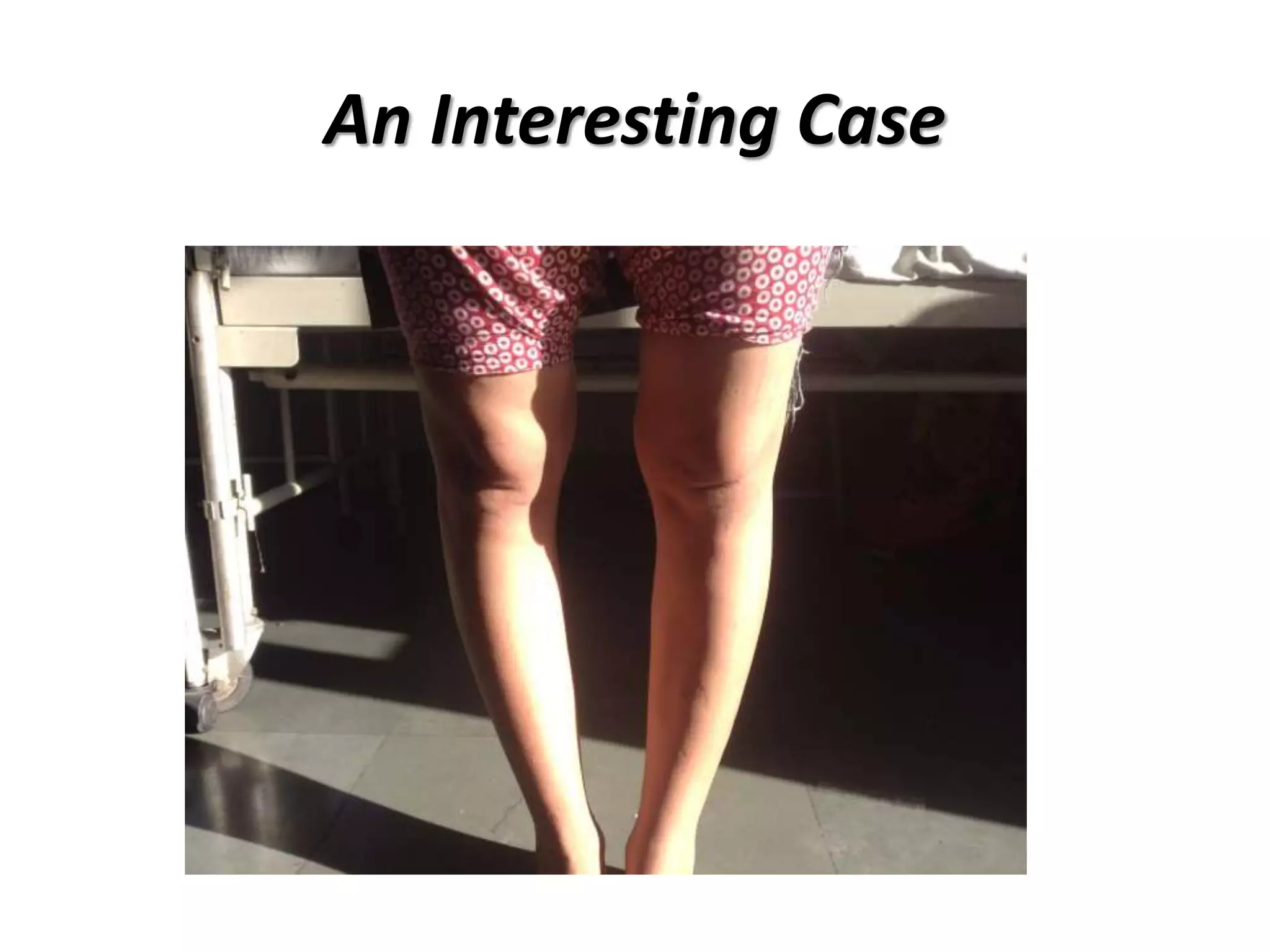
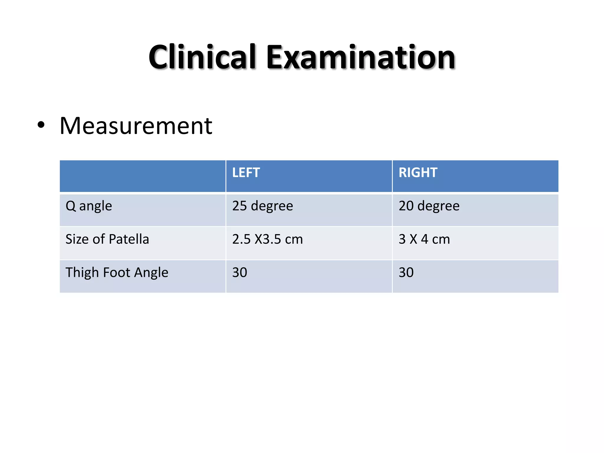
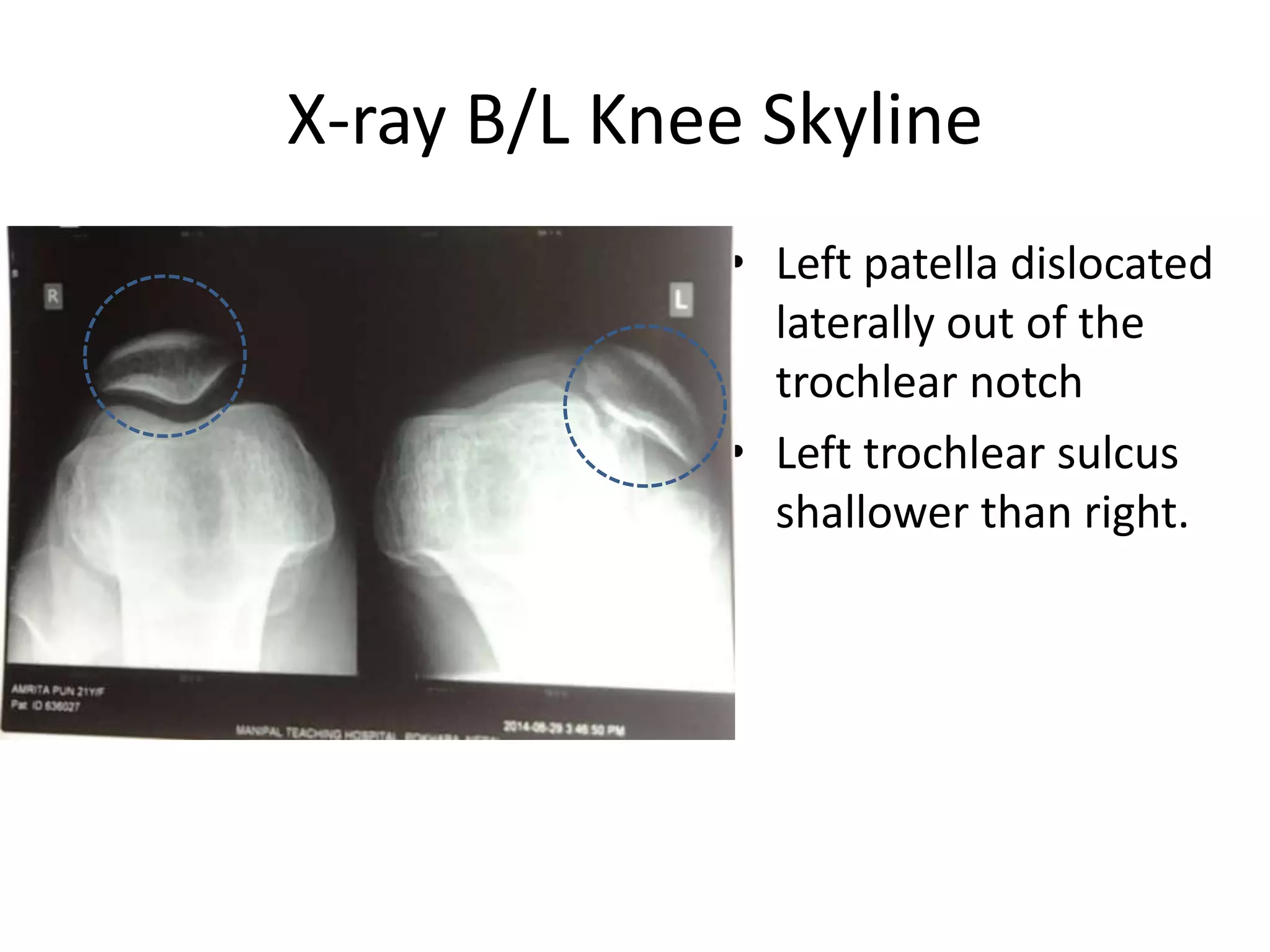
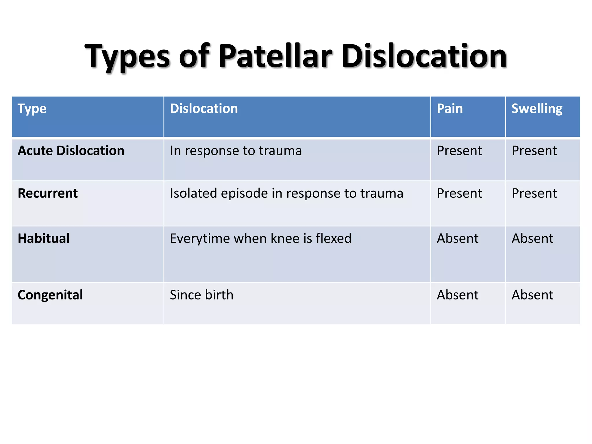
Case Presentation 
Habitual Dislocation of Patella 
Dr Sushil Sharma 
First Year MS Orthopaedic Resident
An Interesting Case
Patient Particulars 
• Name : Amrita Pun 
• Age : 21 
• Sex : F 
• Address : Salyan-3,Yang 
• Date of Admission : 2014 July 28
History 
• Chief Complaints 
– Difficulty in walking 
– Giving way of left knee for past 10 years 
• History of Present Illness 
– Fall injury 10 years back, sustained injury to left 
knee following which she had pain in left knee & 
difficulty in walking. Giving way of left knee.
Clinical Examination 
• Gait : Normal 
• Inspection 
– No swelling, discoloration, 
scar marks 
– Wasting of left quadriceps 
muscle 
– Position of patella: Centrally 
placed (In extension) & 
laterally dislocated in flexion 
– Size of patella : Left appears 
small than right 
– Attitude of leg: B/L varus
Clinical Examination 
• Palpation: 
– No rise in superficial temperature 
– No superficial & joint line tenderness 
– No tenderness over patella 
– Patellar movement restricted medially 
– No patello femoral tenderness and crepitus
Clinical Examination 
• Movement 
– Range of movement 
• Flexion 
– Left Knee : 0-135 
– Right Knee : 0-135 
• Loss of Extension : Not present 
• Internal rotation : 10 degree 
• External rotation : 10 degree
Clinical Examination 
• Measurement 
LEFT RIGHT 
Q angle 25 degree 20 degree 
Size of Patella 2.5 X3.5 cm 3 X 4 cm 
Thigh Foot Angle 30 30
Clinical Examination - Special Tests 
– Apprehension test : 
Negative 
– Patellar grinding test: 
Negative 
– Patellar tracking : Positive J 
Sign (Lateral subluxation of 
patella in flexion) 
– Patellar glide test: 
• 1 quadrant medially 
• 3 quadrant laterally 
– Patellar tilt test : Negative
Investigation 
• Pre-operative investigation: 
– CBC (Within normal limit) 
– Serum Na, Serum K, Serum Urea, Serum 
Creatinine (Within normal limit) 
– Serology : Non reactive 
– RBS : Normal 
– Urine RME : Normal
X-Ray B/L Knee AP 
• Both leg in varus 
position
X-Ray B/L Knee Lateral 
• Blumensaat line : Lower 
pole of patella on line 
with intercondylar 
notch. 
• Insall-Salvati Ratio (N : 1) 
• Right: 1 
• Left: 0.8 (patella baja)
X-ray B/L Knee Skyline 
• Left patella dislocated 
laterally out of the 
trochlear notch 
• Left trochlear sulcus 
shallower than right.
SPECIAL AXIAL VIEWS OF PATELLA 
HUGHSTON MERCHANT LAURIN 
•Patellar Index : 14 
(N: M – 15, F – 17) 
•Sulcus angle : 120 
(N : 118) 
•Patellofemoral Index: 2.6 
(N:1.6) 
•Sulcus angle : 145 
(N:138)
Management 
• Surgical realignment is the treatment of choice. 
• Principle : 
– Medialization of Patella 
– Maintenance of proximal & distal alignment 
• Surgery performed 
– Insall (Suprapatellar realignment) 
– Roux Goldthwait operation (Infrapatellar soft-tissue 
realignment)
Management - Operative 
• Proximal realignment 
• Release of tight lateral 
patellar retinaculum & vastus 
lateralis completely 
• Plication of medial capsule & 
patellar retinaculum to 
strengthen the lax medial 
structures. 
• Vastus medialis obliqus (VMO) 
was advanced & sutured to 
lateral border of patella & 
quadriceps, after locating 
patella in trochlear notch in 
70 degree flexion. 
LATERAL 
MEDIAL
Management - Operative 
Distal realignment 
• Lateral third of patellar 
ligament was released 
from tibial tubersoity 
and passed underneath 
medial portion of 
patellar tendon & 
sutured upwards & 
medially to pes 
anserinus tendon 
LATERAL 
MEDIAL
Post Operative 
• Above knee posterior slab with knee in 5 degree 
of flexion was given for first 5 days 
• A long-leg hinged knee brace was applied later 
with the knee in 20° of flexion 
• Partial weight bearing with crutches for four 
weeks was advised, during which the patient was 
encouraged to do static quadriceps strengthening 
exercises 
• Knee mobilization and full weight bearing was 
started after four weeks.
Post Operative Results 
• Position of patella : Centrally placed 
& no lateral dislocation on flexion. 
• Q angle : 20 
• Range of movement : 0 - 135 
• Extensor Lag : Not Present
Discussion
Anatomy
Introduction 
• Habitual dislocation of the patella is a rare condition 
among adults, where the patella dislocates during 
flexion and relocates during extension without pain 
and swelling unlike the recurrent patellar dislocation. 
• Predisposing factors 
– ligamentous laxity (in women, connective tissue disorder) 
– contracture of the lateral patellar soft tissues 
– patella alta 
– quadriceps contractures 
– hypoplasia of the lateral femoral condyle 
– genu valgum
Patho Anatomy 
• Q angle 
– Male (8-10) 
– Female (15±5) 
• Lateral pull : Vastus 
lateralis, Iliotibial band 
• Medial pull : Vastus 
medialis obliqus (VMO) 
• Increased Q angle : 
Patellar dislocation
Patho Anatomy 
• Genu valgum 
• Increased femoral 
anteversion 
• External tibial torsion 
• Internal femoral torsion 
• Tight lateral retinaculum
Types of Patellar Dislocation 
Type Dislocation Pain Swelling 
Acute Dislocation In response to trauma Present Present 
Recurrent Isolated episode in response to trauma Present Present 
Habitual Everytime when knee is flexed Absent Absent 
Congenital Since birth Absent Absent
Pathology 
First episode of traumatic dislocation 
Tear of capsule on medial side of patella 
If improper healing 
Persistent laxity 
Recurrent dislocation 
Damage to contiguous surface of patella & fem. 
Condyles 
Flattening & then further dislocation
Clinical Features 
• Symptoms 
– Feeling of insecurity in knee (Giving way of knee) 
• Signs 
– Patellofemoral crepitus 
– Postive J sign 
– Increased Q angle
Management 
• Proximal realignment 
– Lateral release 
– Reconstruction of vastus medialis obliquus 
• Distal realignment 
– partial medialization of the ligamentum patella 
– Medialization of tibial tuberosity. 
• always lateral release is combined with medial 
augmentation
Typical Procedure
Acknowledgement 
• Prof Dr S.K. Biswas (HOD, Dept of Orthopaedics) 
• Asst Prof Dr Niraj Ranjeet 
• Dr Krishna Sapkota 
• Dr Pratyunta Raj Onta 
• Dr Alind Kishore 
• Dr Pabin Thapa 
• Dr Upendra Jung Thapa 
• Dr Manoj Prasad Gupta 
• Dr Prakash Dware 
• Department of Anesthesiology 
• Operation Theatre Staffs
References 
• Campbell’s Operative Orthopaedics, 12th 
Edition 
• Apley’s System of Orthopaedics & Fractures, 
9th Edition 
• Post Graduate Companion in Orthopaedics 
• Handbook of Fractures, 4th Edition
Thank You 
Happy Dashain 2071

Habitual dislocation of patella

  • 1.
    Case Presentation HabitualDislocation of Patella Dr Sushil Sharma First Year MS Orthopaedic Resident
  • 2.
  • 3.
    Patient Particulars •Name : Amrita Pun • Age : 21 • Sex : F • Address : Salyan-3,Yang • Date of Admission : 2014 July 28
  • 4.
    History • ChiefComplaints – Difficulty in walking – Giving way of left knee for past 10 years • History of Present Illness – Fall injury 10 years back, sustained injury to left knee following which she had pain in left knee & difficulty in walking. Giving way of left knee.
  • 5.
    Clinical Examination •Gait : Normal • Inspection – No swelling, discoloration, scar marks – Wasting of left quadriceps muscle – Position of patella: Centrally placed (In extension) & laterally dislocated in flexion – Size of patella : Left appears small than right – Attitude of leg: B/L varus
  • 6.
    Clinical Examination •Palpation: – No rise in superficial temperature – No superficial & joint line tenderness – No tenderness over patella – Patellar movement restricted medially – No patello femoral tenderness and crepitus
  • 7.
    Clinical Examination •Movement – Range of movement • Flexion – Left Knee : 0-135 – Right Knee : 0-135 • Loss of Extension : Not present • Internal rotation : 10 degree • External rotation : 10 degree
  • 8.
    Clinical Examination •Measurement LEFT RIGHT Q angle 25 degree 20 degree Size of Patella 2.5 X3.5 cm 3 X 4 cm Thigh Foot Angle 30 30
  • 9.
    Clinical Examination -Special Tests – Apprehension test : Negative – Patellar grinding test: Negative – Patellar tracking : Positive J Sign (Lateral subluxation of patella in flexion) – Patellar glide test: • 1 quadrant medially • 3 quadrant laterally – Patellar tilt test : Negative
  • 10.
    Investigation • Pre-operativeinvestigation: – CBC (Within normal limit) – Serum Na, Serum K, Serum Urea, Serum Creatinine (Within normal limit) – Serology : Non reactive – RBS : Normal – Urine RME : Normal
  • 11.
    X-Ray B/L KneeAP • Both leg in varus position
  • 12.
    X-Ray B/L KneeLateral • Blumensaat line : Lower pole of patella on line with intercondylar notch. • Insall-Salvati Ratio (N : 1) • Right: 1 • Left: 0.8 (patella baja)
  • 13.
    X-ray B/L KneeSkyline • Left patella dislocated laterally out of the trochlear notch • Left trochlear sulcus shallower than right.
  • 14.
    SPECIAL AXIAL VIEWSOF PATELLA HUGHSTON MERCHANT LAURIN •Patellar Index : 14 (N: M – 15, F – 17) •Sulcus angle : 120 (N : 118) •Patellofemoral Index: 2.6 (N:1.6) •Sulcus angle : 145 (N:138)
  • 15.
    Management • Surgicalrealignment is the treatment of choice. • Principle : – Medialization of Patella – Maintenance of proximal & distal alignment • Surgery performed – Insall (Suprapatellar realignment) – Roux Goldthwait operation (Infrapatellar soft-tissue realignment)
  • 16.
    Management - Operative • Proximal realignment • Release of tight lateral patellar retinaculum & vastus lateralis completely • Plication of medial capsule & patellar retinaculum to strengthen the lax medial structures. • Vastus medialis obliqus (VMO) was advanced & sutured to lateral border of patella & quadriceps, after locating patella in trochlear notch in 70 degree flexion. LATERAL MEDIAL
  • 17.
    Management - Operative Distal realignment • Lateral third of patellar ligament was released from tibial tubersoity and passed underneath medial portion of patellar tendon & sutured upwards & medially to pes anserinus tendon LATERAL MEDIAL
  • 18.
    Post Operative •Above knee posterior slab with knee in 5 degree of flexion was given for first 5 days • A long-leg hinged knee brace was applied later with the knee in 20° of flexion • Partial weight bearing with crutches for four weeks was advised, during which the patient was encouraged to do static quadriceps strengthening exercises • Knee mobilization and full weight bearing was started after four weeks.
  • 19.
    Post Operative Results • Position of patella : Centrally placed & no lateral dislocation on flexion. • Q angle : 20 • Range of movement : 0 - 135 • Extensor Lag : Not Present
  • 20.
  • 21.
  • 22.
    Introduction • Habitualdislocation of the patella is a rare condition among adults, where the patella dislocates during flexion and relocates during extension without pain and swelling unlike the recurrent patellar dislocation. • Predisposing factors – ligamentous laxity (in women, connective tissue disorder) – contracture of the lateral patellar soft tissues – patella alta – quadriceps contractures – hypoplasia of the lateral femoral condyle – genu valgum
  • 23.
    Patho Anatomy •Q angle – Male (8-10) – Female (15±5) • Lateral pull : Vastus lateralis, Iliotibial band • Medial pull : Vastus medialis obliqus (VMO) • Increased Q angle : Patellar dislocation
  • 24.
    Patho Anatomy •Genu valgum • Increased femoral anteversion • External tibial torsion • Internal femoral torsion • Tight lateral retinaculum
  • 25.
    Types of PatellarDislocation Type Dislocation Pain Swelling Acute Dislocation In response to trauma Present Present Recurrent Isolated episode in response to trauma Present Present Habitual Everytime when knee is flexed Absent Absent Congenital Since birth Absent Absent
  • 26.
    Pathology First episodeof traumatic dislocation Tear of capsule on medial side of patella If improper healing Persistent laxity Recurrent dislocation Damage to contiguous surface of patella & fem. Condyles Flattening & then further dislocation
  • 27.
    Clinical Features •Symptoms – Feeling of insecurity in knee (Giving way of knee) • Signs – Patellofemoral crepitus – Postive J sign – Increased Q angle
  • 28.
    Management • Proximalrealignment – Lateral release – Reconstruction of vastus medialis obliquus • Distal realignment – partial medialization of the ligamentum patella – Medialization of tibial tuberosity. • always lateral release is combined with medial augmentation
  • 29.
  • 30.
    Acknowledgement • ProfDr S.K. Biswas (HOD, Dept of Orthopaedics) • Asst Prof Dr Niraj Ranjeet • Dr Krishna Sapkota • Dr Pratyunta Raj Onta • Dr Alind Kishore • Dr Pabin Thapa • Dr Upendra Jung Thapa • Dr Manoj Prasad Gupta • Dr Prakash Dware • Department of Anesthesiology • Operation Theatre Staffs
  • 31.
    References • Campbell’sOperative Orthopaedics, 12th Edition • Apley’s System of Orthopaedics & Fractures, 9th Edition • Post Graduate Companion in Orthopaedics • Handbook of Fractures, 4th Edition
  • 32.
    Thank You HappyDashain 2071

Editor's Notes

  • #30 Staheli, Lynn T. Title: Practice of Pediatric Orthopedics, 2nd Edition Copyright ©2006 Lippincott Williams & Wilkins > Table of Contents > Chapter 6 - Knee and Tibia