1. Journal Reading
CLINICAL AND RADIOLOGICAL CHARACTERISTICS OF LUNG DISEASE IN
INFLAMMATORY BOWEL DISEASE
Oleh:
Aulia Dwi Juanita, S.Ked
NPM:
21360331
Preseptor:
dr. Silman Hadori, Sp.Rad., MH.Kes
KEPANITERAAN KLINIK RADIOLOGI
RUMAH SAKIT PERTAMINA BINTANG AMIN
FAKULTAS KEDOKTERAN UNIVERSITAS MALAHAYATI
BANDAR LAMPUNG
2023
2. LEMBAR PENGESAHAN
Journal Reading
CLINICAL AND RADIOLOGICAL CHARACTERISTICS OF LUNG DISEASE
IN INFLAMMATORY BOWEL DISEASE
Penyaji Preseptor
(Aulia Dwi, S.Ked) (dr. Silman Hadori, Sp.Rad., MH.Kes)
KEPANITERAAN KLINIK
RADIOLOGI RUMAH SAKIT
PERTAMINA BINTANG AMIN
FAKULTAS KEDOKTERAN UNIVERSITAS
MALAHAYATIBANDAR LAMPUNG
2023
3. Eur Respir J 2000; 15: 41±48
Printed in UK ± all rights reserved
Copyright #ERS Journals Ltd 2000
European Respiratory Journal
ISSN 0903-1936
Clinical and radiological characteristics of lung disease in
inflammatory bowel disease
R. Mahadeva*, G. Walsh**, C.D.R. Flower**, J.M. Shneerson*
Clinical and radiological characteristics of lung disease in inflammatory bowel disease. R.
Mahadeva, G. Walsh, C.D.R. Flower, J.M. Shneerson. #ERS Journals Ltd 2000.
ABSTRACT: The pulmonary associations of inflammatory bowel disease (IBD) are
poorly characterized. The clinical, physiological and high-resolution computed
tomographic thorax characteristics of the lung disease in patients with IBD presenting
with respiratory symptoms are described.
Detailed clinical information was obtained and standard pulmonary physiological
tests and thorax high-resolution computed tomography performed on 14 patients with
ulcerative colitis (UC) and three with Crohn's disease (CD), 10 male, aged 38±83 yrs.
Respiratory symptoms had been present for 2±50 yrs and extraintestinal mani-
festations were present in three (17.6%). Normal pulmonary physiology (six patients)
was associated with the high resolution computed tomographic changes of bronchi-
ectasis, mosaic perfusion and air trapping suggestive of obliterative bronchiolitis and
a pattern of centrilobular nodules and branching linear opacities ("tree in bud"
appearance) suggestive of either cellular bronchiolitis or bronchiolectasis with mucoid
secretions. Bronchiectasis was found in 13 patients (11 UC, 2 CD), 11 had air trapping
and five had a "tree in bud" appearance on computed tomography. One patient had a
predominantly peripheral reticular pattern at the lung bases similar to that found in
cryptogenic fibrosing alveolitis and one patient had a mixed reticular and ground-
glass pattern in the midzones with a patchy distribution in the central and peripheral
portions of the lungs with air trapping. Eleven patients (three with alveolitis) exhibited
a clinical and/or physiological response to steroids.
Pulmonary abnormalities in ulcerative colitis and Crohn's disease can present years
after the onset of the bowel disease and can affect any part of the lungs. Early
recognition is important as they can be strikingly steroid-responsive.
Eur Respir J 2000; 15: 41±18.
*Dept of Chest Medicine, Papworth Hos-
pital, Cambridge CB3 8RE, **Dept of
Radiology, Addenbrookes Hospital, Cam-
bridge CB2 2QQ, UK.
Correspondence: R. Mahadeva
Respiratory Medicine Unit
Clinic 2A
Box 40
Addenbrookes Hospital
Hills Road
Cambridge CB2 2QQ UK
Fax: 44 1223 216953
Keywords: Bronchiectasis
bronchiolitis
Crohn's disease
high-resolution computed tomography
ulcerative colitis
Received: June 19 1998
Accepted after revision June 7 1999
Ulcerative colitis (UC) and Crohn's disease (CD) are ass-
ociated with a variety of systemic manifestations [1]. UC
has been associated with upper airway stenosis [2], tra-
cheobronchitis [3, 4], bronchiectasis [5±10], constrictive
bronchiolitis [3], panbronchiolitis [11], necrobiotic nod-
ules [8], lung bullae [12], interstitial lung disease [13, 14],
bronchiolitis obliterans organizing pneumonia (BOOP)
[15], sarcoidosis [16], pulmonary vasculitis [17, 18], pul-
monary eosinophilia [8], Wegener's granulomatosis with-
out renal involvement [19] and apical fibrosis [20].
Lung involvement with CD has been less often reported
but has been associated with granulomatous oedema of the
larynx, trachea and bronchi [21], chronic bronchial supp-
uration [5] and chronic bronchitis [8], bronchiectasis [9,
22], BOOP [8], pulmonary oedema [23], interstitial lung
disease [24], alveolar consolidation [25], granulomatous
interstitial involvement [26] and lung infiltrates with
peripheral eosinophilia [8].
The presence of subclinical disease in patients without
respiratory symptoms was suggested in one study because
result of abnormal pulmonary function found in 38% of
UC patients and 54% of CD patients, significantly greater
pro-portions than in a healthy control populations [27].
For editorial comments see page 5
Pul-monary function abnormalities include a decrease in
gas transfer factor [28±30], an elevated functional
residual capacity (FRC) and raised residual volume
(RV) during periods of active bowel disease [30±32] and
an increased frequency of bronchial hyperresponsiveness
[33]. Further-more, alveolar lymphocytosis is evident in
bronchoalveolar lavage fluid from CD patients without
respiratory symptoms [34].
The chest radiograph is often normal in patients with
respiratory symptoms and inflammatory bowel disease
(IBD) [6, 8] and as a result the radiological characteristics
remain poorly characterized. Two studies described the
high resolution computed tomographic findings in seven
patients (in each study) with IBD who presented with
cough and sputum production. However, these reports did
not correlate the computed tomographic appearances with
symptoms or pulmonary physiology or evaluate air trap-
ping with scans performed during expiration [9, 35].
Although respiratory symptoms and physiological and
high resolution computed tomographic abnormalities in
patients with UC and CD have been described separately,
little is known about the associations between these fac-
tors. In this study the relationship between the clinical
features and the physiological and high resolution com-
puted tomographic abnormalities in a group of patients
4. 42 R. MAHADEVA ET AL. 42
LUNG DISEASE AND INFLAMMATORY BOWEL DISEASE
with UC and CD with otherwise unexplained pulmonary
disease is described.
Methods
All of the subjects with CD or UC were identified by
one author (J.M. Shneerson) as they presented at the chest
clinic with respiratory symptoms between 1981 and 1995.
They were all interviewed and their case notes scrutinized
for details of their bowel disease. Characteristics recorded
included the date of diagnosis, the presence of confirma-
tory histological results, the extent of the disease on the
basis of radiological studies and treatment history, e.g.
sulphasalazine, mesalazine or surgery. Features of the pul-
monary disease, including presenting symptoms, date of
diagnosis, smoking history, the presence of other factors
that could account for the clinical features and the response
to steroid treatment (an improvement in symptoms (clinical
response) or forced expiratory volume in one second
(FEV1) and/or forced vital capacity (FVC) of >200 mL
(physiological response)) were also documented. Symp-
toms of cough, sputum, wheeze and breathlessness were
each scored out of a maximum of 2; 0=no symptoms,
1=intermittent symptoms and 2=regular symptoms. The
total symptom score (maximum of 8) for each patient was
derived from the sum of the individual symptom scores.
Each patient underwent standard pulmonary function
tests for FEV1, FVC, total lung capacity (TLC), RV and
transfer coefficient for carbon monoxide (KCO) measured
by means of the single-breath test. The results were com-
pared with those of age-and sex-matched controls [36] and
expressed as a percentage of the predicted value. The
individual results were classified as normal, restrictive or
obstructive. Normal physiology was defined by all meas-
urements being >70% pred, a restrictive defect as a
reduced (<70% pred) FEV1 and FVC with an FEV1/FVC
ratio of >70% pred and an obstructive defect as a reduced
FEV1 with a normal FVC and a low FEV1/FVC ratio
(<70% pred).
Thorax high resolution CT (HRCT) was performed as
close as possible in time to the pulmonary function tests,
1996±1997. Images were acquired during inspiration and
expiration from lung apices to bases (1-mm collimation,
10-mm interval (inspiration) or 30-mm interval (expira-
tion), 5606560 matrix, high spatial frequency reconstruc-
tion algorithm). Images were reviewed at a window level
of -700 and width of 1200. Abnormalities were charac-
terized by a consensus of two radiologists who were un-
aware of the patient symptoms and physiological findings.
Bronchiectasis was defined as bronchi greater in diameter
than the homologous artery together with evidence of a
lack of tapering on sequential slices. Bronchiectasis in each
lobe including the lingula was graded according to the ratio
of the bronchial diameter to that of the adjacent vessel. It
was scored as grade 1 when the ratio was >1 but <2, grade
2 when the ratio was >2 but <3, and grade 3 when the ratio
was >3. Thus, each patient had a maximum possible score
of 18. Each lobe was also assessed for the presence of
centrilobular nodules and branching linear opacities ("tree
in bud" appearance) suggesting cellular bronchiolitis [37],
or dilated small airways filled with secretions (bronch-
iolectasis), and for mosaic perfusion with evidence of air
trapping during expiration suggesting obliterative bronch-
iolitis [38]. The "tree in bud" appearance and air trapping
were each scored out of a maximum of 6 (1 point for each
lobe involved) for each patient.
The relationship between clinical characteristics (sex,
smoking status, colectomy, duration of bowel disease/lung
disease), individual symptom scores, the results of the
individual pulmonary physiological tests listed above (in
% pred), and the severity of bronchiectasis, the extent of
"tree in bud" changes and air trapping on HRCT was
assessed by means of bivariate correlation for all patients.
The relationship between the clinical characteristics, indi-
vidual symptom scores, individual physiological test res-
ults and extent of "tree in bud" changes and air trapping
were also separately analysed by bivariate correlation for
the group of patients with bronchiectasis. A p-value <0.05
was considered statistically significant.
Results
Seventeen patients (10 male) aged 38±83 yrs were
investigated (table 1). Fourteen had UC and three had CD.
Respiratory symptoms had been present for 2±50 yrs and
other extraintestinal manifestations were present in three
subjects (17.6%; one seronegative arthropathy, one mixed
connective tissue disease, one iridocyclitis and ankylos-
ing spondylitis). In 16 patients, the onset of the bowel
disease predated presentation with pulmonary disease
(range 1±25 yrs). Exacerbations of UC and pulmonary
disease occurred concurrently in two patients.
No patients had active bowel disease at the time of the
study and seven patients (six UC) had undergone colec-
tomy for severe disease. Of these, one had a mixed reti-
cular and ground-glass pattern in the midzones with a
patchy distribution in the central and peripheral portions of
the lungs and air trapping, similar to those appearances
seen in extrinsic allergic alveolitis and sarcoidosis. One
patient had a peripheral reticular pattern at the lung bases,
characteristic of cryptogenic fibrosing alveolitis, and five
had bronchiectasis. Four of the seven colectomy patients
developed pulmonary symptoms within 2 yrs of their
surgery (table 1).
Lung histology results were obtained for two patients,
both of whom have previously been reported [13]. One
(No. 4) had cellular interstitial inflammation, little colla-
gen formation and extensive cellular desquamation on
transbronchial biopsy, which responded completely to
corticosteroids.The other (No. 8) had interstitial fibrosis
with fibroblasts and chronic inflammatory cells in the
alveolar walls. Interestingly, having completely respon-
ded to prednisolone therapy, this patient developed new
symptoms some years later and HRCT now shows bron-
chiectasis and air trapping without evidence of interstitial
disease.
Seven individuals were never smokers and seven were
exsmokers who had stopped smoking prior to the onset of
respiratory symptoms and the HRCT scan. It was unlikely
that smoking contributed to the pulmonary disease in the
three current smokers as one had a minimal intake (No. 10)
and the other two (Nos. 6 and 7) had HRCT abnormalities
that are not characteristically associated with smoking.
Five patients were taking sulphasalazine at the time of
diagnosis of the pulmonary disease, one had mild upper
lobe emphysema probably related to smoking and the other
5. 43 R. MAHADEVA ET AL. 43
LUNG DISEASE AND INFLAMMATORY BOWEL DISEASE
Table 1. ± Patient characteristics
Patient
No.
Age
yrs
Sex IBD Age at IBD
diagnosis yrs
Age at PD
diagnosis yrs
Colectomy
(age yrs)
Sulphasalazine Smoking
Hemicolectomy and terminal
1 66 F CD 37 60 ileal resection (49) No Never
2 70 M UC 26 62 Panproctocolectomy (52) No Never
3 58 M UC (distal colitis) 32 56 No No Never
4* 64 M UC 51 50 Panproctocolectomy (51) No Exsmoker
5 71 M UC (total colitis) 65 67 No No Exsmoker
6 76 F UC (distal colitis) 50 62 No 1 g b.i.d. Current
7 68 M UC (distal colitis) 62 65 No 500 mg b.i.d. Current
8* 74 M UC (distal colitis) 59 66 No 1 g b.i.d. Exsmoker
9 81 M UC (proctitis) 67 80 No No Exsmoker
10 51 M UC 15 38 Panproctocolectomy (40) No Current
11 76 M CD 60 73 No No Exsmoker
12 76 F UC 59 62 Panproctocolectomy (64) 500 mg b.i.d. Never
13 55 M UC (distal colitis) 32 45 No 500 mg b.i.d. Exsmoker
14 79 M CD (sigmoid colitis) 40 64 No No Exsmoker
15 83 F UC 31 35 Subtotal colectomy (65) No Never
16 38 F UC (distal colitis) 23 32 No No Never
17 44 F UC 29 30 Panproctocolectomy (30) No Never
*: previously reported [13]. IBD: inflammatory bowel disease; PD: pulmonary disease; F: female; M: male; UC: ulcerative colitis; CD:
Crohn's disease.
four had bronchiectasis, two in association with air trap-
ping. No patients on sulphasalazine showed evidence to
suggest an alveolitis.
Symptoms
Breathlessness was a common presenting symptom in
patients with all patterns of disease. Sputum production
was not present in three of 13 patients with bronchiectasis,
and two of these (Nos. 2 and 5) did not have cough at
presentation.
Eleven patients (three interstitial disease, eight bronch-
iectasis and/or "tree in bud" appearances or air trapping)
including all of the colectomy patients showed clinical and/
or physiological improvement in response to oral or
inhaled corticosteroids (table 2).
Pulmonary function
The six patients with normal pulmonary physiology all
had a combination of bronchiectasis, air trapping and "tree
in bud" appearances of varying severity and extent on
HRCT (table 2).
The KCO was reduced in four patients, one in associ-
ation with fibrosis, one with emphysema which was prob-
ably related to smoking and the other two with the most
extensive and severe bronchiectasis, "tree in bud" appear-
ances and air trapping of all the patients studied. In three
patients, the KCO was elevated, including the patient with
peripheral lung fibrosis who showed no evidence of a
response to bronchodilators to suggest asthma and two
patients with grade 1 bronchiectasis.
High resolution computed tomographic abnormalities
Thirteen (76%) patients had bronchiectasis (11 UC, two
CD) (table 3). Nine patients showed evidence of air
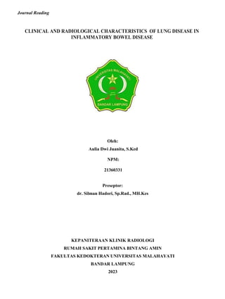
trapping (fig. 1) and five had "tree in bud" changes (fig.
2). Bronchiectasis was present alone in only three patients
and two patients had lower lobes affected by both bron-
chiectasis and air trapping. One patient had bronchiectasis
in all lobes in association with "tree in bud" changes but
no air trapping and three patients had bronchiectasis with
air trapping and "tree in bud" appearances.
Characterization of these abnormalities on a lobar basis
showed that air trapping occurred with bronchiectasis in
four patients and in the absence of bronchiectasis in two,
one of whom showed air trapping in all lobes and grade 1
bronchiectasis in only two lobes (No. 13). In only one lobe
of one patient did "tree in bud" appearances and air trap-
ping both occur in the absence of bronchiectasis.
Two patients (1 and 9) had HRCT changes suggestive of
fibrosis. One showed a mixed reticular and ground-glass
pattern in the midzones with a patchy distribution in the
central and peripheral portions of the lungs and air trap-
ping, and the other a peripheral reticular pattern (figs 3 and
4).
One patient with regular sputum production suggestive of
chronic bronchial suppuration had mild emphysema and
parenchymal bands suggestive of fibrosis in the left lower
lobe, but showed no HRCTevidence of airway disease. The
patient with no high resolution computed tomographic
abnormalities (No. 4) had had histologically proven alve-
olitis 14 yrs previously, which had responded completely to
treatment with steroids.
The relationship between clinical characteristics, physio-
logical parameters and high resolution computed tomo-
graphic features
There was a strong negative correlation between colect-
omy and smoking (r= -0.73, p=0.004) (five of seven were
never smokers). There was no significant correlation
between smoking status and individual symptoms, pulmon-
ary physiological test results or abnormalities on HRCT. A
longer duration of respiratory symptoms was associated
6. 44 R. MAHADEVA ET AL. 44
LUNG DISEASE AND INFLAMMATORY BOWEL DISEASE
Patient Symptom Lung
No. score function
Patient
Table 2. ± Patients' respiratory symptom scores, lung function characteristics and steroid responsiveness
Response to steroids
Clinical Physiological
1 2 Restrictive Improvement in breathlessness NK
2 5 Restrictive Improvement in cough and reduced sputum production with 15 mg
prednisolone daily
Yes
3 2 Normal No No
4 2 Obstructive Improved breathlessness after 2 weeks and resolution of CXR
changes after 60 mg prednisolone daily
Yes
5 2 Obstructive + restrictive NK NK
6 4 Obstructive + restrictive NK NK
7 4 Restrictive NK NK
8 3 Normal Reduced breathless and cough and resolution of CXR changes when
assessed at 1 month after 40 mg prednisolone daily
Yes
9 3 Restrictive NK NK
10 5 Normal
Reduced breathlessness, sputum production and wheeze after 800
mg beclomethasone daily
Yes
11 7 Obstructive Improved wheeze, cough and breathlessness NK
12 5 Obstructive Improvement in wheeze and cough when assessed at 1 month NK
13 3 Normal NK NK
14 3 Obstructive Reduced breathless and cough when assessed at 2 months after
treatment with beclomethasone 800 mg daily
15 7 Obstructive Reduced wheeze, cough and sputum production when assessed at 1
month after 30 mg prednisolone daily
16 5 Normal Reduced wheeze and breathlessness when assessed at 3 months after
beclomethasone 2 mg daily
No
Yes
Yes
17 5 Normal Reduced cough and wheeze after beclomethasone 800 mg daily Yes
CXR: chest radiographic; NK: not known.
with a significantly higher TLC (p=0.013) and a higher RV,
which just missed significance (p=0.052), for patients with
bronchiectasis, but there was no significant correlation be-
tween the severity of bronchiectasis, "tree in bud" appear-
ances or air trapping on HRCT and the duration of
respiratory symptoms. Sputum production was significantly
Table 3. ± High resolution computed tomographic abnor-
malities in each patient*
associated with more extensive and more severe bronch-
iectasis (a higher bronchiectasis score) (r=0.86, p<0.001),
and more extensive "tree in bud" appearances were
significantly associated with cough (r=0.62, p=0.024).
An increased RV was associated with cough and wheeze
(p=0.04 and 0.007 respectively) and with more extensive
"tree in bud" changes but not with the extent of air trapping
(p=0.01 and 0.11 respectively). It was also associated with
a higher bronchiectasis score, which just missed sig-
nificance (p=0.057). The FEV1, FVC, TLC and KCO were
No.
Bronchiectasis
Lobes Score
"Tree
in bud"
Air
trapping
Other not associated with symptomseverity, bronchiectasis, "tree
in bud" changes or air trapping.
Although only five patients had "tree in bud" appear-
ances, more extensive changes were associated with more
1 0 0 0 5 Bronchocentric
ground-glass
change
severe and extensive bronchiectasis (r=0.89, p<0.001). The
extent of air trapping was not significantly linked to the
extent of "tree in bud" changes (r=0.471, p=0.104) nor
2 5 8 5 0
3 2 3 0 2
4 0 0 0 0
5 2 2 0 1
6 5 5 0 0
7 0 0 0 0 Emphysema
8 2 2 0 2
9 0 0 0 0 Peripheral
ground-glass
change
10 5 5 0 2
11 6 9 5 6
12 2 2 0 0
13 2 2 0 6
14 2 2 0 0 Emphysema
15 6 9 6 5
16 4 6 4 5
17 2 3 2 2
*: for scoring system, see Methods.
with the severity of bronchiectasis (p=0.17). The KCO was
not associated with the severity of bronchiectasis or the
extent of air trapping or "tree in bud" changes.
Discussion
Despite the known systemic manifestations of IBD [1]
and a number of reports linking lung disease and IBD,
this association is often overlooked as an extraintestinal
manifestation of either UC or CD. The true prevalence of
the association remains unknown, and although it was
only found in three of 1,400 cases in one study [39],
various factors suggest that this figure is an underestimate
of the true prevalence of the association. Firstly, clinicians
may not consider an association as patients often present
with pulmonary symptoms years after the bowel disease
7. 45
LUNG DISEASE AND INFLAMMATORY BOWEL DISEASE
45 R. MAHADEVA ET AL.
a)
b)
Fig. 1. ± High resolution computed tomography. a) At full inspiration
minor differences in lung attenuation in both lower lobes and bronchial
wall thickening are revealed. b) On expiration, there is evidence of
extensive air trapping in keeping with obliterative bronchiolitis.
and after being discharged from gastrointestinal follow-
up [8]. Asymptomatic patients can have abnormal pulmo-
nary function [27±30] and an alveolar lymphocytosis [34]
and therefore may not present to a respiratory physician.
Patients with bronchiectasis who have a normal chest
radiograph [6, 9] may also be overlooked. Copious spu-
tum production is often reported as a characteristic fea-
ture of the bronchiectasis associated with UC; however,
in the present study it was found that the sputum pro-
ducers were those with the most extensive and severe
bronchiectasis and those with bronchiectasis of lesser
extent and severity did not expectorate sputum. In ad-
dition, patients with normal standard pulmonary function
test results can show HRCT evidence of bronchiectasis,
"tree in bud" changes and evidence of air trapping.
Compared to other series, the present patients had a
lower prevalence of extraintestinal manifestations [8], and
there was a lower proportion of females with bronchial
disease [6, 7]. In agreement with other series [5±9],
colectomy was found to be a risk factor for the develop-
ment of pulmonary disease, with the development of
pulmonary symptoms often occurring close to surgery. It
is important to consider whether therapy with sulphasa-
lazine or mesalazine may have been responsible for the
pulmonary disease. The most common abnormality des-
Fig. 2. ± High resolution computed tomography revealing micronod-
ules in the right lower lobe. Many have a centrilobular distribution and
some are associated with a branching pattern ("tree in bud" changes; A).
There is bronchial wall thickening (B).
cribed in association with sulphasalazine therapy is upper
lobe peripheral opacities, although lower lobe opacities,
eosinophilic pneumonia, interstitial pneumonitis, BOOP
and cavitating nodules have also been reported [40±44].
Four of the present patients taking sulphasalazine showed
evidence of bronchiectasis and air trapping and one had
empysema. None of these patients had eosinophilia,
Fig. 3. ± High resolution computed tomography showing a mixed reti-
cular and ground-glass pattern in the midzones, with a patchy distri-
bution in the central and peripheral portions of the lung; these changes
were seen in association with air trapping on expiratory images.
8. 46
LUNG DISEASE AND INFLAMMATORY BOWEL DISEASE
46 R. MAHADEVA ET AL.
Fig. 4. ± High resolution computed tomography of the lungs bases
revealing a predominantly reticular pattern with the peripheral distri-
bution characteristic of fibrosing alveolitis.
which is usually present in lung disease caused by sulph-
asalazine. These features make it unlikely that the drug
contributed to the pulmonary abnormalities seen in the
patients.
Previous association studies have suggested that smo-
king is a protective factor for UC [45, 46]. The present
study supports this as smoking was significantly nega-
tively correlated with previous colectomy and hence more
severe colitis.
Four patients showed evidence of alveolar disease, three
of whom received significant benefit from steroid treat-
ment, with a complete response in two of these. Inter-
estingly, one of these patients (previously reported as case
2 [11]) subsequently developed new respiratory symp-
toms some years later and was found to have bronchi-
ectasis in association with air trapping. This suggests that
the pulmonary disease can recur in different forms in
individual patients, something that has not previously
been recognized. The other two patients had different
patterns of disease, one with HRCT changes similar to
those in cryptogenic fibrosing alveolitis and the other
with a nonspecific appearance consisting of a mixed reti-
cular and ground-glass pattern.
Although chronic bronchial suppuration [5] and chronic
bronchitis [8] have been reported in association with CD,
until recently there have been no reports of CD in asso-
ciation with bronchiectasis [9, 22]. In the present study,
two patients were found with this association, one of
whom also showed widespread "tree in bud" changes and
air trapping. Interestingly, colectomy had not been per-
formed in either.
Previous studies have shown a correlation between
HRCT and appearances detection of air trapping and
obliterative bronchiolitis [38] and of "tree in bud" ap-
pearances with cellular bronchiolitis [37, 38], although, in
the presence of bronchiectasis, the "tree in bud" ap-
pearance may also be due to dilated small airways filled
with secretions. Published reports have suggested that the
bronchioles are only rarely affected in IBD [8], but in the
present study it was found that nine (53%) patients
showed air trapping and five (29%) a "tree in bud"
appearance. In the two other studies [9, 35] to use HRCT,
expiratory scans were not performed and air trapping
would probably have been underestimated. Whilst air
trapping, in keeping with obliterative bronchiolitis, was
often present in the current patients with bronchiectasis, it
was not associated with the severity or the extent of
bronchiectasis. Interestingly, one patient showed exten-
sive air trapping with only minor bronchiectasis in two
lobes. "Tree in bud" appearances tended to be associated
with extensive and severe bronchiectasis, but were not
linked to the extent of air trapping. Air trapping, "tree in
bud" changes and bronchiectasis can be present within
lobes in individual patients either alone or in combina-
tion, suggesting a common pathogenesis for bronchial
and bronchiolar injury and a continuum of the tissue
response to the same underlying mechanism.
The RV was the only physiological parameter assessed
which was associated with symptoms (cough and sputum
production) and HRCT abnormalities (bronchiectasis and
"tree in bud" appearances). This suggests that it may be the
most useful index of disease severity and may be useful in
monitoring the disease. This also raises the possibility that
the transient hyperinflation noted during attacks of CD
[32] and the elevated FRC and RV associated with active
IBD [30, 31] could reflect bronchial or bronchiolar in-
flammation with subsequent resolution. Indeed, there
were two patients (Nos. 5 and 12) in whom exacerbations
of colitis were paralleled by the development of cough
and sputum production, one of whom had intrapulmonary
shadowing. In both cases, the symptoms and chest radio-
graphic shadowing improved after treatment of the colitis.
No correlation was found between the extent of air trap-
ping and physiological measures of RV or the FEV1,
which contrasts with previous reports of correlations be-
tween the extent of expiratory air trapping and the degree
of airflow obstruction in patients with constrictive bron-
chiolitis and chronic airway disease [47, 48]. This may be
because a semiquantitative scoring system was employed
in these studies or due to the insensitivity of standard
pulmonary function tests for the detection of small air-
ways disease.
Histologically, the reported case of bronchiolar involve-
ment in UC [3] resembled the bronchiolar changes seen
after organ transplantation. The patients' appearances on
HRCT were indistinguishable from the radiological feat-
ures that are often seen in rheumatoid arthritis and graft
versus host disease after bone marrow transplantation and
after lung transplantation, all of which are thought to have
an immunological basis. A large proportion of the pat-
ients had pulmonary disease that was responsive to
steroids either orally, in the case of alveolar disease, or
inhaled or orally or both, in airway disease. These
observations suggest that the pulmonary disease in CD
and UC has an inflammatory basis. One proposed mech-
anism for explaining this association is the common
embryological derivation of the lungs and gastrointestinal
tract from the primitive foregut, and the similarity in the
immune systems in the pulmonary and intestinal mucosa
[6]. This might explain the development of bronchial
disease after colectomy, which implies a shift in the target
for the abnormal inflammatory responses.
When assessing the relationship between two diseases, it
should be considered that the conditions may have occur-
red together by chance. Against this are the temporal asso-
ciation, other reports linking the two conditions and the
9. 47
LUNG DISEASE AND INFLAMMATORY BOWEL DISEASE
47 R. MAHADEVA ET AL.
lack of any alternative causative factors to explain the
pulmonary pathology in these patients. Similarly, with the
small number of patients in the present study, it is possible
that some of the correlations between clinical character-
istics, pulmonary physiological factors and high resolution
computed tomographic characteristics could have occurred
by chance or that correlations may have been missed.
In summary, pulmonary disease in the form of bron-
chiectasis, bronchiolitis and interstitial disease of different
patterns can occur in patients with both ulcerative colitis
and Crohn's disease. A high degree of suspicion is nec-
essary to detect the disease as it may present years after the
bowel disease and the patient with airway disease may lack
the classical symptoms and yield a normal chest radio-
graph and physiological test results. Early detection is im-
portant as both the alveolar and airway disease often
respond well to steroid treatment.
References
17. Isenberg JI, Goldstein H, Korn AR, Ozelaw RS, Rosen V.
Pulmonary vasculitis: an uncommmon complication of
ulcerative colitis. N Engl J Med 1968; 279: 1376±1377.
18. Sargent D, Sessions JT, Fairman RP. Pulmonary vasculitis
complicating ulcerative colitis. South Med J 1985; 78:
624±625.
19. Kedziora JA, Wolff M, Chang J. Limited form of Weg-
eners granulomatosis in ulcerative colitis. Am J Roent-
genol 1975; 125: 127±133.
20. Meadway J. Ulcerative colitis, colitic spondylitis and
associated apical pulmonary fibrosis. Proc Roy Soc Med
1974; 67: 16±17.
21. Lemann M, Messing B, D'Agay F, Modigliani R. Crohn's
disease with respiratory involvement. Gut 1987; 28:
1669±1672.
22. Eaton TE, Lambert N, Wells AU. Bronchiectasis follow-
ing colectomy for Crohn's disease. Thorax 1998; 53: 529±
531.
23. Bradshaw MJ, Harvey RF, Burns-Cox CJ. Crohn's
disease presenting as recurrent pulmonary oedema.
1. Kirsner JB, Shorter RG. Recent developments in non- 24.
BMJ 1981; 283: 1437±1438.
Hotermans G, Benard A, Guenanen H, Demarcq-Delerne
2.
specific inflammatory bowel disease. N Engl J Med 1982;
306: 775±785.
Rickli H, Fretz C, Hoffman M, Walser A, Knoblauch A.
G, Malart T, Wallaert B. Non-granulomatous interstitial
lung disease and Crohn's disease. Eur Respir J 1996; 9:
380±382.
3.
Severe inflammatory upper airway stenosis in ulcerative
colitis. Eur Respir J 1994; 7: 1899±1902.
Wilcox P, Miller R, Miller G, et al. Airway involvement
25. Puntis JWL, Tarlow MJ, Raafat F, Weller PH, Booth IW.
Crohn's disese of the lung. Arch Dis Child 1990; 65:
1270±1271.
4.
in ulcerative colitis. Chest 1987; 92: 18±22.
Vasishta S, Wood JR, McGinty F. Ulcerative tracheo-
26. Calder CJ, Lacy D, Raafat F, Weller PH, Booth IW.
Crohn's disease with pulmonary involvement in a 3 yr old
bronchitis years after colectomy for ulcerative colitis.
Chest 1994; 106: 1279±1281. 27.
boy. Gut 1993; 34: 1636±1638.
Sommer H, Schmidt M, Gruber KD. Lungenfunk-
5. Kraft SC, Earle RH, Roesler M, Esterley JR. Unexplained
bronchopulmonary disease in patients with inflammatory
bowel disease. Arch Intern Med 1976; 136: 454±459. 28.
tionssto
Èrungen bei Colitis ulcerosa und Morbus Crohn.
Dtsch Med Wschr 1986; 111: 812±815.
Heatley RV, Thomas P, Prokipchuk EJ, Gauldie J,
6.
7.
Higgenbottam TW, Cochrane GM, Clark TJH, Turner D,
Millis R, Seymour W. Bronchial disease in ulcerative
colitis. Thorax 1980; 35: 581±585.
Butland RJA, Cole P, Citron KM, Tumer-Warwick M. 29.
Sjenjewicz DJ, Bienenstock J. Pulmonary function ab-
normalities in patients with inflammatory bowel disease.
Q J Med 1982; 203: 241±250.
Munck A, Murciano D, Pariente R, Cezard JP, Navarro J.
8.
Chronic bronchial suppuration and inflammatory bowel
disease. Q J Med 1981; 197: 63±75.
Camus P, Piard F, Ashcroft T, Gal AA, Colby TV. The 30.
Latent pulmonary function abnormalities in children with
Crohn's disease. Eur Respir J 1995; 8: 377±380.
Neilly JB, Main ANH, McSharry C, MurrayJ, Russell RI,
lung in inflammatory bowel disease. Medicine 1993; 72:
151±183.
Moran F. Pulmonary abnormalities in Crohn's disease.
Respiratory Medicine 1989; 83: 487±491.
9. Spira A, Grossman R, Balter M. Large airway disease 31. Douglas JG, McDonald CF, Leslie MJ, Gillon J,
associated with inflammatorybowel disease. Chest 1998; Crompton GK, McHardy GJR. Respiratory impaiment
113: 1723±1726. in inflammatory bowel disease: does it vary with disease
10. Moles KW, Varghese G, Hayes JR. Pulmonary involve- activity? Respiratory Medicine 1989; 83: 389±394.
ment in ulcerative colitis. Br J Dis Chest 1988; 82: 79±83. 32. Pasquis P, Colin R, Denis P, Baptiste P, Galmiche JP,
11. Desai SJ, Gephardt GN, Stoller JK. Diffuse panbron- Hecketswener P. Transient pulmonary impaiment in dur-
chiolitis preceding ulcerative colitis. Chest 1989; 95: ing attacks of Crohn's disease. Respiration 1981; 41: 56±
1342±1344. 59.
12. Shneerson JM. Lung bullae, bronchiectasis, and Hash- 33. Louis E, Louis R, Drion V, et al. Increased frequency of
imoto's disease associated with ulcerative colitis treated
by colectomy. Thorax 1981; 36: 313±314.
bronchial hyperresponsiveness in patients with inflam-
matory bowel disease. Allergy 1995; 50: 729±733.
13. Shneerson JM. Steroid-responsive alveolitis associated 34. Bonniere P, Wallaert B, Cortot A, et al. Latent pulmonary
with ulcerative colitis. Chest 1992; 101: 585±586. involvement in Crohn's disease: biological, functional,
14. McCulloch AJ, McEvoy A, Jackson JD, Jarvis EH. bronchoalveolar lavage and scintigraphic studies. Gut
Severe steroid responsive pneumonitis associated with 1986; 27: 919±925.
pyoderma gangrenosum and ulcerative colitis. Thorax 35. Garg K, Lynch DA, Newell JD. Inflammatory airways
1985; 40: 314±315. disease in ulcerative colitis: CT and high resolution CT
15. Swinburn CR, Jackson GJ, Ashcroft T, Morritt GN, features. J Thorac Imaging 1993; 8: 159±163.
Corris PA. Bronchiolitisobliterans organising pneumonia 36. Cotes J. Lung function. 1975 Blackwell Scientific Publi-
in a patient with ulcerative colitis. Thorax 1988; 43: 735± cations.
736. 37. Garg K, Lynch DA, Newell JD, King TE. Proliferative
16. Rubenstein I, Baum GL. Association of ulcerative colitis and constrictive bronchiolitis: classification and radio-
and sarcoidosis? Letter. Chest 1986; 89: 618. logic features. AJR 1994; 162: 803±808.
10. 48
LUNG DISEASE AND INFLAMMATORY BOWEL DISEASE
48 R. MAHADEVA ET AL.
38. Mu
Èller NL, Miller RR. Diseases of the bronchioles:
CT and histopathologic findings. Radiology 1995; 196:
toxicity in ulcerative colitis mimicking clinical features
of Wegener's granulomatosis. Chest 1996; 110: 556±559.
3±12. 44. Reinoso MA, Schroeder KW, Pisani RJ. Lung disease
39. Rodgers BHG, Clark IM, Kirsner JB. The epidemiologic associated with orally administered mesalazine for ulcer-
and demographic characteristics of inflammatory bowel ative colitis. Chest 1992; 101: 1469±1471.
disease: analysis of a computerised file of 1400 patients. J 45. Harries AD, Baird A, Rhodes J. Nonsmoking: a featurein
Chron Dis 1971; 24: 743±773. ulcerative colitis. BMJ 1982; 284: 706.
40. Williams T, Eidus L, Thomas P. Fibrosing alveolitis, 46. Calkins BM. A meta-analysis of the role of smoking in
bronchiolitis obliterans and sulphasalazinetherapy. Chest inflammatory bowel disease. Dig Dis Sci 1989; 34: 1841±
1982; 81: 766±768. 1854.
41. Jones GR, Malone DNS. Sulphasalazine induced lung 47. Hansell DM. Obliterative bronchiolitis: individual CT
disease. Thorax 1972; 27: 713±717. signs of small airways disease and functional correlation.
42. Davies D, Macfarlane A. Fibrosing alveolitis and Radiology 1997; 203: 721±726.
treatment with sulphasalazine. Gut 1974; 15: 185±188. 48. Lucidarme O. Expiratory CT scans for chronic airway
43. Salerno SM, Ormseth EJ, Roth BJ, Meyer CA, disease: correlation with pulmonary function test results.
Christensen ED, Dillard ED. Sulfasalazine pulmonary AJR 1998; 170: 301±307.
11. 49
LUNG DISEASE AND INFLAMMATORY BOWEL DISEASE
49 R. MAHADEVA ET AL.
Eur Respir J 2000; 15: 41±48 Dicetak di Inggris ±
semua hak dilindungi undang-undang
Hak Cipta #ERS Journals Ltd 2000
Jurnal Pernapasan Eropa
ISSN 0903-1936
Karakteristik klinis dan radiologis penyakit paru-paru di
penyakit radang usus
R. Mahadeva*, G. Walsh**, CDR Flower**, JM Shneerson*
Karakteristik klinis dan radiologis penyakit paru-paru pada penyakit radang usus.
R. Mahadeva, G. Walsh, CDR Flower, JM Shneerson. #ERS Jurnal Ltd 2000.
ABSTRACT: Asosiasi paru penyakit radang usus (IBD) ditandai dengan buruk.
Karakteristik thorax tomografi terkomputasi klinis, fisiologis, dan beresolusi
tinggi dari penyakit paru-paru pada pasien dengan IBD yang menunjukkan
gejala pernapasan dijelaskan.
Informasi klinis terperinci diperoleh dan tes fisiologis paru standar dan tomografi
terkomputasi resolusi tinggi toraks dilakukan pada 14 pasien dengan kolitis ulserativa
(UC) dan tiga dengan penyakit Crohn (CD), 10 laki-laki, berusia 38 ± 83 tahun.
Gejala pernapasan telah ada selama 2 ± 50 tahun dan manifestasi ekstraintestinal
muncul pada tiga (17,6%). Fisiologi paru normal (enam pasien) dikaitkan dengan perubahan
tomografi terkomputasi resolusi tinggi dari bronkiektasis, perfusi mosaik dan jebakan
udara yang menunjukkan bronkiolitis obliteratif dan pola nodul sentrilobular dan kekeruhan
linier bercabang ("penampilan pohon dalam kuncup") yang menunjukkan baik seluler
bronkiolitis atau bronkiolektasis dengan sekresi mukoid. Bronkiektasis ditemukan pada 13
pasien (11 UC, 2 CD), 11 memiliki air trapping dan lima memiliki gambaran "tree in bud"
pada computed tomography. Satu pasien memiliki pola retikuler perifer yang dominan di
dasar paru-paru yang mirip dengan yang ditemukan pada alveolitis fibrosa kriptogenik dan
satu pasien memiliki pola campuran retikuler dan groundglass di zona tengah dengan
distribusi tidak rata di bagian tengah dan perifer paru-paru dengan perangkap udara.
Sebelas pasien (tiga dengan alveolitis) menunjukkan respons klinis dan/atau fisiologis
terhadap steroid.
Kelainan paru pada kolitis ulserativa dan penyakit Crohn dapat muncul bertahun-tahun
setelah timbulnya penyakit usus dan dapat mempengaruhi bagian paru mana pun.
Pengenalan dini penting karena mereka sangat responsif terhadap steroid.
Eur Respir J 2000; 15: 41±18.
* Departemen Kedokteran Dada, Rumah Sakit
Papworth, Cambridge CB3 8RE, **Departemen
Radiologi, Rumah Sakit Addenbrookes,
Cambridge CB2 2QQ, Inggris.
Korespondensi: Klinik Unit
Pengobatan Pernapasan R.
Mahadeva 2A
Kotak 40
Rumah Sakit Addenbrookes
Jalan Bukit
Faks Inggris Cambridge CB2
2QQ: 44 1223 216953
Kata kunci: Bronkiektasis
bronkiolitis
Penyakit Crohn
kolitis ulseratif tomografi terkomputasi
resolusi tinggi
Diterima: 19 Juni 1998 Diterima
setelah revisi 7 Juni 1999
Kolitis ulserativa (UC) dan penyakit Crohn (CD)
berhubungan dengan berbagai manifestasi sistemik [1].
UC telah dikaitkandengan stenosis saluran napas atas
[2], trakeobronkitis [3, 4], bronkiektasis [5±10],
bronkiolitis konstriktif [3], panbronkiolitis [11], nodul
nekrobiotik [8], bula paru [12], interstitial penyakit paru-
paru [13, 14], bronkiolitis obliterans pengorganisasian
pneumonia (BOOP) [15], sarkoidosis [16], vaskulitis paru
[17, 18], eosinofilia paru [8], granulomatosis Wegener
tanpa keterlibatan ginjal [19] dan fibrosis apikal [ 20].
Keterlibatan paru-paru dengan CD lebih jarang
dilaporkan tetapi dikaitkan dengan edema
granulomatosa laring, trakea dan bronkus [21],
supurasi bronkial kronis [5] dan bronkitis kronis [8],
bronkiektasis [9, 22], BOOP [8 ], edema paru [23],
penyakit paru interstisial [24], konsolidasi alveolar
[25], keterlibatan interstisial granulomatosa [26] dan
infiltrat paru dengan eosinofilia perifer [8].
Kehadiran penyakit subklinis pada pasien tanpa gejala
pernapasan disarankan dalam satu penelitian karena
hasil fungsi paru abnormal ditemukan pada 38% pasien
UC dan 54% pasien CD, proporsi yang jauh lebih besar
daripada populasi kontrol yang sehat [27].
Untuk komentar editorial lihat halaman 5
Abnormalitas fungsi paru meliputi penurunan faktor
transfer gas [28±30], peningkatan kapasitas residu
fungsional (FRC) dan peningkatan volume residu (RV)
selama periode penyakit usus aktif [30±32] dan
peningkatan frekuensi bronkial. hiperresponsif [33].
Selanjutnya, limfositosis alveolar terbukti dalam
cairan lavage bronkoalveolar dari pasien CD tanpa
gejala pernapasan [34].
Radiografi dada seringkali normal pada pasien dengan
gejala pernapasan dan penyakitradang usus (IBD) [6, 8] dan
sebagai akibatnya karakteristik radiologi tetap buruk. Dua
penelitian menggambarkan temuan tomografi terkomputasi
resolusi tinggi pada tujuh pasien (dalam setiap penelitian)
dengan IBD yang mengalami batuk dan produksi dahak.
Namun, laporan ini tidak mengkorelasikan penampilan
computed tomographic dengan gejala atau fisiologi paru
atau mengevaluasi air trapping dengan scan yang dilakukan
selama ekspirasi [9, 35].
Meskipun gejala pernapasan dan kelainan tomografi
terkomputasi fisiologis dan resolusi tinggi pada pasien
dengan UC dan CD telah dijelaskan secara terpisah,
sedikit yang diketahui tentang hubungan antara faktor-
faktor ini. Dalam penelitian ini hubungan antara
gambaran klinis dan kelainan tomografi terkomputasi
fisiologis dan resolusi tinggi pada sekelompok pasien
12. 42 R.MAHADEVA ET AL. 42
PENYAKIT PARU DAN PENYAKIT PERADANGAN USUS
dengan UC dan CD dengan penyakit paru yang tidak dapat
dijelaskan dijelaskan.
Metode
Semua subjek dengan CD atau UC diidentifikasi oleh
satu penulis (JM Shneerson) saat mereka hadir di klinik
dada dengan gejala pernapasan antara tahun 1981 dan
1995. Mereka semua diwawancarai dan catatan kasus
mereka diteliti untuk rincian penyakit usus mereka.
Karakteristik yang dicatat termasuk tanggal diagnosis,
adanya hasil histologis konfirmasi, luasnya penyakit
berdasarkan studi radiologis dan riwayat pengobatan,
misalnya sulphasalazine, mesalazine atau pembedahan.
Gambaran penyakit paru, termasuk gejala yang muncul,
tanggal diagnosis, riwayat merokok, adanya faktor lain
yang dapat menjelaskan gambaran klinis dan respons
terhadap pengobatan steroid (perbaikan gejala (respons
klinis) atau volume ekspirasi paksa dalam satu kedua
(FEV1) dan/atau kapasitas vital paksa (FVC) >200 mL
(respon fisiologis)) juga didokumentasikan. Gejala batuk,
dahak, mengi dan sesak napas masing-masing dinilai
dari maksimal 2; 0 = tidak ada gejala, 1 = gejala
intermiten dan 2 = gejala biasa. Skor gejala total
(maksimum 8) untuk setiap pasien berasal dari jumlah
skor gejala individu.
Setiap pasien menjalani tes fungsi paru standar untuk
FEV1, FVC, total kapasitas paru-paru (TLC), RV dan
koefisientransfer untuk karbon monoksida (KCO) yang
diukur dengan tes sekali napas. Hasilnya dibandingkan
dengan kontrol usia dan jenis kelamin yang cocok [36]
dan dinyatakan sebagai persentase dari nilai prediksi.
Hasil individu diklasifikasikan sebagai normal, restriktif
atau obstruktif. Fisiologi normal didefinisikan oleh semua
pengukuran >70% pred, defek restriktifsebagai FEV1
berkurang (<70% pred).1dan FVC dengan FEV1Rasio /FVC
>70% pred dan cacat obstruktif sebagai FEV1 berkurang1
dengan FVC normal dan FEV1 rendah1rasio /FVC (<70%
sebelum).
CT resolusi tinggi Thorax (HRCT) dilakukan
sedekat mungkin dengan tes fungsi paru, 1996 ±
1997. Gambar diperoleh selama inspirasi dan
ekspirasi dari apeks paru ke basis (kolimasi 1 mm,
interval 10 mm (inspirasi) atau interval 30 mm
(ekspirasi), 5606560 matriks, algoritma
rekonstruksi frekuensi spasial tinggi). Gambar
ditinjau pada tingkat jendela -700 dan lebar 1200.
Kelainan ditandai oleh konsensus dua ahli radiologi
yang tidak mengetahui gejala pasien dan temuan
fisiologis. Bronkiektasis didefinisikan sebagai
diameter bronkus yang lebih besar daripada arteri
homolog bersamaan dengan bukti kurangnya
lonjong pada irisan berurutan. Bronkiektasis di
setiap lobus termasuk lingula dinilai berdasarkan
rasio diameter bronkial dengan pembuluh yang
berdekatan. Itu dinilai sebagai kelas 1 ketika
rasionya >1 tetapi <2, kelas 2 ketika rasionya>2
tetapi <3, dan kelas 3 ketika rasionya>3. Dengan
demikian, setiap pasien memiliki skor maksimum
yang mungkin sebesar 18.
iolitis [38]. Penampilan "pohon dalam tunas" dan perangkap udara
masing-masing diberi skor maksimal 6 (1 poin untuk setiap lobus
yang terlibat) untuk setiap pasien.
Hubungan antara karakteristik klinis (jenis kelamin,
status merokok, kolektomi, durasi penyakit usus/
penyakit paru), skor gejala individu, hasil tes fisiologis
paru individu yang tercantum di atas (dalam% pred),
dan tingkat keparahan bronkiektasis, tingkat
perubahan "tree in bud" dan air trapping pada HRCT
dinilai melalui korelasi bivariat untuk semua pasien.
Hubungan antara karakteristik klinis, skor gejala
individu, hasil tes fisiologis individu dan tingkat
perubahan "tree in bud" dan perangkap udara juga
dianalisis secara terpisah dengan korelasi bivariat
untuk kelompok pasien dengan bronkiektasis. Nilai p
<0,05 dianggap signifikan secara statistik.
Hasil
Tujuh belas pasien (10 laki-laki) berusia 38 ± 83 tahun
diselidiki (tabel 1). Empat belas memiliki UC dan tiga
memiliki CD. Gejala pernapasan telah ada selama 2 ± 50
tahun dan manifestasi ekstraintestinal lainnya muncul
pada tiga subjek (17,6%; satu artropati seronegatif, satu
penyakit jaringan ikat campuran, satu iridosiklitis dan
ankylosing spondylitis). Pada 16 pasien, timbulnya
penyakit usus mendahului presentasi dengan penyakit
paru (kisaran 1 ± 25 tahun). Eksaserbasi UC dan penyakit
paru terjadi bersamaan pada dua pasien.
Tidak ada pasien yang memiliki penyakit usus aktif pada saat
penelitian dan tujuh pasien (enam UC) telah menjalani kolektomi
untuk penyakit parah. Dari jumlah tersebut, salah satunya
memiliki pola campuran reticular dan ground-glass di zona
tengah dengan distribusi yang tidak merata di bagian tengah
dan perifer paru-paru dan perangkap udara, mirip dengan yang
terlihat pada alveolitis alergi ekstrinsik dan sarkoidosis. Satu
pasien memiliki pola retikuler perifer di dasar paru, karakteristik
alveolitis fibrosa kriptogenik, dan lima pasien menderita
bronkiektasis. Empat dari tujuh pasien kolektomi mengalami
gejala paru dalam waktu 2 tahun setelah operasi (tabel 1).
Hasil histologi paru diperoleh untuk dua pasien, keduanya
telah dilaporkan sebelumnya [13]. Satu (No. 4) mengalami
peradangan interstitial seluler, sedikit pembentukan kolagen
dan deskuamasi seluler yang luas pada biopsi transbronkial,
yang merespons sepenuhnya terhadap kortikosteroid. Yang
lainnya (No. 8) memiliki fibrosis interstisial dengan fibroblas
dan sel inflamasi kronis di dinding alveolar. Menariknya,
setelah sepenuhnya menanggapi terapi prednisolon, pasien
ini mengembangkan gejala baru beberapa tahun kemudian
dan HRCT sekarang menunjukkan bronkiektasis dan
perangkap udara tanpa bukti penyakit interstitial.
Tujuh orang tidak pernah merokok dan tujuh orang
mantan perokok yang telah berhenti merokok sebelum
timbulnya gejala pernapasan dan pemindaian HRCT. Tidak
mungkin merokok berkontribusi pada penyakit paru pada
tiga perokok saat ini karena satu memiliki asupan minimal
(No. 10) dan dua lainnya (No. 6 dan 7) memiliki kelainan
HRCT yang tidak secara khas terkait dengan merokok.
Lima pasien memakai sulphasalazine pada saat diagnosis
penyakitparu, satu menderita emfisema lobus atas ringan
mungkin terkait dengan merokok dan yang lainnya
13. 43 R.MAHADEVA ET AL. 43
PENYAKIT PARU DAN PENYAKIT PERADANGAN USUS
Tabel 1. ± Karakteristik Pasien
No.
Pasie
n.
Jenis
Kelamin dan
Usia (thn)
I
B
D
Usia di
Diagnosa
IBD
Usia di
Diagnosa PD Kole
ktom
i
(usia
tahu
n)
Meroko
k
Hemikolektomi dan
terminal
1 66 F C
D
3
7
6
0
reseksi ileum
(49)
Tidak pernah
2 70 M U
C
2
6
6
2
Panproktokolektomi
(52)
Tidak pernah
3 58 M UC (kolitis
distal)
3
2
5
6
T
i
d
a
k
k
Tidak pernah
4* 64 M U
C
5
1
5
0
Panproktokolektomi
(51)
Bekas
perokok
5 71 M UC (kolitis
total)
6
5
6
7
Tidak Bekas
perokok
6 76 F UC (kolitis
distal)
5
0
6
2
Tidak Saat
ini
7 68 M UC (kolitis
distal)
6
2
6
5
Tidak Saat
ini
8* 74 M UC (kolitis
distal)
5
9
6
6
Tidak Bekas
perokok
9 81 M UC
(proktitis)
6
7
8
0
Tidak Bekas
perokok
10 51 M U
C
1
5
3
8
Panproktokolektomi
(40)
Saat
ini
11 76 M C
D
6
0
7
3
T
i
d
a
k
Bekas
perokok
*: dilaporkan sebelumnya [13]. IBD: penyakit radang usus; PD: penyakit paru; F: perempuan; G: laki-laki; UC: kolitis ulserativa; CD:
Penyakit Crohn.
empat menderita bronkiektasis, dua berhubungan dengan perangkap udara.
Tidak ada pasien yang menggunakan sulphasalazine yang menunjukkan bukti
yang menunjukkan alveolitis.
Gejala
Sesak napas adalah gejala umum yang muncul pada
pasien dengan semua pola penyakit. Produksi sputum
tidak ada pada tiga dari 13 pasien dengan bronkiektasis,
dan dua di antaranya (No. 2 dan 5) tidak mengalami
batuk.
Sebelas pasien (tiga penyakit interstisial, delapan
bronkiektasis dan/atau penampakan "tree in bud" atau air
trapping) termasuk semua pasien kolektomi menunjukkan
perbaikan klinis dan/atau fisiologis sebagai respons terhadap
kortikosteroid oral atau inhalasi (tabel 2).
Fungsi paru
Keenam pasien dengan fisiologi paru normal semuanya memiliki
kombinasi bronkiektasis, perangkap udara dan penampilan "tree in
bud" dengan berbagai tingkat keparahan dan perluasan pada HRCT
(tabel 2).
KCO berkurang pada empat pasien, satu terkait dengan
fibrosis, satu dengan emfisema yang mungkin terkait
dengan merokok dan dua lainnya dengan bronkiektasis
yang paling luas dan parah, penampilan "pohon dalam
tunas" dan perangkap udara dari semua pasien yang diteliti.
Pada tiga pasien, KCO meningkat, termasuk pasien dengan
fibrosis paru perifer yang tidak menunjukkan bukti respons
terhadap bronkodilator yang menunjukkan asma dan dua
pasien dengan bronkiektasis derajat 1.
Kelainan tomografi terkomputasi resolusi tinggi
Tiga belas (76%) pasien mengalami bronkiektasis (11 UC, dua CD)
(tabel 3). Sembilan pasien menunjukkan bukti adanya udara
perangkap (gbr. 1) dan lima memiliki perubahan "pohon dalam
tunas" (gbr. 2). Bronkiektasis muncul sendirian hanya pada tiga
pasien dan dua pasien memiliki lobus bawah yang terkena
bronkiektasis dan perangkap udara. Satu pasien mengalami
bronkiektasis di semua lobus yang berhubungan dengan perubahan
"tree in bud" tetapi tidak ada air trapping dan tiga pasien mengalami
bronkiektasis dengan tampilan air trapping dan "tree in bud".
Karakterisasi kelainan ini berdasarkan lobar menunjukkan
bahwa air trapping terjadi dengan bronkiektasis pada empat
pasien dan tidak adanya bronkiektasis pada dua pasien, salah
satunya menunjukkan air trapping di semua lobus dan grade 1
bronkiektasis hanya pada dua lobus (No. 13). Hanya pada satu
lobus dari satu pasien tampak "pohon dalam tunas" dan udara
terperangkap keduanya terjadi tanpa adanya bronkiektasis.
Dua pasien (1 dan 9) mengalami perubahan HRCT yang
menunjukkan fibrosis. Satu menunjukkan campuran retikuler dan
pola ground-glass di zona tengah dengan distribusi yang tidak
merata di bagian tengah dan perifer paru-paru dan perangkap
udara, dan yang lainnya menunjukkan pola retikuler perifer (gambar
3 dan 4).
Satu pasien dengan produksi sputum teratur yang
menunjukkan supurasi bronkial kronis memiliki emfisema
ringan dan pita parenkim yang menunjukkan fibrosis di lobus
kiri bawah, tetapi tidak menunjukkan bukti HRCT penyakit
saluran napas. Pasien tanpa kelainantomografi terkomputasi
resolusi tinggi (No. 4) telah terbukti secara histologis alveolitis 14
tahun sebelumnya, yang telah merespon sepenuhnya terhadap
pengobatan dengan steroid.
Hubungan antara karakteristik klinis, parameter
fisiologis, dan fitur tomografi terkomputasi
resolusi tinggi
Terdapat korelasi negatif yang kuat antara kolektomi
dan merokok (r= -0,73, p=0,004) (lima dari tujuh tidak
pernah merokok). Tidak ada korelasi yang signifikan
antara status merokok dan gejala individu, hasil tes
fisiologis paru atau kelainan pada HRCT. Durasi yang
lebih lama dari gejala pernapasan dikaitkan
14. 44 R.MAHADEVA ET AL. 44
PENYAKIT PARU DAN PENYAKIT PERADANGAN USUS
Tabel 2. ± Skor gejala pernapasan pasien, karakteristik fungsi paru, dan respons steroid
No.
Pasien
Gejala
skor
fungsi
paru
Respon terhadap steroid
Klinis Fisiologis
1 2 Bersifat membatasi Peningkatan sesak napas NK
2 5 Bersifat membatasi Batuk membaik dan produksi dahak berkurang dengan prednisolon Ya
15 mg setiap hari
3 2 Normal TIDAK TIDAK
4 2 Obstruktif Peningkatan sesak napas setelah 2 minggu dan resolusi perubahan Ya
CXR setelah 60 mg prednisolon setiap hari
5 2 obstruktif + restriktif NK NK
6 4 obstruktif + restriktif NK NK
7 4 Bersifat membatasi NK NK
8 3 Normal Mengurangi sesak napas dan batuk dan resolusi perubahan CXR ketika Ya
dinilai pada 1 bulan setelah 40 mg prednisolon setiap hari
9
10
3
5
Bersifat
membatasi
Normal
NK
Mengurangi sesak napas, produksi sputum dan mengi setelah 800 Mg
beklometason setiap hari
NK
Ya
11 7 Obstruktif Peningkatan mengi, batuk dan sesak napas Perbaikan NK
12 5 Obstruktif mengi dan batuk bila dinilai pada 1 bulan NK NK
13 3 Normal NK
14 3 Obstruktif Sesak napas dan batuk berkurang saat dinilai 2 bulan setelahnya TIDAK
pengobatan dengan beklometason 800Mg setiap hari
15 7 Obstruktif Mengurangi mengi, batuk dan produksi sputum bila dinilai pada 1 bulan Ya
setelah 30 mg prednisolon setiap hari
16 5 Normal Mengurangi mengi dan sesak napas saat dinilai pada 3 bulan setelah Ya
beklometason 2 mg setiap hari
17 5 Normal Mengurangi batuk dan mengi setelah beklometason 800Mg setiap hari Ya
CXR: radiografi dada; NK: tidak diketahui.
dengan TLC yang secara signifikan lebih tinggi (p = 0,013) dan RV
yang lebih tinggi, yang hanya melewatkan signifikansi (p =
0,052), untuk pasien dengan bronkiektasis, tetapi tidak ada
korelasi yang signifikan antara tingkat keparahan bronkiektasis,
penampilan "tree in bud" atau udara menjebak HRCT dan durasi
gejala pernapasan. Produksi sputum secara signifikan
Tabel 3. ± Kelainan tomografi terkomputasi resolusi
tinggipadasetiappasien*
terkait dengan bronkiektasis yang lebih luas dan lebih
parah (skor bronkiektasis lebih tinggi) (r=0,86, p<0,001),
dan penampakan "tree in bud" yang lebih luas secara
signifikan terkait dengan batuk (r=0,62, p=0,024).
Peningkatan R V dikaitkan dengan batuk dan mengi (masing-
masing p = 0,04 dan 0,007) dan dengan perubahan "tree in bud"
yang lebih luas tetapi tidak dengan tingkat perangkap udara
(masing-masing p = 0,01 dan 0,11). Hal ini juga terkait dengan
skor bronkiektasis yang lebih tinggi, yang tidak signifikan
(p=0,057). FEV1, FVC, TLC danKBERSAMAtidak terkait dengan
No
pasien
Bronkiektasis "Pohon
dalam tunas"
Udara
penangkapan
Lainnya keparahan gejala, bronkiektasis, perubahan "pohon dalam
tunas" atau perangkap udara.
Lobus
1 0
Skor
0 0 5 Bronkosentris
tanah-kaca
mengubah
Meskipun hanya lima pasien yang memiliki gambaran "tree in
bud", perubahan yang lebih luas dikaitkan dengan bronkiektasis
yang lebih parah dan luas (r=0,89, p<0,001). Luasnya perangkap
udara tidak berhubungan secara signifikan dengan luasnya
2 5 8 5
3 2 3 0
4 0 0 0
5 2 2 0
6 5 5 0
7 0 0 0
8 2 2 0
9 0 0 0
10 5 5 0
11 6 9 5
12 2 2 0
13 2 2 0
14 2 2 0
15 6 9 6
16 4 6 4
17 2 3 2
0
2
0
1
0
0 Empisema
2
0 Periferal
tanah-kaca
mengubah
2
6
0
6
0 Empisema
5
5
2
perubahan "tree in bud" (r=0,471, p=0,104) atau dengan tingkat
keparahan bronkiektasis (p=0,17). ItuKBERSAMAtidak terkait
dengan tingkat keparahan bronkiektasis atau luasnya
perangkap udara atau perubahan "tree in bud".
Diskusi
Terlepas dari manifestasi sistemik IBD yang diketahui [1] dan
sejumlah laporan yang menghubungkan penyakit paru-paru dan
IBD, hubungan ini sering diabaikan sebagai manifestasi
ekstraintestinal baik UC atau CD. Prevalensi sebenarnya dari asosiasi
tersebut masih belum diketahui, dan meskipun hanya ditemukan
pada tiga dari 1.400 kasus dalam satu penelitian [39], berbagai faktor
menunjukkan bahwa angka ini terlalu rendah dari prevalensi
sebenarnya dari asosiasi tersebut. Pertama, dokter mungkin tidak
mempertimbangkan hubungan karena pasien sering datang dengan
* : untuk sistem penilaian, lihatMetode. gejala paru bertahun-tahun setelah penyakit usus
15. 45
PENYAKIT PARU DAN PENYAKIT PERADANGAN USUS
45 R.MAHADEVA ET AL.
A)
B)
Gambar 1. ± Computed tomography beresolusi tinggi. a) Pada inspirasi penuh
perbedaan kecil pada atenuasi paru di kedua lobus bawah dan penebalan
dinding bronkus terlihat. b) Saat ekspirasi, terdapat bukti adanya udara yang
terperangkap secara ekstensif sesuai dengan bronkiolitis obliteratif.
dan setelah keluar dari tindak lanjut gastrointestinal [8].
Pasien tanpa gejala dapat memiliki fungsi paru abnormal
[27±30] dan limfositosis alveolar [34] dan oleh karena itu
mungkin tidak datang ke dokter pernapasan. Pasien dengan
bronkiektasis yang memiliki rontgen dada normal [6, 9] juga
dapat diabaikan. Produksi sputum yang banyak sering
dilaporkan sebagai ciri khas bronkiektasis yang
berhubungan dengan UC; namun, dalam penelitian ini
ditemukan bahwa penghasil sputum adalah mereka dengan
bronkiektasis yang paling luas dan parah dan mereka
dengan bronkiektasis dengan tingkat dan keparahan yang
lebih rendah tidak mengeluarkan sputum. Selain itu, pasien
dengan hasil uji fungsi paru standar normal dapat
menunjukkan bukti HRCT bronkiektasis, perubahan "tree in
bud" dan bukti adanya udara yang terperangkap.
Dibandingkan dengan seri lainnya, pasien saat ini
memiliki prevalensi manifestasi ekstraintestinal yang lebih
rendah [8], dan terdapat proporsi wanita yang lebih rendah
dengan penyakit bronkial [6, 7]. Sesuaidengan seri lainnya
[5±9], kolektomi ditemukan sebagai faktor risiko
perkembangan penyakit paru, dengan perkembangan gejala
paru sering terjadi menjelang pembedahan. Penting untuk
mempertimbangkan apakah terapi dengan sulphasalazine
atau mesalazine mungkin bertanggung jawab atas penyakit
paru. Gambaran kelainan yang paling umum
Gambar 2. ± Computed tomography beresolusi tinggi menunjukkan mikronodul
di lobus kanan bawah. Banyak yang memiliki distribusi sentrilobular dan
beberapa terkait dengan pola percabangan ("pohon dalam tunas" berubah; A).
Terdapat penebalan dinding bronkus (B).
berhubungan dengan terapi sulphasalazine adalah opasitas
perifer lobus atas, meskipun opasitas lobus bawah,
pneumonia eosinofilik, pneumonitis interstisial, BOOP dan
nodul kavitasi juga telah dilaporkan [40±44]. Empat dari
pasien yang memakai sulphasalazine menunjukkan bukti
bronkiektasisdan air trapping dan satu mengalami
empisema. Tak satu pun dari pasien ini memiliki eosinofilia,
Gambar 3. ± Computed tomography beresolusi tinggi menunjukkan
campuran retikuler dan pola ground-glass di zona tengah, dengan
distribusi tidak merata di bagian tengah dan perifer paru; perubahan ini
terlihat terkait dengan udara yang terperangkap pada gambar ekspirasi.
16. 46
PENYAKIT PARU DAN PENYAKIT PERADANGAN USUS
46 R.MAHADEVA ET AL.
Gambar 4. ± Computed tomography beresolusi tinggi dari dasar paru
menunjukkan pola retikuler yang dominan dengan karakteristik
distribusi perifer alveolitis fibrosa.
yang biasanya hadir pada penyakit paru-paru yang disebabkan
oleh sulphasalazine. Fitur-fitur ini membuat obat tersebut tidak
mungkin berkontribusi pada kelainan paru yang terlihat pada
pasien.
Studi asosiasi sebelumnya telah menyarankan bahwa merokok
merupakan faktor pelindung untuk UC [45, 46]. Penelitian ini
mendukung hal ini karena merokok berkorelasi negatif secara
signifikan dengan kolektomi sebelumnya dan karenanya kolitis yang
lebih parah.
Empat pasien menunjukkan bukti penyakit alveolar, tiga di
antaranya menerima manfaat yang signifikan dari pengobatan
steroid, dengan respons lengkap pada dua di antaranya.
Menariknya, salah satu pasien ini (sebelumnya dilaporkan
sebagai kasus 2 [11]) kemudian mengembangkan gejala
pernapasan baru beberapa tahun kemudian dan ditemukan
memiliki bronkiektasis yang berhubungan dengan air trapping.
Ini menunjukkan bahwa penyakit paru dapat kambuh dalam
berbagai bentuk pada masing-masing pasien, sesuatu yang
sebelumnya tidak diketahui. Dua pasien lainnya memiliki pola
penyakit yang berbeda, satu dengan perubahan HRCT yang
serupa dengan yang ada pada alveolitis fibrosa kriptogenik dan
yang lainnya dengan penampilan nonspesifik yang terdiri dari
pola campuran reticular dan ground-glass.
Meskipun supurasi bronkial kronis [5] dan bronkitis kronis
[8] telah dilaporkan berhubungan dengan CD, sampai saat
ini belum ada laporan tentang CD yang berhubungan
dengan bronkiektasis [9, 22]. Dalam penelitian ini, dua
pasien ditemukan dengan hubungan ini, salah satunya juga
menunjukkan perubahan "tree in bud" yang meluas dan
perangkap udara. Menariknya, kolektomi juga belum
dilakukan.
Studi sebelumnya telah menunjukkan korelasi antara
HRCT dan pendeteksian penampilan dari perangkap udara
dan bronkiolitis obliteratif [38] dan penampilan "tree in bud"
dengan bronkiolitis seluler [37, 38], walaupun, pada adanya
bronkiektasis, "tree in bud" "Penampilan juga mungkin
karena saluran udara kecil yang melebar penuh dengan
sekresi. Laporan yang diterbitkan menunjukkan bahwa
bronkiolus jarang terkena IBD [8], tetapi dalam penelitian ini
ditemukan bahwa sembilan (53%) pasien menunjukkan
perangkap udara dan lima (29%) penampilan "pohon dalam
tunas". Dalam dua studi lain [9, 35] menggunakan HRCT,
pemindaian ekspirasi tidak dilakukan dan perangkap udara
mungkin diremehkan. Sementara perangkap udara, sesuai
dengan bronkiolitis obliteratif, sering hadir pada pasien
dengan bronkiektasis saat ini, itu tidak terkait dengan
tingkat keparahan atau luasnya bronkiektasis. Menariknya,
satu pasien menunjukkan perangkap udara yang luas
dengan hanya bronkiektasiskecil di dua lobus. Penampilan
"pohon dalam kuncup" cenderung dikaitkan dengan
bronkiektasisyang luas dan parah, tetapi tidak terkait
dengan tingkat perangkap udara. Air trapping, perubahan
"tree in bud" dan bronkiektasisdapat hadir dalam lobus
pada masing-masing pasien baik sendiri atau dalam
kombinasi, menunjukkan patogenesis umum untuk cedera
bronkial dan bronkiolar dan rangkaian respon jaringan
terhadap mekanisme dasar yang sama.
RV adalah satu-satunya parameter fisiologis yang
dinilai yang dikaitkandengan gejala (batuk dan produksi
sputum) dan kelainan HRCT (penampilan bronkiektasis
dan "tree in bud"). Ini menunjukkan bahwa ini mungkin
merupakan indeks keparahan penyakit yang paling
berguna dan mungkin berguna dalam memantau
penyakit. Hal ini juga meningkatkan kemungkinan bahwa
hiperinflasi sementara dicatat selama serangan CD [32]
dan peningkatan FRC dan RV terkait dengan IBD aktif
[30, 31] dapat mencerminkan peradangan bronkial atau
bronkiolar dengan resolusi selanjutnya. Memang, ada
dua pasien (No. 5 dan 12) di mana eksaserbasi kolitis
disejajarkan dengan perkembangan produksi batuk dan
sputum, salah satunya memiliki bayangan
intrapulmoner. Dalam kedua kasus, gejala dan bayangan
radiografi dada membaik setelah pengobatan kolitis.1,
yang kontras dengan laporan sebelumnya tentang
korelasi antara luasnya perangkap udara ekspirasi dan
derajat obstruksi aliran udara pada pasien dengan
bronkiolitis konstriktif dan penyakit saluran napas kronis
[47, 48]. Ini mungkin karena sistem penilaian
semikuantitatif digunakan dalam studi ini atau karena
ketidakpekaan tes fungsi paru standar untuk mendeteksi
penyakit saluran napas kecil.
Secara histologis, kasus keterlibatan bronkiolar yang
dilaporkan di UC [3] menyerupai perubahan bronkiolar yang
terlihat setelah transplantasi organ. Penampilan pasien pada
HRCT tidak dapat dibedakan dari gambaran radiologis yang
sering terlihat pada rheumatoid arthritis dan cangkok
melawanpenyakit inang setelah transplantasi sumsum
tulang dan setelah transplantasi paru-paru, yang semuanya
dianggap memiliki dasar imunologis. Sebagian besar pasien
memiliki penyakit paru yang responsif terhadap steroid baik
secara oral, dalam kasus penyakit alveolar, atau inhalasi
atau oral atau keduanya, pada penyakit saluran napas.
Pengamatan ini menunjukkan bahwa penyakitparu pada CD
dan UC memiliki dasar inflamasi. Salah satu mekanisme
yang diusulkan untuk menjelaskan hubungan ini adalah
derivasi embriologi umum dari paru-paru dan saluran
pencernaan dari usus depan primitif, dan kesamaan dalam
sistem kekebalan pada mukosa paru dan usus [6]. Ini
mungkin menjelaskan perkembangan penyakit bronkial
setelah kolektomi, yang menyiratkan pergeseran target
respons inflamasiabnormal.
Saat menilai hubungan antara dua penyakit, harus
dipertimbangkan bahwa kondisi tersebut mungkin terjadi
bersamaan secara kebetulan. Terhadap ini adalah asosiasi
sementara, laporan lain yang menghubungkan dua kondisi dan
17. 47
PENYAKIT PARU DAN PENYAKIT PERADANGAN USUS
47 R.MAHADEVA ET AL.
kurangnya faktor penyebab alternatif untuk menjelaskan
patologi paru pada pasien ini. Demikian pula, dengan
sejumlah kecil pasien dalam penelitian ini, ada kemungkinan
bahwa beberapa korelasi antara karakteristik klinis, faktor
fisiologis paru dan karakteristik tomografi terkomputasi
resolusi tinggi dapat terjadi secara kebetulan atau bahwa
korelasi mungkin terlewatkan.
Singkatnya, penyakit paru berupa bronkiektasis, bronkiolitis,
dan penyakit interstisial dengan pola berbeda dapat terjadi pada
pasien dengan kolitis ulserativa dan penyakit Crohn. Kecurigaan
tingkat tinggi diperlukan untuk mendeteksi penyakit karena
penyakit ini dapat muncul bertahun-tahun setelah penyakit usus
dan pasien dengan penyakit saluran napas mungkin tidak
memiliki gejala klasik dan menghasilkan radiografi dada normal
dan hasil tes fisiologis. Deteksi dini penting karena penyakit
alveolar dan saluran napas sering merespon dengan baik
terhadap pengobatan steroid.
Referensi
1. Kirsner JB, RG Lebih Pendek. Perkembangan terkini pada
penyakit radang usus nonspesifik.N Engl J Med1982;
306: 775±785.
2. Rickli H, Fretz C, Hoffman M, Walser A, Knoblauch A. Stenosis
saluran napas atas inflamasi parah pada kolitis ulserativa.
Eur Respir J1994; 7: 1899±1902.
3. Wilcox P, Miller R, Miller G,et al.Keterlibatan saluran napas
pada kolitis ulserativa.Dada1987; 92: 18±22.
4. Vasishta S, Wood JR, McGinty F. Tracheobronchitis ulseratif
bertahun-tahun setelah kolektomi untuk kolitis ulserativa.
Dada1994; 106 : 1279±1281.
5. Kraft SC, Earle RH, Roesler M, Esterley JR. Penyakit bronkopulmoner
yang tidak dapat dijelaskan pada pasien dengan penyakit radang
usus.Arch Intern Med1976; 136: 454±459.
6. Higgenbottam TW, Cochrane GM, Clark TJH, Turner D,
Millis R, Seymour W. Penyakit bronkial pada kolitis
ulserativa.Dada1980; 35: 581±585.
7. Butland RJA, Cole P, Citron KM, Tumer-Warwick M.
Supurasi bronkial kronis dan penyakit radang usus.
QJ Med1981; 197: 63±75.
8. Camus P, Piard F, Ashcroft T, Gal AA, Colby TV. Paru-
paru pada penyakit radang usus.Obat1993; 72:
151±183.
9. Spira A, Grossman R, Balter M. Penyakit saluran napas besar yang
berhubungan dengan penyakit radang usus.Dada1998; 113:
1723±1726.
10. Tahi Lalat KW, Varghese G, Hayes JR. Keterlibatan paru-paru
pada kolitis ulserativa.Br J Dis Dada1988; 82: 79±83.
11. Desai SJ, Gephardt GN, Stoller JK. Panbronkiolitis difus
sebelum kolitis ulserativa.Dada1989; 95 :
1342±1344.
12. Shneerson JM. Bulla paru, bronkiektasis, dan penyakit Hashimoto
yang berhubungan dengan kolitis ulserativa diobati dengan
kolektomi.Dada1981; 36: 313±314.
13. Shneerson JM. Alveolitis responsif steroid terkait dengan
kolitis ulserativa.Dada1992; 101 : 585±586.
14. McCulloch AJ, McEvoy A, Jackson JD, Jarvis EH.
Pneumonitis responsif steroid berat yang terkait
dengan pioderma gangrenosum dan kolitis ulserativa.
Dada 1985; 40: 314±315.
15. Swinburn CR, Jackson GJ, Ashcroft T, Morritt GN, Corris
PA. Bronkiolitis obliterans mengatur pneumonia pada
pasien dengan kolitis ulserativa.Dada1988; 43: 735±
736.
16. Rubenstein I, Baum GL. Asosiasi kolitis ulserativa dan
sarkoidosis? Surat.Dada1986; 89: 618.
17. Isenberg JI, Goldstein H, Korn AR, Ozelaw RS, Rosen V.
Vaskulitis paru: komplikasi kolitis ulserativa yang
jarang terjadi.N Engl J Med1968; 279: 1376±1377.
18. Sersan D, Sesi JT, Fairman RP. Vaskulitis paru yang
mempersulit kolitis ulserativa.Medi Selatan J1985; 78:
624±625.
19. Kedziora JA, Wolff M, Chang J. Bentuk granulomatosis
Wegeners terbatas pada kolitis ulserativa.Am J
Roentgenol1975; 125 : 127±133.
20. Meadway J. Kolitis ulseratif, spondilitis kolitis, dan
fibrosis paru apikal terkait.Proc Roy Soc Med 1974;
67: 16±17.
21. Penyakit Lemann M, Messing B, D'Agay F, Modigliani R.
Crohn dengan keterlibatan pernapasan.Usus1987; 28:
1669±1672.
22. Eaton TE, Lambert N, Wells AU. Bronkiektasis setelah
kolektomi untuk penyakit Crohn.Dada1998; 53 :
529±531.
23. Bradshaw MJ, Harvey RF, Burns-Cox CJ. Penyakit Crohn
bermanifestasi sebagai edema paru berulang.
BMJ1981; 283: 1437±1438.
24. Hotermans G, Benard A, Guenanen H, Demarcq-Delerne
G, Malart T, Wallaert B. Penyakit paru interstisial non-
granulomatosa dan penyakit Crohn.Eur Respir J1996; 9:
380±382.
25. Puntis JWL, Tarlow MJ, Raafat F, Weller PH, Booth IW.
Penyakitparu-paru Crohn.Anak Arch Dis1990; 65 :
1270±1271.
26. Calder CJ, Lacy D, Raafat F, Weller PH, Booth IW. Penyakit Crohn
dengan keterlibatan paru pada anak laki-laki berusia 3 tahun.
Usus1993; 34: 1636±1638.
27. Sommer H, Schmidt M, Gruber KD. Paru-paru berfungsi
untuk Èrungen bei Colitis ulcerosa und Morbus Crohn.
Dtsch Med Wschr1986; 111: 812±815.
28. Heatley RV, Thomas P, Prokipchuk EJ, Gauldie J,
Sjenjewicz DJ, Bienenstock J. Kelainan fungsi paru
pada pasien dengan penyakit radang usus.
QJ Med1982; 203: 241±250.
29. Munck A, Murciano D, Pariente R, Cezard JP, Navarro
J. Kelainan fungsi paru laten pada anak dengan
penyakit Crohn.Eur Respir J1995; 8: 377±380.
30. Neilly JB, ANH Utama, McSharry C, Murray J, Russell RI,
Moran F. Kelainan paru pada penyakit Crohn.
Kedokteran Pernafasan1989; 83 : 487±491.
31. Douglas JG, McDonald CF, Leslie MJ, Gillon J, Crompton GK,
McHardy GJR. Gangguan pernapasan pada penyakit
radang usus: apakah itu berbeda dengan aktivitas
penyakit?Kedokteran Pernafasan1989; 83 : 389±394.
32. Pasquis P, Colin R, Denis P, Baptiste P, Galmiche JP,
Hecketswener P. Gangguan paru sementara selama
serangan penyakit Crohn.Pernafasan1981; 41: 56 ± 59.
33. Louis E, Louis R, Drion V,et al.Peningkatan frekuensi
hiperresponsif bronkus pada pasien dengan
penyakit radang usus.Alergi1995; 50: 729±733.
34. Bonniere P, Wallaert B, Cortot A,et al.Keterlibatan paru
laten pada penyakit Crohn: studi biologi, fungsional,
lavage bronkoalveolar, dan scintigrafi.Usus 1986; 27:
919±925.
35. Garg K, Lynch DA, Newell JD. Penyakit saluran napas
inflamasipada kolitis ulserativa: CT dan fitur CT
resolusi tinggi.Pencitraan J Thorac1993; 8: 159±163.
36. Cotes J. Fungsi paru-paru. 1975 Publikasi Ilmiah
Blackwell.
37. Garg K, Lynch DA, Newell JD, Raja TE. Bronkiolitis
proliferatif dan konstriktif: klasifikasi dan gambaran
radiologis.AJR1994; 162: 803±808.
18. 38. MuÈ ller NL, Miller RR. Penyakit bronkiolus: CT dan temuan histopatologis.Radiologi1995; 196: 3±12.
39. Rodgers BHG, Clark IM, Kirsner JB. Karakteristik epidemiologis dan demografis penyakit radang usus: analisis file
terkomputerisasi dari 1400 pasien.J Chron Dis1971;24: 743±773.
40. Williams T, Eidus L, Thomas P. Fibrosing alveolitis, bronchiolitis obliterans dan terapi sulphasalazine.Dada
1982; 81: 766±768.
41. Jones GR, Malone DNS. Sulphasalazine menginduksi penyakit paru-paru.Dada1972; 27: 713±717.
42. Davies D, Macfarlane A. Fibrosing alveolitis dan pengobatan dengan sulphasalazine.Usus1974; 15: 185±188.
43. Salerno SM, Ormseth EJ, Roth BJ, Meyer CA,
Christensen ED, Dillard ED. Sulfasalazine paru
toksisitas pada kolitis ulserativa meniru gambaran klinis granulomatosis Wegener.Dada1996; 110 : 556±559.
44. Reinoso MA, Schroeder KW, Pisani RJ. Penyakit paru-paru yang terkait dengan mesalazine yang diberikan secara oral untuk kolitis
ulserativa.Dada1992; 101 : 1469±1471.
45. Harries AD, Baird A, Rhodes J. Tidak Merokok: fitur
kolitis ulserativa.BMJ1982; 284: 706.
46. Calkins BM. Sebuah meta-analisis dari peran merokokpada penyakit radang usus.Gali Dis Sci1989; 34: 1841± 1854.
47. Hansel DM. Bronkiolitis obliteratif: tanda CT individu penyakit saluran udara kecil dan korelasi fungsional. Radiologi1997; 203:
721±726.
48. Lucidarme O. CT scan ekspirasi untuk penyakit saluran napas kronis:korelasi dengan hasil tes fungsi paru. AJR1998; 170 :
301±307.