2. Introduction
• Vestigeal organ
• Surgical importance: Propensity for
inflammation
• Most important cause of “Acute Abdomen” in
young adults
• Appendicectomy is most common surgery
performed worldwide
3. 3
History
• Recognition as clinical entity
Reginald Fitz ‘perforating
inflammation of the vermiform
appendix’
• Charles McBurney described
the clinical manifestation the
point of max. tenderness in the
right iliac fossa
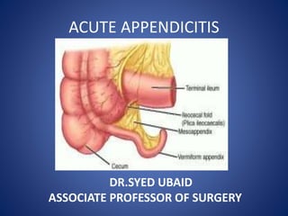
4. Anatomy
• Present only in humans,
• At birth: Short & broad at its junction with the
Caecum
• Typical tubular structure produced by 02 years
of age
• Results from differential growth of caecum
5. Anatomy
• Position: constant at the confluence of
the 03 taenia coli of caecum
• Mesoappendix : Arises from the lower surface
of the mesentery of terminal ileum
• Appendicular Artery: Branch of Ileo-colic
artery – End Artery
• 04-06 Lymphatic channels traverse
Mesoappendix ----------> Ileo-caecal LNs
6. Anatomy
• The three taeniae coli converge at the junction
of the cecum with the appendix and can be a
useful landmark to identify the appendix.
• The appendix can vary in length from <1 cm to
>30 cm; most appendices are 6 to 9 cm long.
13. Etiology
• No unifying hypothesis
• Bacterial proliferation
• Initiating event is controversial
• Obstruction of appendix lumen
• Faecolith or fibrotic stricture
• Worm infestations: Oxyuris vermicularis
• Neoplasms: Ca caecum, carcinoids
• Viral
• No luminal obstruction
• Low dietary fibre
14. • A faecolith (sometimes referred to as an
‘appendicolith’) is composed of inspissated
faecal material, calcium phosphates,
bacteria and epithelial debris
18. Pathology
• Continued mucous secretion and inflammatory
exudation intraluminal pressure
obstructing lymphatic drainage.
• Edema and mucosal ulceration develop with
bacterial translocation to submucosa .
• Further distention may cause venous obstruction
and ischcemia of the appendix wall.
• Bacterial invasion through muscularis externea
producing acute appendicitis.
• Ischaemic necrosis of appendicular wall
gangrenous appendix bacterial contamination of
peritoneal cavity.
19. Pathology cont.
• Greater omentum becomes
adherent to inflamed
appendix paracaecal abscess
• Extremes of age
• Immunosuppression
• Faecolith obstruction
• Pelvic appendix
• Previous abdominal surgery
26. Bacteriology of perforated appendicitis
TYPE OF BACTERIA PATIENTS (%)
ANAEROBIC
B. fragilis 80
B. thetiaotaomicron 61
Bilophila wadsworthia 55
Peptostrptococcus spp 46
AEROBIC
E.coli 77
S.viridans 43
Group D streptococcus 27
P.aeruginosa 18
28. Two clinical syndromes of acute appendicitis can
be discerned, acute catarrhal (non-
obstructive) appendicitis and acute
obstructive appendicitis, the latter
characterised by a more acute course.
31. Visceral abdominal pain
• Visceral abdominal pain, organs, visceral
peritoneum, mesentery
1-distention of a hollow viscus
2-ischemia to a viscus
3-inflammation
Referred to midline embryological development
33. 3/23/2017 33
Colicky pain
• Colicky pain form of visceral pain rhythmic
pain resulting from smooth muscle spasm as
a reaction to luminal obstruction
• intestinal obstruction, passage of gallstone in
biliary ducts, ureteric colic
• Often thought to be indigestion, constipation
and is frequently ignored.
• Lasts for a variable period, usually few hours
/ 2 or 3 days, rarely longer
34. 3/23/2017 34
Referred pain
• The vague referred pain in the periumblical
• stretching of the lumen or spasm.
• The visceral innervation comes from the 10th
thoracic spinal segment.
• Accompany the sympathetic nerves through the
superior mesenteric plexus and the lesser
splanchnic nerve
• If the visceral innervation is higher, then the
mid-line pain will be higher.
• Testicular pain due to visceral referral of
afferent nerve impulses to the same spinal
segment
35. 3/23/2017 35
Somatic pain
• Skin, fascia, muscle and parietal peritoneum
• Sever and precisely localized
• Inflamed parietal peritoneum cutaneous
hyperesthesia and tenderness
• The right iliac fossa pain is due to the irritation
of parietal peritoneum
• somatic pain as opposed to earlier visceral.
36. 3/23/2017 36
Guarding and rigidity
• Guarding is the protective phenomenon
• abdominal muscles increase in tone
• attempts to localize the inflammation
• There is tenderness causing the patient to
constantly tense the abdominal wall
muscles in palpation
• Voluntary guarding /apparent guarding
• Involuntary guarding /true guarding
• Muscular spasm rigidity
• localized initially progressing to generalized
with perforation or increasing peritonitis
37. 3/23/2017 37
Tenderness
• Tenderness:
–localised over McBurney's point
–not evident before later inflammation of
serosa and parietal peritoneum
–often masked in obese due to inability to
displace viscus
• Blumberg’s sign The pain is elicited with
pressing the abdominal wall deeply with
fingers and abruptly releasing it.
38. 3/23/2017 38
Reflexes
• Visceral afferent fibers participate in reflex
activities. Reflex sweating, salivation, nausea,
vomiting and tachycardia may accompany
visceral pain.
• Carried by autonomic nerve fibers
39. 3/23/2017 39
Signs
• lying still, with shallow breaths and reluctant to cough
• fever 37.5-38.5C, worsening with perforation
• Foetor oris - halitosis
• Furred tongue
• Flushed
• infrequently, diarrhoea:
– early and transient as a result of visceral pain
– later if retroileal or pelvic involvement appendix; this is
typically prolonged and mucoid
• constipation - sometimes for a few days before the
attack
40. 3/23/2017 40
Signs to elicit in appendicitis
• Pointing sign
• Rovsing’s sign
• Psoas sign
• Obturator sign
45. Clinical features
• Risk factors for perforation:
1. Extremes of age
2. Immunosuppression
3. Diabetes mellitus
4. Pelvic appendix
5. Previous abdominal surgery
46. “Typical” Presentation
• Dull, crampy central abdo pain
• Malaise/vomiting/anorexia/low grade fevers
• Pain worsens & localises to RIF with
cough/movement tenderness
• Systemic symptoms
54. Special clinical scenarios
• As per age:
1. Infants
- Uncommon <36 mths
- Difficult to diagnose
- Diffuse peritonitis common
- High incidence of perforation
55. Special clinical scenarios
2. Children
- Vomiting
- Marked anorexia
3. Elderly
- High incidence of gangrene & perforation
- Features of SAIO
4. Obese
- Diminished signs/ delayed dignosis
- Midline/ Laparoscopic approach
56. Special clinical scenarios
5. Pregnancy
- Most common extra-uterine cause of acute
abdomen
- Delayed presentation
- Fetal loss
• 3-5%
• Upto 20% : Perforation
57. Going through the sea of DDX
• Mesenteric lymphadenitis
• Acute appendicitis
• Henoch Schonlein purpura
• lobar pneumonia
• Meckel's diverticulitis
• Intussusception
• Ureteric stone
• Enterocolitis
• Familial Mediterranean fever
67. Tzanakis Score
1. Rt lower abdominal tenderness = 4
2. Rebound tenderness = 3
3. WBC’s> 12,000 in the blood = 2
4. Positive USS findings of appendicitis = 6
• Total score = 15
• > 8 = 96% chances
68. X-ray signs
• Reported signs include:
• increased soft tissue density in the right lower
quadrant
• a faecolith in the right iliac fossa
– the majority are radio-opaque
– occur in about 10% of those with appendicitis
– often mistaken for ureteric calculus or gallstones
• a gas-filled appendix
• free intraperitoneal gas
69. Plain Abdominal X-Rays
• Low sensitivity
• Appendicoliths picked up in only 10-15% cases
• Can be combined with Barium enema
• Failure of appendix to “Fill up”
• Low specificity 20% of normal Appendices
do not fill up
84. Diagnostic Laparoscopy
• Small fraction of pts
• Women of child bearing age
• Prompt intervention ------- Implications on
future fertility
85. Laboratory Examinations
• WBC’s elevated
• Normal in 10% cases
• TLC > 20,000 s/o PERFORATION
• Polymorphs > 75%
• Minimal pyuria Common
• Microscopic haematuria
86. Appendiceal Rupture
Immediate appendectomy has long been the recommended treatment for acute appendicitis
because of the presumed risk of progression to rupture.
The overall rate of perforated appendicitis is 25.8%. Children <5 years of age and patients >65
years of age have the highest rates of perforation (45 and 51%, respectively)
delays in presentation are responsible for the majority of perforated appendices.
Appendiceal rupture occurs most frequently distal to the point of luminal obstruction along
the antimesenteric border of the
appendix. Rupture should be suspected in the presence of fever with a temperature of
>39°C (102°F) and a white blood cell
count of >18,000 cells/mm3
87. Options
• Open / traditional surgery
• Laparoscopic
• Natural orifice surgery (no incision
appendectomy )
88. Appendicectomy - Open
• Incision over McBurney’s point or point of
maximal tenderness
• Straightforward, good exposure, technically
easier
• Longer recovery, risk of hernia & adhesions,
can’t see pelvic structures as well
89. Open appendetomy
• For open appendectomy most surgeons use either a McBurney
(oblique) or Rocky-Davis (transverse) right lower quadrant muscle-
splitting incision in patients with suspected appendicitis. The
incision should be centered over either the point of maximal
tenderness or a palpable mass
90. Open / traditional
• if incision is perpendicular to the line :
• Grid iron incision
• Why called grid iron ?
• if better access is needed , one
can change gridiron to
Rutherford Morrison's incision
• Lanz incision (transverse skin crease incision ,
2cm below umbilicus at mid-clavicular line)
100. Then
• put drain if you find dirty iliac fossa ,
otherwise :
• suture .
101. Laparoscopic appendetomy
• Laparoscopic appendectomy usually requires the use of three
ports. Four ports may occasionally be necessary to mobilize a
retrocecal appendix. The surgeon usually stands to the patient's
left. One assistant is required to operate the camera. One trocar is
placed in the umbilicus (10 mm), and a second trocar is placed in
the suprapubic position. Some surgeons place this second port in
the left lower quadrant. The suprapubic trocar is either 10 or 12
mm, depending on whether or not a linear stapler will be used.
• The placement of the third trocar (5 mm) is variable and usually is
either in the left lower quadrant, epigastrium, or right upper
quadrant.
103. Appendicectomy - Laparoscopic
• “Keyhole” surgery
• Lower complication rate, quicker
recovery
• Sometimes difficulty in mobilisation
requiring open procedure
104.
105. Natural orifice surgery
• No incision appendectomy
• Using the natural orifices like
anal canal endoscopically or
trans-vaginally .
• Less pain
• No scar
• Less hospital stay
• Fewer complications
• It takes about 50 minutes
107. Management of Appendicular
abscess/Lump
• Late presentation
• Clinically mass & fever
• Subject to imaging studies to ascertain:
- Presence
- Size
• > 4-6cms Antibiotics+Drainage
• < 4 cms Conservative mgt(Oschner
Sherren’s Regime)
108. Management of Appendicular
abscess/Lump
• Criteriae for stopping conservative mgt:
- Rising pulse rate
- Increase in the size of the mass
- Increasing/ spreading abdominal pain