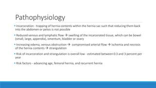
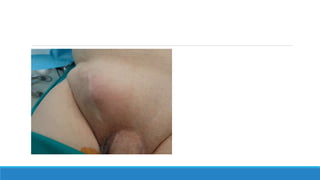
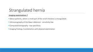
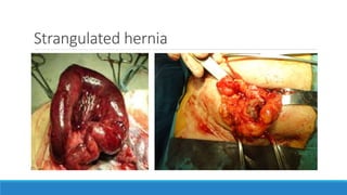
Stragulated Inguinal
hernia
DR CARUNYA MANNAN
INGUINAL ANATOMY
Clinical classification
 Reducible
 Irreducible - adhesions
 Obstructed / Incarcerated – irreducibility with obstruction
 Strangulated – irreducibility + obstruction + venous/arterial compromise
 Inflammed hernia
Pathophysiology
 Incarceration - trapping of hernia contents within the hernia sac such that reducing them back
into the abdomen or pelvis is not possible
 Reduced venous and lymphatic flow  swelling of the incarcerated tissue, which can be bowel
(small, large, appendix), omentum, bladder or ovary
 Increasing edema, venous obstruction  compromised arterial flow  ischemia and necrosis
of the hernia contents  strangulation
 Risk of incarceration and strangulation is overall low - estimated between 0.3 and 3 percent per
year
 Risk factors - advancing age, femoral hernia, and recurrent hernia
Clinical presentation
 Irreducible swelling, erythema of groin skin
 Strangulated hernias  symptoms of bowel obstruction  nausea, vomiting, abdominal pain
and distention
 Systemic symptoms – fever, with onset of sepsis
 Generalized peritonitis - does not occur since the ischemic or necrotic tissue is trapped within
the hernia sac.
 Strangulated segment of bowel is reduced (spontaneously or unwittingly) generalized
peritoneal signs may be present
Hernia Repair Techniques
Non-mesh repairs
Shouldice repair
 Anterior approach that is commonly used for open repair of inguinal hernias that is performed
without mesh
 Lowest hernia recurrence
 Division of all of the layers of the floor of the inguinal canal and reduction of the hernia,
followed by reconstruction of the inguinal canal with a four-layer overlap technique using
continuous fine wire sutures to obliterate the hernia defect
Non-mesh repairs
Desarda repair
 A flap of the external oblique muscle aponeurosis is used to “patch” the defect in a manner
similar to a Lichtenstein repair, but without prosthetic material
Bassini repair
 Primary tissue approximation approach to inguinal hernia repair in which the weakened
inguinal floor is strengthened by suturing the conjoined tendon to the inguinal ligament from
the pubic tubercle medially to the area of the internal ring laterally
Non-mesh repairs
McVay repair
 Involves incising the transversalis fascia in the region of Hesselbach's triangle to enter the preperitoneal space to
expose the pectineal ligament (Cooper's ligament)
 Conjoined tendon is then sutured to Cooper's ligament from the pubic tubercle laterally as far as the vicinity of
the femoral sheath as it crosses Cooper's ligament
 Transition stitch is placed incorporating the conjoined tendon, Cooper's ligament, the femoral sheath at the
medial aspect of the femoral vein, and the inguinal ligament
 Inguinal floor is repaired by approximating the conjoined tendon to the inguinal ligament extending laterally to
the area of the internal ring
 Generates considerable tension  requires a relaxing incision
 Anterior rectus sheath behind the external oblique aponeurosis should be exposed from the pubic tubercle
cephalad for several centimeters and it is then incised from the pubic tubercle extending cephalad for
approximately 6 centimeters along the fusion of the external oblique aponeurosis with the sheath's other
components
Mesh repairs
 Lichtenstein repair
 Plug and patch repair
 Kugel repair
 Bilayer mesh repair
Lichtenstein repair
Strangulated hernia
Imaging examinations ?
 Obese patients, where a small part of the small intestine is strangulated.
 Ultrasonography of the lower abdomen - sensitivity low
 Computed tomography - low specificity
 Imaging findings / combination with physical examination
Surgical diagnostics
 Definitive diagnosis of strangulation of the intestine can only be made through surgical
exploration
 Midline laparotomy incision - possible intestinal resection
 In some studies, almost the half of overall of midline laparotomies were performed without any
intestinal resection
 Elderly patients + co-morbidities  Diagnostic laparoscopy
 Laparoscopy could help to diagnose bowel ischemia thus decreasing both negative and
nontherapeutic laparotomy rates
Romain B, Chemaly R, Meyer N, Brigand C, Steinmetz JP, Rohr S. Prognostic factors of postoperative morbidity and mortality in strangulated groin hernia. Hernia. 2012 16: 405-10.
Strangulated hernia
Surgical techniques
 Reverse trendelenburg position during induction of anesthesia – avoid spontaneous reduction
 Bassini and Shouldice techniques - are preferred from contemporary tension-free techniques,
due to high possibility of mesh infection, in tension free techniques
 Viability of bowel - bowel resection and anastomosis will be needed/ frequently be performed
through the groin incision
 Abdominal exploration (open or laparoscopic)
Tension-free techniques in Strangulated
hernia?
 Presence of a strangulated inguinal hernia cannot be considered a contraindication for the use
of a prosthetic mesh
 Lichtenstein hernioplasty - successfully used not only as an elective operation but also as an
emergency operation for incarcerated inguinal hernia with a good outcome, with a low risk of
the local infectious complications and low rate of postoperative complications
 Polypropylene meshes - ideal for use in contaminated or potentially contaminated fields
 Macroporous structure - with pores of diameter larger than 70 micronmeters  allows contact
among the bacteria, which measures almost one micrometer in diameter, and the cells of the
immune system, granulocytes and macrophages, with a diameter of 15–20 micronmeters, which
is significant for the recovery from infections
Bessa SS, Katri KM, Abdel Salam WN, Abdel-Baki NA.. Early results from the use of the Lichtenstein repair in the management of strangulated groin hernia. Hernia. 2007
11: 239-42.

Obstructed & stragulated hernia1

  • 1.
  • 2.
  • 3.
    Clinical classification  Reducible Irreducible - adhesions  Obstructed / Incarcerated – irreducibility with obstruction  Strangulated – irreducibility + obstruction + venous/arterial compromise  Inflammed hernia
  • 4.
    Pathophysiology  Incarceration -trapping of hernia contents within the hernia sac such that reducing them back into the abdomen or pelvis is not possible  Reduced venous and lymphatic flow  swelling of the incarcerated tissue, which can be bowel (small, large, appendix), omentum, bladder or ovary  Increasing edema, venous obstruction  compromised arterial flow  ischemia and necrosis of the hernia contents  strangulation  Risk of incarceration and strangulation is overall low - estimated between 0.3 and 3 percent per year  Risk factors - advancing age, femoral hernia, and recurrent hernia
  • 5.
    Clinical presentation  Irreducibleswelling, erythema of groin skin  Strangulated hernias  symptoms of bowel obstruction  nausea, vomiting, abdominal pain and distention  Systemic symptoms – fever, with onset of sepsis  Generalized peritonitis - does not occur since the ischemic or necrotic tissue is trapped within the hernia sac.  Strangulated segment of bowel is reduced (spontaneously or unwittingly) generalized peritoneal signs may be present
  • 7.
  • 8.
    Non-mesh repairs Shouldice repair Anterior approach that is commonly used for open repair of inguinal hernias that is performed without mesh  Lowest hernia recurrence  Division of all of the layers of the floor of the inguinal canal and reduction of the hernia, followed by reconstruction of the inguinal canal with a four-layer overlap technique using continuous fine wire sutures to obliterate the hernia defect
  • 9.
    Non-mesh repairs Desarda repair A flap of the external oblique muscle aponeurosis is used to “patch” the defect in a manner similar to a Lichtenstein repair, but without prosthetic material Bassini repair  Primary tissue approximation approach to inguinal hernia repair in which the weakened inguinal floor is strengthened by suturing the conjoined tendon to the inguinal ligament from the pubic tubercle medially to the area of the internal ring laterally
  • 10.
    Non-mesh repairs McVay repair Involves incising the transversalis fascia in the region of Hesselbach's triangle to enter the preperitoneal space to expose the pectineal ligament (Cooper's ligament)  Conjoined tendon is then sutured to Cooper's ligament from the pubic tubercle laterally as far as the vicinity of the femoral sheath as it crosses Cooper's ligament  Transition stitch is placed incorporating the conjoined tendon, Cooper's ligament, the femoral sheath at the medial aspect of the femoral vein, and the inguinal ligament  Inguinal floor is repaired by approximating the conjoined tendon to the inguinal ligament extending laterally to the area of the internal ring  Generates considerable tension  requires a relaxing incision  Anterior rectus sheath behind the external oblique aponeurosis should be exposed from the pubic tubercle cephalad for several centimeters and it is then incised from the pubic tubercle extending cephalad for approximately 6 centimeters along the fusion of the external oblique aponeurosis with the sheath's other components
  • 11.
    Mesh repairs  Lichtensteinrepair  Plug and patch repair  Kugel repair  Bilayer mesh repair
  • 12.
  • 13.
    Strangulated hernia Imaging examinations?  Obese patients, where a small part of the small intestine is strangulated.  Ultrasonography of the lower abdomen - sensitivity low  Computed tomography - low specificity  Imaging findings / combination with physical examination
  • 14.
    Surgical diagnostics  Definitivediagnosis of strangulation of the intestine can only be made through surgical exploration  Midline laparotomy incision - possible intestinal resection  In some studies, almost the half of overall of midline laparotomies were performed without any intestinal resection  Elderly patients + co-morbidities  Diagnostic laparoscopy  Laparoscopy could help to diagnose bowel ischemia thus decreasing both negative and nontherapeutic laparotomy rates Romain B, Chemaly R, Meyer N, Brigand C, Steinmetz JP, Rohr S. Prognostic factors of postoperative morbidity and mortality in strangulated groin hernia. Hernia. 2012 16: 405-10.
  • 15.
  • 16.
    Surgical techniques  Reversetrendelenburg position during induction of anesthesia – avoid spontaneous reduction  Bassini and Shouldice techniques - are preferred from contemporary tension-free techniques, due to high possibility of mesh infection, in tension free techniques  Viability of bowel - bowel resection and anastomosis will be needed/ frequently be performed through the groin incision  Abdominal exploration (open or laparoscopic)
  • 17.
    Tension-free techniques inStrangulated hernia?  Presence of a strangulated inguinal hernia cannot be considered a contraindication for the use of a prosthetic mesh  Lichtenstein hernioplasty - successfully used not only as an elective operation but also as an emergency operation for incarcerated inguinal hernia with a good outcome, with a low risk of the local infectious complications and low rate of postoperative complications  Polypropylene meshes - ideal for use in contaminated or potentially contaminated fields  Macroporous structure - with pores of diameter larger than 70 micronmeters  allows contact among the bacteria, which measures almost one micrometer in diameter, and the cells of the immune system, granulocytes and macrophages, with a diameter of 15–20 micronmeters, which is significant for the recovery from infections Bessa SS, Katri KM, Abdel Salam WN, Abdel-Baki NA.. Early results from the use of the Lichtenstein repair in the management of strangulated groin hernia. Hernia. 2007 11: 239-42.