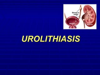
This document discusses urolithiasis (kidney stones). It covers theories of stone formation, epidemiology, risk factors, types of stones, clinical manifestations, diagnostic imaging techniques including ultrasound, CT scans, and cystoscopy, as well as treatments including pain relief, stone removal procedures like ESWL, ureteral stenting, and open surgeries. It also discusses bladder stones including diagnostic evaluation, cystoscopy, and surgical treatment options.
This document discusses bladder diverticula. It defines bladder diverticula as a herniation of bladder mucosa between detrusor muscle fibers. Bladder diverticula can be congenital or acquired secondary to conditions causing bladder outlet obstruction. The document classifies and describes the epidemiology, presentations, evaluations, imaging findings, and management options for bladder diverticula. Common imaging modalities discussed are cystoscopy, cystography, intravenous urography, and cross-sectional imaging. The document also addresses complications of bladder diverticula such as malignancy and challenges in surgical management.
The document discusses various conditions affecting the urinary bladder including stones, fistulas, and diverticulums. It describes the types, causes, symptoms, examinations, and treatments for bladder stones. It also discusses fistulas that can occur between the bladder and other organs, such as vesicovaginal fistulas from prolonged labor or surgery. Diverticulums of the bladder are described as occurring due to high bladder pressures from outflow obstruction.
The document discusses acute urinary retention and acute renal failure. It defines the conditions and outlines their causes, which can be pre-renal, renal, or post-renal in nature. The evaluation and management of the conditions is also described. Causes include issues like decreased perfusion, obstruction, toxicity, and inflammation. Treatment involves supportive care, addressing the underlying cause, and dialysis in some situations to manage electrolyte and acid-base imbalances.
This document provides an overview of benign prostatic hyperplasia (BPH) including its etiology, pathology, clinical findings, and investigation. It notes that BPH begins as microscopic nodules in the transitional zone of the prostate that can grow and compress surrounding tissue. Common symptoms include urinary frequency, urgency, and nocturia. Evaluation involves assessment of lower urinary tract symptoms, digital rectal exam, urinalysis, post-void residual measurement, and in some cases urodynamic testing. BPH is a common condition among older men that results from changes in hormone levels and growth factors.
This document provides an overview of urolithiasis (kidney stones). It discusses the epidemiology, classification, pathogenesis, clinical features, investigations, treatment modalities, complications, and prevention of kidney stones. Treatment depends on the location and size of the stone and includes extracorporeal shock wave lithotripsy, percutaneous nephrolithotomy, ureteroscopy, and open surgery. The goal is to remove stones while minimizing complications such as infection, obstruction, and loss of renal function. Prevention focuses on adequate fluid intake, dietary modifications, and medical management for certain stone types.
This document discusses ectopic ureters and ureteroceles. Some key points:
1. Ectopic ureters and ureteroceles are congenital abnormalities that occur due to abnormal development of the ureter and urinary tract.
2. Clinical presentations can include urinary tract infections, incontinence, pain, and obstruction. Evaluation involves ultrasound, voiding cystourethrogram, nuclear scans, and possibly MRI.
3. Management depends on factors like obstruction, reflux, and renal function. Options include observation, acute decompression, definitive surgery like reimplantation, and in some cases total reconstruction or upper pole nephrectomy. Complications
This document discusses staghorn calculi, which are large infection stones that fill the renal pelvis and calyces. It covers the etiology, pathogenesis, epidemiology, clinical presentation, evaluation, and various treatment modalities for staghorn calculi. The primary treatment is percutaneous nephrolithotomy (PCNL), which has stone-free rates of around 80% but risks include injury to adjacent organs and sepsis. Other options include extracorporeal shockwave lithotripsy, ureteroscopy, open surgery, dissolution therapy, antibiotics, urease inhibitors, urinary acidification, and dietary modification, but PCNL generally provides the best chance of complete stone clearance.
Post-obstructive diuresis refers to high urine output that can occur after relief of urinary tract obstruction. It is caused by accumulation of water, sodium, and urea during the period of obstruction. There are two main types - physiological diuresis which is self-limiting as fluid balance returns to normal, and pathological diuresis where inappropriate water loss continues beyond normalization of volume status. Treatment involves careful fluid management to replace losses based on urine output and electrolyte monitoring, as most cases will resolve spontaneously once homeostasis is restored. However, those with risk factors like edema may require closer monitoring and intravenous fluids.
The document discusses the management of haematuria in the emergency department. It covers the common causes of haematuria, appropriate investigations in the ED including imaging and labs, goals of treatment focusing on resuscitation, ensuring free urine drainage, safe discharge, and prompt follow up. It provides guidance on managing macroscopic haematuria including bladder washouts and irrigation. Indications for admission or safe discharge are outlined along with special circumstances. Three case examples are presented and management steps discussed.
This document provides an overview of bladder anatomy, function, and bladder outlet obstruction. It describes the key parts of the bladder including relations to other organs. Normal micturition and factors that assist storage are explained. Causes of bladder outlet obstruction include anatomical and functional issues. Common symptoms include hesitancy, weak stream, and urinary tract infections. Investigations help locate the site of obstruction and assess kidney and bladder function.
The document discusses two causes of obstructive uropathy: nephrolithiasis and hydronephrosis. Nephrolithiasis is the formation of urinary calculi (stones) in the kidneys or urinary tract. Calculi most commonly occur in middle-aged men and can cause pain. Hydronephrosis is the dilation of the renal pelvis and calyces due to partial or intermittent blockage of urine flow. It is usually caused by obstruction in the ureter but can also be congenital or due to conditions affecting the bladder. Advanced hydronephrosis leads to compression of the renal cortex. Both conditions are demonstrated in photographs showing enlarged kidneys with dilated pelvis and
Post-obstructive diuresis occurs after relief of a urinary tract obstruction, where large amounts of salt, water, and urea are excreted in the urine. It is caused by accumulation of fluids and solutes during obstruction and impairment of tubular reabsorption capabilities. Risk factors include edema and azotemia. The pathophysiology involves derangements in urinary concentrating ability due to disrupted aquaporin channels and sodium transport, as well as insensitivity to ADH. Treatment focuses on complete relief of obstruction, fluid replacement, electrolyte correction, and monitoring.
The document discusses renal stones (nephrolithiasis), including their definition, etiology, risk factors, pathophysiology, types, clinical manifestations, diagnostic studies, management, nursing management, prevention, and questions. Renal stones are formed by crystallization of substances in the urine, and can be caused by metabolic, lifestyle, or genetic factors. Common types include calcium oxalate, calcium phosphate, uric acid, cystine, and struvite stones. Treatment may involve medical management, extracorporeal shock wave lithotripsy (ESWL), or surgical procedures like ureteroscopy. Nursing focuses on pain management, education, and preventing infection and recurrence.
Femoral hernia is the third common hernia after inguinal and incisional hernias. The swelling in femoral hernia is below and lateral to pubic tubercle. It is more common in females. Strangulation is very common in this hernia.
Please find the power point on Renal and bladder stones. I tried present it on understandable way and all the contents are reviewed by experts and from very reliable references. Thank you
The document summarizes the management of upper urinary tract obstruction. Relieving the obstruction can be done through ureteral stenting or nephrostomy tubing, both of which come with potential side effects. Symptoms are treated with analgesics, antipyretics, and antibiotics. Underlying causes like stones may pass spontaneously, be treated with extracorporeal shockwave lithotripsy, percutaneous nephrolithotomy, ureteroscopy, or open surgery. Recurrence is prevented through increased fluid intake, reduced calcium intake, correcting metabolic abnormalities, treating infections, and urinary alkalization.
This document provides an overview of the evaluation and management of ureteric stones. It discusses the typical signs and symptoms of ureteric stones including flank pain radiating to the groin. Imaging options like ultrasound, KUB, CT are outlined. Treatment depends on factors like stone size and location, and may include pain control, conservative management, medical expulsive therapy, or active stone removal procedures like ESWL, URS, or PCNL. Prevention emphasizes adequate hydration and dietary modifications.
This document defines and discusses different types of hematuria including visible and non-visible hematuria. It outlines the renal, ureteral, bladder, and urethral causes of hematuria such as kidney stones, renal cell carcinoma, cystitis, prostate cancer, and trauma. The document provides diagnostic features of different causes of hematuria including loin pain and clots with kidney stones, flank mass and fever with renal cell carcinoma, and intermittent painless hematuria in workers exposed to chemicals indicating possible bladder cancer. Key points emphasize that hematuria always requires investigation and that location and characteristics of hematuria can provide clues to underlying causes.
Here are the key steps in my approach for a female patient referred for recurrent UTIs:
1. Take a detailed history regarding symptoms, number and timing of previous UTIs, results of urine cultures, potential complicating factors.
2. Perform a physical exam paying attention to the abdomen and genitourinary system.
3. Order urinalysis and urine culture to confirm current infection.
4. Consider further imaging like renal ultrasound if history suggests possibility of structural abnormalities.
5. Review medications for potential interactions.
6. Discuss lifestyle and hygiene modifications that may help reduce risk of recurrence.
7. Consider prophylactic antibiotics if recurrent infections are severe or frequent. The choice would
The ureters are tubular structures that transport urine from the kidneys to the bladder. They have multiple layers including epithelium, smooth muscle, and adventitia. Sites of natural narrowing include the ureteropelvic junction (UPJ) and ureterovesical junction. UPJ obstruction is most common in boys and on the left side. It can be caused by intrinsic narrowing at the UPJ or extrinsic compression. Surgical intervention is considered if renal function declines or symptoms develop. Treatment options include open or laparoscopic pyeloplasty, endopyelotomy, or ureterocalycostomy depending on the specifics of each case.
This document provides an overview of initial investigations and radiological investigations in urology. It discusses urinalysis, urine culture, cytology, biochemistry, ultrasound, and prostate-specific antigen as initial investigations. Ultrasound uses include evaluating the kidneys, bladder, prostate, and scrotum. Urodynamics tests lower urinary tract function. Radiological investigations include plain x-rays, retrograde urethrograms, intravenous urography, CT scans, MRI, PET scans, and nuclear medicine tests. The document provides details on the procedures and clinical applications of each test.
This document discusses the evaluation and management of urolithiasis or kidney stones. It begins with an overview of the burden of kidney stones, noting the prevalence and high recurrence rates. It then covers the clinical, radiological, and metabolic evaluation of patients with suspected kidney stones. The majority of the document discusses various treatment approaches for kidney stones including medical expulsive therapy, shockwave lithotripsy, ureteroscopy, percutaneous nephrolithotomy, and dietary and fluid management recommendations. Complications of different surgical procedures are also summarized.
Kidney stones form when dietary minerals in urine become concentrated enough to crystallize. They are typically classified by location and chemical composition, with calcium salts and uric acid being most common. Risk factors include diet, medical conditions, and family history. Stones form through supersaturation when urine is too concentrated for minerals to remain in solution. Symptoms include flank pain, nausea, and blood in urine. Diagnosis involves history, physical exam, and imaging tests like ultrasound or CT. Treatment focuses on resolving underlying causes and preventing stone recurrence.
A 58-year old man who works in a dye factory and smokes cigarettes is experiencing hematuria for 3 months. Urine cytology would be performed to screen for bladder cancer given his risk factors of occupational exposure to carcinogens and smoking history. Proper collection of the second morning voided urine sample is important for accurate cytology results. The patient is most likely to have transitional cell carcinoma of the bladder due to his risk factors, and this type of bladder cancer has different grades based on how abnormal the cells appear.
This document discusses alcohol withdrawal, including its goals, evaluation and treatment. It describes the symptoms of withdrawal which can range from minor (insomnia, anxiety) to severe (delirium tremens). It recommends correcting fluid/electrolyte abnormalities and using a tapering regimen of benzodiazepines like chlordiazepoxide or diazepam to safely manage withdrawal symptoms. Adjunct medications like clonidine may also help. Close monitoring is needed as untreated severe withdrawal can be life-threatening.
Urothelial carcinoma biomarkers can be detected in urine samples to aid in the diagnosis and monitoring of bladder cancer. Nuclear matrix protein 22 (NMP22) and bladder tumor antigen (BTA) tests detect proteins released by dying tumor cells with reasonable sensitivity and specificity. Urine cytology examines cells for morphological changes, having high sensitivity for high-grade tumors but low sensitivity for low-grade tumors. Newer tests like CxBladder, CertNDx, and microsatellite analysis analyze genetic markers and show promise in improving detection rates, especially for low-grade tumors. No single test is perfect and cystoscopy remains the gold standard, but urine biomarkers provide a non-invasive method to supplement standard evaluation and monitoring of patients
This document discusses kidney stones (urolithiasis), including:
- Types of stones are classified by composition, x-ray appearance, or size and shape. Common types include calcium oxalate, uric acid, struvite, and cystine stones.
- Risk factors for stone formation include age, sex, family history, diet, fluid intake, medical conditions, and anatomical abnormalities.
- Evaluation of stone patients includes analyzing the stone composition and performing metabolic testing to identify predisposing factors.
- Treatment options depend on the stone characteristics and include watchful waiting, extracorporeal shockwave lithotripsy (ESWL), ureteroscopy, percutaneous nephrolith
This document summarizes urine cytology and various urinary markers for detecting bladder cancer. Urine cytology is the gold standard but has low sensitivity of 40-62%. Newer urinary markers like BTA, ImmunoCyt, NMP-22, and UroVysion have higher sensitivities than cytology of 50-86%, but lower specificities and can be affected by benign conditions. No single marker currently has a sensitivity of 90%, which patients indicate is needed to replace cystoscopy. Therefore, the best approach is a combination of cystoscopy and urinary markers for non-muscle-invasive bladder cancer surveillance.
The patient is a 63-year-old male with a history of alcohol abuse presenting with symptoms of acute alcohol withdrawal including tremors, anxiety, tachycardia and hypokalemia. Initial treatment involves monitoring, managing withdrawal symptoms, addressing nutritional deficits and providing counseling and referrals to support ongoing sobriety and prevent relapse.
Alcohol withdrawal symptoms and management are discussed. Symptoms include tachycardia, hypertension, fever, and agitation and occur 48 hours after last drink. The CIWA protocol monitors for withdrawal symptoms and manages with IV fluids, vitamins, and lorazepam prn. Electrolyte abnormalities should also be monitored and treated. Withdrawal risk is greatest 12-24 hours after last drink and complications like seizures or delirium tremens may require ICU care.
Retention of urine occurs when one is unable to completely empty the bladder. It can be acute, occurring suddenly due to obstruction, or chronic, developing over time. Acute retention requires immediate catheterization to drain the bladder, while chronic retention involves identifying and treating the underlying cause of the partial obstruction. A thorough history, exam, and testing is needed to determine if the retention is due to issues in the bladder, prostate, urethra, or other causes and select the appropriate management.
Mr. XYZ, age 39, presented with tremors, vomiting blood, seizures, and confusion. He has a history of alcohol dependence and multiple relapses. On examination, he was confused, tachycardic, and jaundiced. Lab tests showed elevated liver enzymes. He was diagnosed with delirium tremens during acute alcohol withdrawal. He was treated with benzodiazepines, thiamine, and other supportive care. Benzodiazepines are the primary treatment for alcohol withdrawal to prevent progression to severe withdrawal states like delirium tremens and seizures.
This document discusses screening and treatment for alcohol withdrawal. It recommends using the CAGE assessment tool to screen for alcohol dependency and risk of withdrawal symptoms. For patients who screen positive, it suggests making referrals to social services and informing physicians. It also describes the CIWA-Ar scale for assessing withdrawal symptoms and guiding medication administration to safely manage alcohol withdrawal syndrome.
This document provides an overview of alcohol withdrawal syndromes. It defines alcohol withdrawal, describes the pathophysiology and timeline of symptoms. Minor withdrawal can occur within 6-12 hours and includes autonomic hyperactivity and insomnia. Alcoholic hallucinosis may occur in the first 24-48 hours. Withdrawal seizures typically occur within 8-48 hours. Delirium tremens occurs 2-14 days later and has a mortality rate of 5% with treatment. Benzodiazepines are the main treatment, with fixed dose or symptom-triggered regimens using medications like diazepam or lorazepam. Delirium tremens requires ICU care and treatment until the patient is alert and
The document describes the anatomy and physiology of the urinary tract and kidney, risk factors and types of kidney stones, and methods for diagnosing and treating stones, including increasing fluid intake, altering diet, using medications to change urine composition, and surgically removing stones with procedures like ureteroscopy and lithotripsy. Kidney stones form when substances like calcium, oxalate, and uric acid become supersaturated in the urine and crystallize into solid masses.
Urinary calculi are composed of substances normally found in urine that precipitate out of solution to form stones. The most common types of stones found are calcium oxalate, calcium phosphate, uric acid, and cystine. Risk factors for stone formation include dehydration, diet, medical conditions, and anatomical abnormalities. Management involves dietary changes to reduce stone-forming substances in urine, increased fluid intake, and surgical procedures like ESWL or ureteroscopy for stone removal.
Peptic ulcer disease is caused by gastric or duodenal ulcers that form lesions in the stomach or duodenal mucosa. Risk factors include H. pylori infection, smoking, NSAID use, and genetic factors. Symptoms include epigastric pain relieved by food and antacids. Treatment aims to relieve pain, eradicate H. pylori infection, heal ulcers, and prevent recurrence through lifestyle changes and medication like PPIs or H2 blockers. Surgery was more common historically but is now rare due to H. pylori treatments, though it may be used for complications like perforation.
This document discusses urolithiasis (kidney stones). It begins by defining urolithiasis and noting its prevalence and cost. It then covers the epidemiology, types, symptoms, diagnosis, and management of kidney stones. The main points are that kidney stones can form anywhere in the urinary tract, have a lifetime risk of 2-20% depending on location, and are most commonly treated through active medical expulsion or minimally invasive surgeries like ESWL or ureteroscopy. Surgical intervention is indicated for large or obstructing stones, infection, or if conservative measures fail.
Urolithiasis refers to the formation of stones in the urinary tract. Stones can form in the kidneys (nephrolithiasis), ureters (ureterolithiasis), or bladder (cystolithiasis). Risk factors include disorders of the urinary tract, liver, endocrine system, infections, and metabolic disorders. Patients may experience flank pain radiating to the groin due to obstruction. Diagnosis involves urinalysis, blood tests, imaging like ultrasounds or CT scans. Treatment depends on stone size and location but may include pain medication, increasing fluid intake, stone removal procedures like ESWL, ureteroscopy, or open surgery. Preventing recurrence involves
Renal transplantation involves selecting recipients by diagnosing their primary kidney disease, ruling out active infections or malignancies, and assessing operative risks. Suitable living donors undergo evaluation with CT angiography and surgery to remove a kidney, with efforts to minimize warm ischemia time and preserve vessel length. The donated kidney is preserved through simple cold storage before transplantation via vascular anastomosis and ureter reimplantation during the recipient's operation. Lifelong immunosuppression is required post-operatively to prevent rejection, while infection risk remains.
Group 3 presented on nephrolithiasis, urolithiasis, and hydronephrosis. The presentation discussed the conditions, including that nephrolithiasis involves stone formation in the kidney. Diagnostic tests including IV pyelogram, retrograde pyelogram, x-rays, and urine tests were covered. Potential complications from the conditions include UTI, hydronephrosis, stone recurrence, and kidney damage. Nursing interventions focus on relieving pain, increasing fluid intake, educating the patient, and monitoring for complications.
The document discusses ureteroceles, which are saccular dilations of the terminal portion of the ureter. Ureteroceles are birth defects that occur in about 1 in 500 to 4,000 people and are more common in females. They can be caused by delayed or incomplete development of the ureteral bud leading to early prenatal obstruction. Ureteroceles are classified as ectopic or intravesical and may cause urinary tract infections, masses, incontinence or obstruction. Diagnosis involves imaging tests like ultrasound, VCUG, IVP or CT scan. Treatment depends on symptoms but may include antibiotics, endoscopic incision, upper pole nephrectomy, u
This document discusses various imaging techniques for evaluating the urinary system, including plain films, intravenous urography, ultrasound, CT, MRI, and angiography. It provides details on the principles, preparations, procedures and indications for each method. Common congenital anomalies of the kidneys are also listed such as horseshoe kidney, pelvic kidney, and duplicate collecting systems.
The document discusses various disorders of the genitourinary system including urological obstructions, disorders of the kidney, and disorders of the ureters, urinary bladder and urethra. It covers the etiology, risk factors, clinical presentation, diagnostic evaluation, and management of various conditions like urethral strictures, renal calculi, nephrotic syndrome, acute glomerulonephritis and more. Nursing management is also described which involves monitoring vitals, intake/output, administering medications as ordered, and educating patients.
This document discusses radiology of the urinary tract system and disorders. It describes how different imaging modalities like CT, ultrasound, and IVU can be used to identify urinary tract calculi, nephrocalcinosis, and obstruction at different levels of the urinary tract. Causes of obstruction include calculi, tumors, strictures, congenital issues, and extrinsic compression that are best evaluated using CT, ultrasound, or IVU depending on the level and nature of obstruction.
The document discusses the kidney, ureter, and various urological conditions involving obstruction of urine flow from the kidneys to the bladder such as hydronephrosis and pyonephrosis. It describes the anatomy and function of the kidneys and ureters. Causes of urinary obstruction include kidney stones, tumors, strictures, and congenital abnormalities. Patients present with flank pain, urinary tract infections, and renal impairment depending on severity and chronicity of obstruction. Investigations include ultrasound, IVU, CT urogram and MAG-3 scans. Management involves relieving obstruction through stenting, nephrostomy or surgery.
This document discusses the evaluation and management of urinary stones. Key points include:
- Clinical history is important to determine type of pain, risk factors, and if it is a first-time or recurrent stone.
- NCCT is the preferred initial imaging test for evaluation of suspected renal or ureteral stones.
- Conservative management involves increased fluid intake of at least 3 liters per day, a low-sodium diet moderate in animal protein and high in fruits/vegetables, and weight loss if overweight.
- For recurrent stone formers, a metabolic evaluation can identify underlying causes and guide targeted medical therapy like potassium citrate for calcium stones.
- Small distal ureteral stones
This document discusses various acute urological conditions that commonly present as emergencies. It covers non-traumatic conditions like urinary retention, hematuria, renal colic, and infections as well as traumatic injuries. For each condition, it describes the typical presentation, important distinguishing features, investigations, differential diagnoses, and initial management approach. It provides a useful overview of how to evaluate and treat many urgent urological problems.
This document discusses urinary catheterization. It describes catheters as plastic tubes inserted into the bladder via the urethra to drain urine or inject liquids for treatment or diagnosis. Critically ill patients requiring strict urine output monitoring are often catheterized. The document describes different catheter types including Foley, Robinson, Coude, and hematuria catheters.
This document provides an overview of intravenous urography (IVU), including indications, contraindications, technique, projections, and modifications. IVU involves intravenous injection of contrast medium to visualize the urinary tract. It is declining in use due to newer modalities like CT and MRI, adverse effects of contrast, and cost. The document outlines patient preparation, standard projections including nephrogram, 5-minute, compression, and post-void films. It also discusses non-routine projections, contrast agents, and radiation protection. Complications and aftercare are briefly mentioned.
The document describes a case presentation of a 10-year-old male child who presented with abdominal pain. An ultrasound and intravenous pyelogram (IVP) were performed. The ultrasound found right pyonephrosis, right and left renal calculi, and left hydroureteronephrosis. The IVP confirmed these findings and also found a right ureteric calculus and question of a left ureteric stricture. The document then provides details on IVP, including how it is performed, indications, advantages, limitations, normal findings, and examples of various abnormalities that can be seen on an IVP.
Hydronephrosis is the dilation of the renal pelvis and calyces caused by urinary outflow obstruction. It can result from anatomical or functional issues anywhere along the urinary tract. Chronic or severe hydronephrosis can lead to permanent kidney damage if not treated. Treatment depends on the cause and severity but may include ureteral stents, percutaneous nephrostomy tubes, or open surgery to bypass or remove the obstruction. The goal is to relieve obstruction and preserve kidney function.
Radiological methods such as intravenous urograms (IVUs), ultrasound, CT, MRI, and retrograde/antegrade urography are used to study the urinary system and diagnose diseases. IVUs provide anatomical images of the kidneys and pelvicalyceal systems while ultrasound is useful for differentiating renal cysts from tumors. CT and MRI are valuable for detecting small renal cell carcinomas, urinary tract stones, and other conditions. Emergent conditions like testicular torsion, bladder rupture, renal trauma, and ureteral obstruction can be diagnosed using these radiological techniques.
The document provides information on renal diseases. It discusses that kidney disease can be a silent killer and childhood nephrotic syndrome is mostly curable. It also notes that acute post-streptococcal glomerulonephritis mostly recovers and does not recur, and that hematuria in small children is usually harmless. Risk of kidney disease increases with age. Acute renal failure can often be prevented.
This document provides information on the anatomy, physiology, diseases, and surgical treatment of the large intestine. It discusses the segments of the large intestine, including the cecum, ascending colon, transverse colon, descending colon, and sigmoid colon. Common conditions that may require surgery include obstruction of the colon due to carcinoma, diverticular disease, or inflammatory disorders. Surgical options for treating an obstructed colon include resection with primary anastomosis or resection with a temporary or permanent colostomy. Staging of colon cancer is also outlined from Stage 0 through Stage IV.
This document discusses various urological investigations including radiological investigations like renal ultrasound, transrectal ultrasound, scrotal ultrasound, intravenous urogram, retrograde ureteropyelography, and non-radiological investigations like urine analysis, urine cytology, and urodynamic studies. Flexible ureterorenoscopy allows visualization of the ureters and renal pelvis while rigid cystoscopy is better for detecting lesions that may require resection. Together these investigations help evaluate issues like hematuria, urinary tract infections, and voiding symptoms.
The document discusses the anatomy, physiology, imaging, and conditions of the large intestine. It provides details on:
1) The anatomy of the large intestine including its layers, divisions of the colon, fixation and mobility of parts.
2) Common conditions like obstruction, which can be caused by cancer, volvulus, or diverticulitis. Diagnosis involves imaging and symptoms like abdominal pain and distension.
3) Methods of examination including colonoscopy, barium enema, and CT scans. Colonoscopy allows for direct visualization and biopsy while CT provides details on location and cause of issues like tumors or obstruction.
Giloy in Ayurveda - Classical Categorization and SynonymsPlanet Ayurveda
Giloy, also known as Guduchi or Amrita in classical Ayurvedic texts, is a revered herb renowned for its myriad health benefits. It is categorized as a Rasayana, meaning it has rejuvenating properties that enhance vitality and longevity. Giloy is celebrated for its ability to boost the immune system, detoxify the body, and promote overall wellness. Its anti-inflammatory, antipyretic, and antioxidant properties make it a staple in managing conditions like fever, diabetes, and stress. The versatility and efficacy of Giloy in supporting health naturally highlight its importance in Ayurveda. At Planet Ayurveda, we provide a comprehensive range of health services and 100% herbal supplements that harness the power of natural ingredients like Giloy. Our products are globally available and affordable, ensuring that everyone can benefit from the ancient wisdom of Ayurveda. If you or your loved ones are dealing with health issues, contact Planet Ayurveda at 01725214040 to book an online video consultation with our professional doctors. Let us help you achieve optimal health and wellness naturally.
The biomechanics of running involves the study of the mechanical principles underlying running movements. It includes the analysis of the running gait cycle, which consists of the stance phase (foot contact to push-off) and the swing phase (foot lift-off to next contact). Key aspects include kinematics (joint angles and movements, stride length and frequency) and kinetics (forces involved in running, including ground reaction and muscle forces). Understanding these factors helps in improving running performance, optimizing technique, and preventing injuries.
The skin is the largest organ and its health plays a vital role among the other sense organs. The skin concerns like acne breakout, psoriasis, or anything similar along the lines, finding a qualified and experienced dermatologist becomes paramount.
Osvaldo Bernardo Muchanga-GASTROINTESTINAL INFECTIONS AND GASTRITIS-2024.pdfOsvaldo Bernardo Muchanga
GASTROINTESTINAL INFECTIONS AND GASTRITIS
Osvaldo Bernardo Muchanga
Gastrointestinal Infections
GASTROINTESTINAL INFECTIONS result from the ingestion of pathogens that cause infections at the level of this tract, generally being transmitted by food, water and hands contaminated by microorganisms such as E. coli, Salmonella, Shigella, Vibrio cholerae, Campylobacter, Staphylococcus, Rotavirus among others that are generally contained in feces, thus configuring a FECAL-ORAL type of transmission.
Among the factors that lead to the occurrence of gastrointestinal infections are the hygienic and sanitary deficiencies that characterize our markets and other places where raw or cooked food is sold, poor environmental sanitation in communities, deficiencies in water treatment (or in the process of its plumbing), risky hygienic-sanitary habits (not washing hands after major and/or minor needs), among others.
These are generally consequences (signs and symptoms) resulting from gastrointestinal infections: diarrhea, vomiting, fever and malaise, among others.
The treatment consists of replacing lost liquids and electrolytes (drinking drinking water and other recommended liquids, including consumption of juicy fruits such as papayas, apples, pears, among others that contain water in their composition).
To prevent this, it is necessary to promote health education, improve the hygienic-sanitary conditions of markets and communities in general as a way of promoting, preserving and prolonging PUBLIC HEALTH.
Gastritis and Gastric Health
Gastric Health is one of the most relevant concerns in human health, with gastrointestinal infections being among the main illnesses that affect humans.
Among gastric problems, we have GASTRITIS AND GASTRIC ULCERS as the main public health problems. Gastritis and gastric ulcers normally result from inflammation and corrosion of the walls of the stomach (gastric mucosa) and are generally associated (caused) by the bacterium Helicobacter pylor, which, according to the literature, this bacterium settles on these walls (of the stomach) and starts to release urease that ends up altering the normal pH of the stomach (acid), which leads to inflammation and corrosion of the mucous membranes and consequent gastritis or ulcers, respectively.
In addition to bacterial infections, gastritis and gastric ulcers are associated with several factors, with emphasis on prolonged fasting, chemical substances including drugs, alcohol, foods with strong seasonings including chilli, which ends up causing inflammation of the stomach walls and/or corrosion. of the same, resulting in the appearance of wounds and consequent gastritis or ulcers, respectively.
Among patients with gastritis and/or ulcers, one of the dilemmas is associated with the foods to consume in order to minimize the sensation of pain and discomfort.
Know the difference between Endodontics and Orthodontics.Gokuldas Hospital
Your smile is beautiful.
Let’s be honest. Maintaining that beautiful smile is not an easy task. It is more than brushing and flossing. Sometimes, you might encounter dental issues that need special dental care. These issues can range anywhere from misalignment of the jaw to pain in the root of teeth.
How to Control Your Asthma Tips by gokuldas hospital.Gokuldas Hospital
Respiratory issues like asthma are the most sensitive issue that is affecting millions worldwide. It hampers the daily activities leaving the body tired and breathless.
The key to a good grip on asthma is proper knowledge and management strategies. Understanding the patient-specific symptoms and carving out an effective treatment likewise is the best way to keep asthma under control.
Histololgy of Female Reproductive System.pptxAyeshaZaid1
Dive into an in-depth exploration of the histological structure of female reproductive system with this comprehensive lecture. Presented by Dr. Ayesha Irfan, Assistant Professor of Anatomy, this presentation covers the Gross anatomy and functional histology of the female reproductive organs. Ideal for students, educators, and anyone interested in medical science, this lecture provides clear explanations, detailed diagrams, and valuable insights into female reproductive system. Enhance your knowledge and understanding of this essential aspect of human biology.
2. Theories of Stone FormationTheories of Stone Formation
A. Nucleation Theory
B. Stone Matrix Theory
C. Inhibitor of Crystallization Theory
3. EpidemiologyEpidemiology
Men > WomenMen > Women
4 % of population (Germany)4 % of population (Germany)
Maximum rate at 30 – 50 y.o.Maximum rate at 30 – 50 y.o.
In children - rareIn children - rare
GeographyGeography
EthnologyEthnology
4. RISK FACTORSRISK FACTORS
••Start of disease early in life: <25 yearsStart of disease early in life: <25 years
••Stone containing brushiteStone containing brushite
••Only one functioning kidneyOnly one functioning kidney
••Disease associated with stone formationDisease associated with stone formation::
-- hyperparathyroidismhyperparathyroidism
- renal tubular acidosis (partial/complete)- renal tubular acidosis (partial/complete)
- jejunoileal bypass- jejunoileal bypass
- Crohn’s disease- Crohn’s disease
- intestinal resection- intestinal resection
- malabsorptive conditions- malabsorptive conditions
- sarcoidosis- sarcoidosis
- hyperthyroidism- hyperthyroidism
5. Renal CalculiRenal Calculi
1 Coral calculus1 Coral calculus
2 Coral calculi fragment2 Coral calculi fragment
3 Calculi, which are impregnated with blood pigments3 Calculi, which are impregnated with blood pigments
6. TYPES OF KIDNEY STONESTYPES OF KIDNEY STONES
There are 4 basic kinds of kidney stones:There are 4 basic kinds of kidney stones:
Calcium stones (composed of calcium oxalate).Calcium stones (composed of calcium oxalate).
Uric acid stones.Uric acid stones.
Struvite stones (composed of magnesiumStruvite stones (composed of magnesium
ammonium phosphate).ammonium phosphate).
Cystine stone.Cystine stone.
7. Clinical ManifestationsClinical Manifestations
Acute obstruction of the urinaryAcute obstruction of the urinary
tract may cause renal colic, a formtract may cause renal colic, a form
of severe abdominal pain oftenof severe abdominal pain often
accompanied by nausea andaccompanied by nausea and
vomiting due to celiac ganglionvomiting due to celiac ganglion
stimulation.stimulation.
Onset is sudden, often during theOnset is sudden, often during the
night or in the early morningnight or in the early morning
8. Clinical ManifestationsClinical Manifestations
Obstructing calculi in the upper urinaryObstructing calculi in the upper urinary
tract cause an extreme crescendo liketract cause an extreme crescendo like
pain in the flank that generally radiatespain in the flank that generally radiates
laterally around the abdomen to thelaterally around the abdomen to the
corresponding groin and testicles incorresponding groin and testicles in
males and labia major in females.males and labia major in females.
9. Laboratry InvestigationsLaboratry Investigations
Stone analysisStone analysis: In every patient one stone should: In every patient one stone should
be analysed.be analysed.
Blood analysisBlood analysis: Calcium Albumin Creatinine Urate: Calcium Albumin Creatinine Urate
Urinalysis:Urinalysis: Fasting morning spot urine sampleFasting morning spot urine sample
Dip-stick test: pH, Leucocytes/BacteriaDip-stick test: pH, Leucocytes/Bacteria
Cystine test, Ca, P, citrate, urateCystine test, Ca, P, citrate, urate
11. Diagnostic imagingDiagnostic imaging
Routine examination involves a plain abdominal film of the kidneys, uretersRoutine examination involves a plain abdominal film of the kidneys, ureters
and bladder (KUB)and bladder (KUB)..
At least 80% of all renal stones are radiopaque and therefore readily visible onAt least 80% of all renal stones are radiopaque and therefore readily visible on
a plain film of the abdomena plain film of the abdomen
13. ExcretoryExcretory urourographygraphy
Excretory pyelography must not be carried out in the following patients - those:Excretory pyelography must not be carried out in the following patients - those:
With an allergy to contrast mediaWith an allergy to contrast media
With S-creatinine level > 200 µmol/LWith S-creatinine level > 200 µmol/L
On medication with metforminOn medication with metformin
With myelomatosisWith myelomatosis
14. Retrograde pneumopyelographyRetrograde pneumopyelography
Special examinations that can beSpecial examinations that can be
carried out include:carried out include:
Retrograde or antegradeRetrograde or antegrade
pyelographypyelography
Retrograde pneumo-pyelographyRetrograde pneumo-pyelography
or cystographyor cystography
Spiral (helical) unenhancedSpiral (helical) unenhanced
computed tomography (CT)computed tomography (CT)
ScintigraphyScintigraphy..
16. Diagnostic imagingDiagnostic imaging
CCystoscopia shows swallowing of the ureter orifice in lower location ofystoscopia shows swallowing of the ureter orifice in lower location of
the stone, it may also partially project out to the orificethe stone, it may also partially project out to the orifice..
20. Pain reliefPain relief
Pain relief involves the administration by various routes ofPain relief involves the administration by various routes of
the following agents:the following agents:
Diclofenac sodiumDiclofenac sodium
IndomethacinIndomethacin
Hydromorphone hydrochloride + atropine sulphateHydromorphone hydrochloride + atropine sulphate
BaralginBaralgin
No-spae + AnalgineNo-spae + Analgine
TramadolTramadol
21. Pain reliefPain relief
Warm bathWarm bath
Spasmolytic “cocktails” (with papaverine,Spasmolytic “cocktails” (with papaverine,
spasmalgone, no-spanum, promedole) should bespasmalgone, no-spanum, promedole) should be
taken.taken.
A high dosage of the cystenal or urolesan (20 dropsA high dosage of the cystenal or urolesan (20 drops
on the piece of sugar) is rather effective at the starton the piece of sugar) is rather effective at the start
of the renal colic.of the renal colic.
Physical method.Physical method.
22. Pain reliefPain relief
When pain relief cannot beWhen pain relief cannot be
obtained by medical means,obtained by medical means,
drainage by stenting ordrainage by stenting or
percutaneous nephrostomy (PN)percutaneous nephrostomy (PN)
or stone removal should beor stone removal should be
carried out.carried out.
percutaneous nephrostomypercutaneous nephrostomy
23. Stone removalStone removal
The overall passage rate of ureteral stones is:The overall passage rate of ureteral stones is:
Proximal ureteral stones: 25%Proximal ureteral stones: 25%
Mid-ureteral stones: 45%Mid-ureteral stones: 45%
Distal ureteral stones: 70%Distal ureteral stones: 70%
24. Indications for ActiveIndications for Active Stone removalStone removal
Active stone removal is stronglyActive stone removal is strongly
recommended in patients fulfilling therecommended in patients fulfilling the
following criteria:following criteria:
- p- persistent pain despite adequateersistent pain despite adequate
medicationmedication;;
- p- persistent obstruction with risk ofersistent obstruction with risk of
impaired renal functionimpaired renal function;;
-- stone with urinary tract infectionstone with urinary tract infection;;
- r- risk of pyonephrosis or urosepsisisk of pyonephrosis or urosepsis;;
- b- bilateral obstructionilateral obstruction;;
- obstructing calculus in a solitary- obstructing calculus in a solitary
functioning kidney.functioning kidney.
25.
26. Percutaneous ProceduresPercutaneous Procedures
Percutaneous nephrostomyPercutaneous nephrostomy..
Because of this technique, urologists canBecause of this technique, urologists can
now perform operative procedures withinnow perform operative procedures within
the kidney without using the standard largethe kidney without using the standard large
flank incisions and mobilization of theflank incisions and mobilization of the
kidney.kidney.
30. Closed Surgical ProceduresClosed Surgical Procedures
Cystoscopic techniqueCystoscopic technique
With the patient under anesthesia and withWith the patient under anesthesia and with
fluoroscopic control, stones in the distalfluoroscopic control, stones in the distal
ureter can sometimes be removed with aureter can sometimes be removed with a
wire stone basket.wire stone basket.
UreteropyeloscopyUreteropyeloscopy
Manipulation of small ureteral stonesManipulation of small ureteral stones
under direct vision with a ureteroscope isunder direct vision with a ureteroscope is
a major advance in the management ofa major advance in the management of
ureteral calculi. With this technique, smallureteral calculi. With this technique, small
stones can be easily trapped in a stonestones can be easily trapped in a stone
basket and safely extracted through thebasket and safely extracted through the
dilated ureter.dilated ureter.
31.
32. Extracorporeal Shock WaveExtracorporeal Shock Wave
LithotripsyLithotripsy
An extracorporeal noninvasive technique that uses shock waves toAn extracorporeal noninvasive technique that uses shock waves to
disintegrate urinary calculi while the patient is immersed in a water bathdisintegrate urinary calculi while the patient is immersed in a water bath
has been tested extensively and is now in clinical use.has been tested extensively and is now in clinical use.
With this technique, calculi in the upper urinary tract are reduced toWith this technique, calculi in the upper urinary tract are reduced to
fragments, which pass spontaneously from the collecting system andfragments, which pass spontaneously from the collecting system and
bladder in most patients.bladder in most patients.
34. ESWLESWL
2500 - 3000 impulses per procedure
Complications:
Cardiac Arrhythmia, Haematoma,
Infection, Colic
Re-ESWL
about 20 %
35. Indications to surgical operationIndications to surgical operation
Frequent attacks of the renal colic or persistentFrequent attacks of the renal colic or persistent
pain that disables the patient.pain that disables the patient.
Disorder of the urine outflow causing theDisorder of the urine outflow causing the
hydronephrotic degeneration of the kidney.hydronephrotic degeneration of the kidney.
Obturative anuria.Obturative anuria.
Frequent attacks of the acute pyelonephritis,Frequent attacks of the acute pyelonephritis,
progress of the chronic pyelonephritis thatprogress of the chronic pyelonephritis that
causes renal insufficiency.causes renal insufficiency.
Total hematuria.Total hematuria.
Calculous pyonephrosis, apostematousCalculous pyonephrosis, apostematous
pyelonephritis or carbuncle of the kidney.pyelonephritis or carbuncle of the kidney.
Stone at the sole kidney that causes obstruction.Stone at the sole kidney that causes obstruction.
Stone in the ureter of the sole kidney that won’tStone in the ureter of the sole kidney that won’t
pass away spontaneously.pass away spontaneously.
36. Open Surgical ProceduresOpen Surgical Procedures
Pyelolithotomy:Pyelolithotomy:
Simple pyelolithotomy is used forSimple pyelolithotomy is used for
removal of calculi confined to theremoval of calculi confined to the
renal pelvis.renal pelvis.
Minimal dissection of the renalMinimal dissection of the renal
sinus is usually needed, andsinus is usually needed, and
exposure of the entire kidney isexposure of the entire kidney is
not required.not required.
This procedure is not indicated forThis procedure is not indicated for
the removal of entrapped calicealthe removal of entrapped caliceal
stones or large, branched renalstones or large, branched renal
calculicalculi..
38. Open Surgical ProceduresOpen Surgical Procedures
Ureterolithotomy.Ureterolithotomy.
There are retroperitoneal,There are retroperitoneal,
transperitoneal and combined surgicaltransperitoneal and combined surgical
accesses. It depends on stone location.accesses. It depends on stone location.
To remove stone from the superiorTo remove stone from the superior
ureter the Fedorov’s access is used,ureter the Fedorov’s access is used,
from medial ureter – Cuckulidze’s orfrom medial ureter – Cuckulidze’s or
Derev’yanko access is performed, theDerev’yanko access is performed, the
inferior ureter – Pyrogov’s access isinferior ureter – Pyrogov’s access is
needed, the pelvic portion of ureterneeded, the pelvic portion of ureter
may be accessed through themay be accessed through the
suprapubic arcuate incision.suprapubic arcuate incision.
41. BLADDER STONESBLADDER STONES
The composition of bladder stonesThe composition of bladder stones
varies according to the urinary pH andvaries according to the urinary pH and
the concentration of stone-formingthe concentration of stone-forming
elements in the urine.elements in the urine.
In the USA, calcium oxalate is theIn the USA, calcium oxalate is the
most common constituent, whereas inmost common constituent, whereas in
European countries, uric acid andEuropean countries, uric acid and
urate stones predominate.urate stones predominate.
42. Diagnostic EvaluationDiagnostic Evaluation
Patients with bladder stones frequently give aPatients with bladder stones frequently give a
history of hesitancy, frequency, dysuria,history of hesitancy, frequency, dysuria,
hematuria, dribbling, or chronic urinary tracthematuria, dribbling, or chronic urinary tract
infection unresponsive to antimicrobial druginfection unresponsive to antimicrobial drug
therapy.therapy.
43.
44. Diagnostic EvaluationDiagnostic Evaluation
Most vesical stones are radiopaque and apparent on a plain film of the pelvis.Most vesical stones are radiopaque and apparent on a plain film of the pelvis.
Oblique films may be helpful in differentiating bladder stones from calcifications inOblique films may be helpful in differentiating bladder stones from calcifications in
ovaries, lymph nodes, or uterine fibroids.ovaries, lymph nodes, or uterine fibroids.
47. TreatmentTreatment
Small bladder stones may be removed bySmall bladder stones may be removed by
transurethral irrigation.transurethral irrigation.
Larger stones may be crushed by one of a varietyLarger stones may be crushed by one of a variety
of different manual lithotrities and removed fromof different manual lithotrities and removed from
the bladder by irrigation.the bladder by irrigation.
Ultrasonic and electrohydraulic lithotriptors areUltrasonic and electrohydraulic lithotriptors are
available to fragment large bladder calculi.available to fragment large bladder calculi.
48. TreatmentTreatment
Stones that are too large to manage transurethrallyStones that are too large to manage transurethrally
and stones associated with prostatic hypertrophyand stones associated with prostatic hypertrophy
should be removed by a suprapubic surgicalshould be removed by a suprapubic surgical
procedure which allows for contemporaneousprocedure which allows for contemporaneous
prostatectomy.prostatectomy.
49. Preventive treatment in calcium stone diseasePreventive treatment in calcium stone disease
Preventive treatment in patients with calcium stone disease shouldPreventive treatment in patients with calcium stone disease should
be started with conservative measures.be started with conservative measures.
Pharmacological treatment should be instituted only when thePharmacological treatment should be instituted only when the
conservative regimen fails. Patients should be encouraged to haveconservative regimen fails. Patients should be encouraged to have
a high fluid intake.a high fluid intake.
50. Preventive treatment in calcium stone diseasePreventive treatment in calcium stone disease
Diet should be of a 'common sense' type - a mixed balanced diet withDiet should be of a 'common sense' type - a mixed balanced diet with
contributions from all food groups but without excesses of any kind.contributions from all food groups but without excesses of any kind.
The intake of fruits and vegetables should be encouraged because of theThe intake of fruits and vegetables should be encouraged because of the
beneficial effects of fibre. Care must be taken, however, to avoid fruits andbeneficial effects of fibre. Care must be taken, however, to avoid fruits and
vegetables that are rich in oxalate. Wheat bran is rich in oxalate and should bevegetables that are rich in oxalate. Wheat bran is rich in oxalate and should be
avoided. In order to avoid an oxalate load, the excessive intake of productsavoided. In order to avoid an oxalate load, the excessive intake of products
rich in oxalate should be limited or avoided. This is of particular importance inrich in oxalate should be limited or avoided. This is of particular importance in
patients in whom high excretion of oxalate has been demonstrated.patients in whom high excretion of oxalate has been demonstrated.
The following products have a high content of oxalateThe following products have a high content of oxalate ::
Rhubarb 530 mg oxalate/100 gRhubarb 530 mg oxalate/100 g
Spinach 570 mg oxalate/100 gSpinach 570 mg oxalate/100 g
Cocoa 625 mg oxalate/100 gCocoa 625 mg oxalate/100 g
Tea leaves 375-1450 mg oxalate/100 gTea leaves 375-1450 mg oxalate/100 g
Nuts 200-600 mg oxalate/100 g.Nuts 200-600 mg oxalate/100 g.
51. Preventive treatment in calcium stone diseasePreventive treatment in calcium stone disease
Vitamin C in doses up to 4 g/day can be taken without increasing the risk ofVitamin C in doses up to 4 g/day can be taken without increasing the risk of
stone formation.stone formation.
Animal protein should not be ingested in excessive amounts. It isAnimal protein should not be ingested in excessive amounts. It is
recommended that the animal protein intake is limited to approximately 150recommended that the animal protein intake is limited to approximately 150
g/day.g/day.
Calcium intake should not be restricted unless there are very strong reasonsCalcium intake should not be restricted unless there are very strong reasons
for such advice. The minimum daily requirement for calcium is 800 mg and thefor such advice. The minimum daily requirement for calcium is 800 mg and the
general recommendation is 1000 mg/day. Supplements of calcium are notgeneral recommendation is 1000 mg/day. Supplements of calcium are not
recommended except in cases of enteric hyperoxaluria, in which additionalrecommended except in cases of enteric hyperoxaluria, in which additional
calcium should be ingested with meals.calcium should be ingested with meals.
52. Preventive treatment in uric acid stone stone diseasePreventive treatment in uric acid stone stone disease
The intake of foodstuffs particularly rich in urate shouldThe intake of foodstuffs particularly rich in urate should
be restricted in patients with hyperuricosuric calciumbe restricted in patients with hyperuricosuric calcium
oxalate stone disease , as well as in patients with uricoxalate stone disease , as well as in patients with uric
acid stone disease. The intake of urate should not beacid stone disease. The intake of urate should not be
more than 500 mg/day.more than 500 mg/day.
Below are examples of food rich in urate :Below are examples of food rich in urate :
Calf thymus 900 mg urate/100 gCalf thymus 900 mg urate/100 g
Liver 260-360 mg urate/100 gLiver 260-360 mg urate/100 g
Kidneys 210-255 mg urate/100 gKidneys 210-255 mg urate/100 g
Poultry skin 300 mg urate/100 gPoultry skin 300 mg urate/100 g
Herring with skin, sardines, anchovies, sprats 260-500Herring with skin, sardines, anchovies, sprats 260-500
mg urate/100 g.mg urate/100 g.