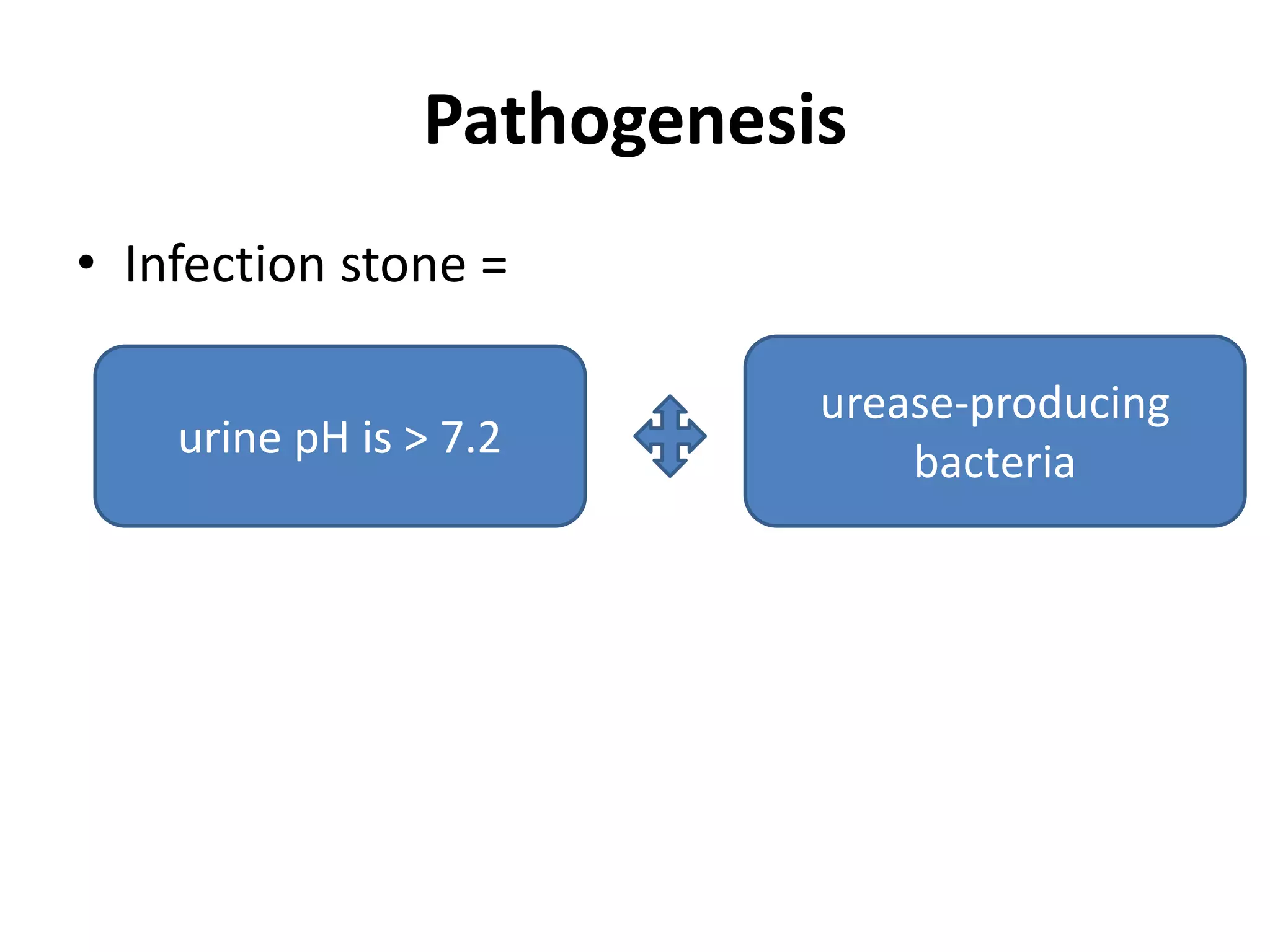
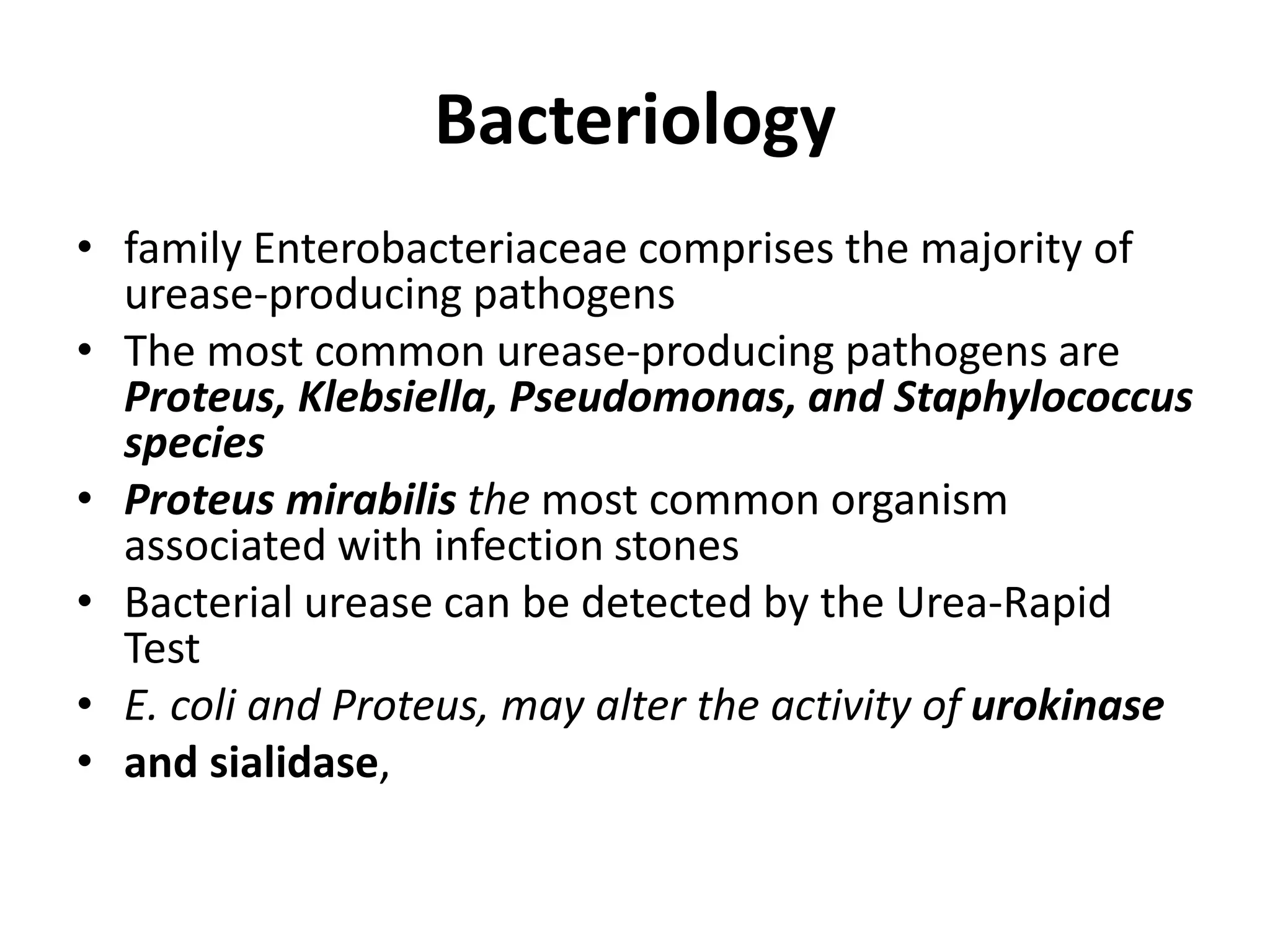
Staghorn Calculus – Etiology,
Management & Prevention
BACKGROUND
• infection stones, struvite, triple phosphate stones,
staghorn calculus.
• Magnesium Ammonium Phosphate
(MgNH4PO4·6H2O) + calcium carbonate apatite
crystals (Ca10(PO4)6·CO3)
Potential for morbidity and mortality:
1. Untreated infection stones – progressive renal demise
2. Inadequately treated struvite stone – niduses for
recurrent UTI and recurrent struvite stone formation.
3. bacteria reside within these stones – life-threatening
sepsis
Pathogenesis
• Infection stone =
urine pH is > 7.2
urease-producing
bacteria
• (NH2 )2CO + H2O → 2NH3 + CO2
• NH3 + H2O → NH4
+ + OH− pK = 9.0
• presence of urease, ammonia continues to be
produced despite alkaline urine, further
increasing urinary pH.
• Promotes the hydration of carbon dioxide to
carbonic acid
• CO2 +H2O→ H2CO3 pK = 4.5
• H2CO3 → H+ + HCO3
− pK = 6.3
• HCO3- → H+ + CO3
2- pK= 10.2
• The dissociation of hydrogen phosphate under
alkaline conditions provides phosphate
OTHER FACTORS:
• The relative decrease in stone inhibitors(citrate) may also
play a role in struvite physicochemistry.
• GAGs theory
• Stasis
Bacteriology
• family Enterobacteriaceae comprises the majority of
urease-producing pathogens
• The most common urease-producing pathogens are
Proteus, Klebsiella, Pseudomonas, and Staphylococcus
species
• Proteus mirabilis the most common organism
associated with infection stones
• Bacterial urease can be detected by the Urea-Rapid
Test
• E. coli and Proteus, may alter the activity of urokinase
• and sialidase,
Epidemiology
• Infection stones comprise 5% to 15% of all stones
• More often in women (ratio of 2 : 1)
Increased risk for infection calculi:
1. Elderly
2. premature infants
3. diabetics
4. urinary stasis as a result of urinary tract obstruction,
urinary diversion, or neurologic disorders.
5. Spinalcord–injured patients
6. use of indwelling catheters
CLINICAL FINDINGS
A complete history
• of chronic flank pain, malaise, fever,
• dysuria, and intermittent hematuria
• immunosuppressed state (diabetes mellitus,
steroid intake, etc.),
• history of previous stone disease
• past surgical history - for urological procedures
• history of using multiple, alternating antibiotics
Physical Examination
• a chronically ill-appearing patient
• Body habitus,
• presence of vertebral kyphoscoliosis
• In acute pyelonephritis or pyonephrosis -
1. toxic appearance
2. costovertebral angle tenderness
Laboratory studies
• complete blood count
• basic metabolic panel
• Urinalysis
• Urine culture
Imaging Modalities
• Renal sonography
• X ray KUB
• IVP
• CT urography
• Nuclear renography
NATURAL HISTORY OF INFECTION
STAGHORN CALCULI
• Pyonephrosis
• xanthogranulomatous pyelonephritis
• end stage hydronephrotic kidney
• severe pyelonephritic changes
• Perinephric abscess
• Carcinoma
• the overall rate of renal deterioration was 28%
– Solitary, previous, recurrent, hypertension, complete,
diversion, neurogenic bladder, refused treatment
• asymptomatic
TREATMENT
• The primary goal of staghorn stone management is
complete stone eradication.
• Various modalities of treatments are:
– Surgical
• PCNL
• ESWL
• OPEN
• SANDWICH THERAPY
– Non surgical
• Dissolution therapy
• Antibiotics
• urease inhibitors,
• urinary acidification,
• dietary modification.
Percutaneous Nephrostolithotomy (PCNL)
the treatment of choice
• superior stone-free outcomes
• acceptably low morbidity.
• Stone free rate of ~80%
• overall risk of transfusion was 18%
• serious complications was 15%. i.e.
– injury to adjacent organs (colon, spleen, liver),
– hydropneumothorax,
– collecting system perforations,
– sepsis,
– vascular injury,
– renal loss.
Technical advances in PCNL
1. flexible nephroscopy is mandatory after
debulking the dominant stone
2. to establish multiple percutaneous tracts
3. second look nephroscopy.
Extracorporeal Shockwave Lithotripsy
(Monotherapy)
• SWL is the least invasive of the operative
approaches
• SWL monotherapy had the lowest success rate.
• Risks included
– colic requiring admission,
– significant perirenal hematoma,
– obstruction including steinstrasse,
– pyelonephritis,
– renal loss.
• “sandwich therapy”- pcnl -> eswl -> pcnl
Ureteroscopy
• flexible ureteroscopy has been used in
combination with PCNL
– to avoid multiple access tracts
– to access calyces that would be difficult to access
in an antegrade manner
Open & Laparoscopic Surgery
• Anatrophic nephrolithotomy and pyelolithotomy
operations
• alternative in patients who require concomitant
heminephrectomy, pyeloplasty
• in those with ectopic kidneys that cannot be
safely accessed percutaneously
• Other indications:
– morbid obesity,
– large symptomatic anterior caliceal diverticular
stones,
– large stone volume with infundibular stenosis
– massive collecting system dilation
Dissolution therapy
• Boric acid and permanganate
• Suby’s solution G
• Hemiacidrin or Renacidin® adding D-gluconic acid.
• following precautions must be exercised during intrarenal
chemolysis:
– Low intrarenal pressures must be maintained (<30 cm water),
– Serum magnesium and phosphate must be monitored closely,
– The urine must be sterile. Broad-spectrum antibiotics are given for 14
days in the perioperative period,
– The collecting system must be unobstructed and there must be no
extravasation.
• Indication: in high-risk patients, with residual calculi after
percutaneous renal surgery.
• Demerits : prolonged hospital stay, cost and risk of complications.
Antibiotics
• Culture-specific preoperative and
perioperative antibiotics are critical to prevent
sepsis
• Long-term, low-dose, culture specific
antimicrobials are important to prevent new
stone growth and progression after surgery.
• AUA Guidelines Panel stated emphatically that
treatment with antibiotics alone is not
standard of care.
Urease Inhibitors
• Acetohydroxamic acid (AHA) is the only FDA-approved
urease inhibitor.
• Irreversibly inhibits bacterial urease
• High renal clearance,
• Penetrate the bacterial cell wall,
• Acts synergistically with several antibiotics
• Adverse effects- tremulousness, thrombophlebitis,
neurologic, hematologic, and dermatologic.
• Contraindicated in patients with serum creatinine
greater than 2.5 mg/dL
Urinary acidification
• L-methionine to acidify urine
• oral intake of 1,500–3,000 mg daily of L-
methionine
• gastric patch pyeloplasty (animal model)
Dietary modification
Aim :
To deplete the substrates of struvite calculi,
including urinary phosphate, magnesium, and
ammonia.
• (Shorr regimen) a regimen of a low-phosphorous,
low-calcium diet with oral estrogens and
aluminum hydroxide gel
• Adverse effects: constipation, anorexia, lethargy,
bone pain, and hypercalciuria, increased risk of
breast and uterine cancers.
Staghorn calculus – etiology, diagnosis, management

Staghorn calculus – etiology, diagnosis, management

  • 1.
    Staghorn Calculus –Etiology, Management & Prevention
  • 2.
    BACKGROUND • infection stones,struvite, triple phosphate stones, staghorn calculus. • Magnesium Ammonium Phosphate (MgNH4PO4·6H2O) + calcium carbonate apatite crystals (Ca10(PO4)6·CO3) Potential for morbidity and mortality: 1. Untreated infection stones – progressive renal demise 2. Inadequately treated struvite stone – niduses for recurrent UTI and recurrent struvite stone formation. 3. bacteria reside within these stones – life-threatening sepsis
  • 3.
    Pathogenesis • Infection stone= urine pH is > 7.2 urease-producing bacteria
  • 4.
    • (NH2 )2CO+ H2O → 2NH3 + CO2 • NH3 + H2O → NH4 + + OH− pK = 9.0 • presence of urease, ammonia continues to be produced despite alkaline urine, further increasing urinary pH. • Promotes the hydration of carbon dioxide to carbonic acid • CO2 +H2O→ H2CO3 pK = 4.5 • H2CO3 → H+ + HCO3 − pK = 6.3 • HCO3- → H+ + CO3 2- pK= 10.2
  • 5.
    • The dissociationof hydrogen phosphate under alkaline conditions provides phosphate OTHER FACTORS: • The relative decrease in stone inhibitors(citrate) may also play a role in struvite physicochemistry. • GAGs theory • Stasis
  • 6.
    Bacteriology • family Enterobacteriaceaecomprises the majority of urease-producing pathogens • The most common urease-producing pathogens are Proteus, Klebsiella, Pseudomonas, and Staphylococcus species • Proteus mirabilis the most common organism associated with infection stones • Bacterial urease can be detected by the Urea-Rapid Test • E. coli and Proteus, may alter the activity of urokinase • and sialidase,
  • 9.
    Epidemiology • Infection stonescomprise 5% to 15% of all stones • More often in women (ratio of 2 : 1) Increased risk for infection calculi: 1. Elderly 2. premature infants 3. diabetics 4. urinary stasis as a result of urinary tract obstruction, urinary diversion, or neurologic disorders. 5. Spinalcord–injured patients 6. use of indwelling catheters
  • 10.
    CLINICAL FINDINGS A completehistory • of chronic flank pain, malaise, fever, • dysuria, and intermittent hematuria • immunosuppressed state (diabetes mellitus, steroid intake, etc.), • history of previous stone disease • past surgical history - for urological procedures • history of using multiple, alternating antibiotics
  • 11.
    Physical Examination • achronically ill-appearing patient • Body habitus, • presence of vertebral kyphoscoliosis • In acute pyelonephritis or pyonephrosis - 1. toxic appearance 2. costovertebral angle tenderness
  • 12.
    Laboratory studies • completeblood count • basic metabolic panel • Urinalysis • Urine culture
  • 13.
    Imaging Modalities • Renalsonography • X ray KUB • IVP • CT urography • Nuclear renography
  • 15.
    NATURAL HISTORY OFINFECTION STAGHORN CALCULI • Pyonephrosis • xanthogranulomatous pyelonephritis • end stage hydronephrotic kidney • severe pyelonephritic changes • Perinephric abscess • Carcinoma • the overall rate of renal deterioration was 28% – Solitary, previous, recurrent, hypertension, complete, diversion, neurogenic bladder, refused treatment • asymptomatic
  • 16.
    TREATMENT • The primarygoal of staghorn stone management is complete stone eradication. • Various modalities of treatments are: – Surgical • PCNL • ESWL • OPEN • SANDWICH THERAPY – Non surgical • Dissolution therapy • Antibiotics • urease inhibitors, • urinary acidification, • dietary modification.
  • 17.
    Percutaneous Nephrostolithotomy (PCNL) thetreatment of choice • superior stone-free outcomes • acceptably low morbidity. • Stone free rate of ~80% • overall risk of transfusion was 18% • serious complications was 15%. i.e. – injury to adjacent organs (colon, spleen, liver), – hydropneumothorax, – collecting system perforations, – sepsis, – vascular injury, – renal loss.
  • 18.
    Technical advances inPCNL 1. flexible nephroscopy is mandatory after debulking the dominant stone 2. to establish multiple percutaneous tracts 3. second look nephroscopy.
  • 19.
    Extracorporeal Shockwave Lithotripsy (Monotherapy) •SWL is the least invasive of the operative approaches • SWL monotherapy had the lowest success rate. • Risks included – colic requiring admission, – significant perirenal hematoma, – obstruction including steinstrasse, – pyelonephritis, – renal loss. • “sandwich therapy”- pcnl -> eswl -> pcnl
  • 20.
    Ureteroscopy • flexible ureteroscopyhas been used in combination with PCNL – to avoid multiple access tracts – to access calyces that would be difficult to access in an antegrade manner
  • 21.
    Open & LaparoscopicSurgery • Anatrophic nephrolithotomy and pyelolithotomy operations • alternative in patients who require concomitant heminephrectomy, pyeloplasty • in those with ectopic kidneys that cannot be safely accessed percutaneously • Other indications: – morbid obesity, – large symptomatic anterior caliceal diverticular stones, – large stone volume with infundibular stenosis – massive collecting system dilation
  • 22.
    Dissolution therapy • Boricacid and permanganate • Suby’s solution G • Hemiacidrin or Renacidin® adding D-gluconic acid. • following precautions must be exercised during intrarenal chemolysis: – Low intrarenal pressures must be maintained (<30 cm water), – Serum magnesium and phosphate must be monitored closely, – The urine must be sterile. Broad-spectrum antibiotics are given for 14 days in the perioperative period, – The collecting system must be unobstructed and there must be no extravasation. • Indication: in high-risk patients, with residual calculi after percutaneous renal surgery. • Demerits : prolonged hospital stay, cost and risk of complications.
  • 23.
    Antibiotics • Culture-specific preoperativeand perioperative antibiotics are critical to prevent sepsis • Long-term, low-dose, culture specific antimicrobials are important to prevent new stone growth and progression after surgery. • AUA Guidelines Panel stated emphatically that treatment with antibiotics alone is not standard of care.
  • 24.
    Urease Inhibitors • Acetohydroxamicacid (AHA) is the only FDA-approved urease inhibitor. • Irreversibly inhibits bacterial urease • High renal clearance, • Penetrate the bacterial cell wall, • Acts synergistically with several antibiotics • Adverse effects- tremulousness, thrombophlebitis, neurologic, hematologic, and dermatologic. • Contraindicated in patients with serum creatinine greater than 2.5 mg/dL
  • 25.
    Urinary acidification • L-methionineto acidify urine • oral intake of 1,500–3,000 mg daily of L- methionine • gastric patch pyeloplasty (animal model)
  • 26.
    Dietary modification Aim : Todeplete the substrates of struvite calculi, including urinary phosphate, magnesium, and ammonia. • (Shorr regimen) a regimen of a low-phosphorous, low-calcium diet with oral estrogens and aluminum hydroxide gel • Adverse effects: constipation, anorexia, lethargy, bone pain, and hypercalciuria, increased risk of breast and uterine cancers.