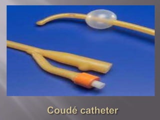
2. One method of examining bladder function is by
injecting dye that is visible on X-rays through a
tube (catheter) to fill the bladder. X-rays are taken
while the bladder is full and while the patient is
urinating (voiding) to determine if fluid is forced
out of the bladder through the urethra (normal) or
up through the ureters into the kidney
(vesicoureteral reflux). This study is usually done
with the patient lying on an X-ray table.
3. This test is commonly performed to diagnose
the cause of:
Urinary tract infections
Abnormal bladder emptying
Present at birth (congenital) problems with
the bladder or urethra
Urethral stricture (in males)
4. The test is performed in a hospital radiology
department or in a health care provider's office by an x-
ray technician.
You will lie on your back on the x-ray table. A thin,
flexible tube called a catheter will be gently inserted
into the urethra (the tube that carries urine from the
bladder to the outside of the body) and passed into the
bladder.
Contrast dye flows through the catheter into the
bladder. This dye helps the bladder show up better on
x-ray images.
The x-rays are taken from various angles while the
bladder is full of contrast dye. The catheter is removed
so that you can urinate. Images are taken while you
empty your bladder.
5. Risks:
You may have some discomfort when urinating after this
test because of irritation from the catheter.
You may have bladder spasms after this test, which may
be a sign of an allergic reaction to the contrast dye.
Abnormal results may indicate the following:
Narrowing of the urethra (urethral stricture)
Neurogenic bladder
Pouch-like sacs (diverticula) on the walls of the bladder or
urethra
Prostatic enlargement
Reflux nephropathy
Ureterocele
6.
7. A retrograde pyelogram is a diagnostic X-ray
procedure used in combination with a cystoscopy
to determine the presence of stones, tumors, or
other obstructions in the kidneys and ureters.
These diseases or medical conditions may be
diagnosed by, screened for, or associated with
Retrograde pyelogram:
Kidney stones
Blood clots
Tumors
Urethral obstruction
Renal stricture
Urethral stricture
8. During the retrograde pyelogram, the patient will
first be asked to remove any clothing or jewelry
that may interfere with the procedure. Some
patients may have an intravenous line (IV) inserted
in their arm or hand to administer a mild sedative
before the procedure. Next, the patient will be
asked to lie face up on the X-ray table with his or
her legs in stirrups.
Then an endoscope will be inserted through the
urethral opening, and then into the bladder. The
endoscope can be a rigid or flexible tube that may
have a light attached to it for increased visibility.
This will be followed by a catheter, through which
the dye will be injected. A series of x rays will be
taken during timed intervals to track the dye as it
travels through the bladder and the kidneys.
9. Having a retrograde pyelogram carries
minimal risk. The amount of radiation in the
dye used during the procedure is very low, but
patients who may be pregnant may be advised
to avoid this procedure. The dye used may also
cause allergic reactions in a small percentage of
patients. Anyone with an allergy or sensitivity
to any types of dyes, iodine, or shellfish should
be careful. Patients with pregnancies or
allergies should discuss these concerns with
their physician before having a retrograde
pyelogram done.
10.
11. An intravenous pyelogram, also called intravenous
urography, is a diagnostic x-ray of the kidneys,
ureters, and bladder. When a contrast dye is
injected intravenously (IV), the urinary tract will
show up very clearly, which is not seen on regular
x-rays. An intravenous pyelogram may be done for
many reasons, including the following:
to detect kidney tumors
to identify blockages or obstructions of the
normal flow of urine
to detect kidney or bladder stones
to establish if the prostate gland is enlarged
to detect injuries to the urinary tract
12. An intravenous pyelogram procedure follows this process:
The patient will be positioned on the x-ray table.
A preliminary x-ray will be taken.
An intravenous (IV) line will be started in the hand or arm for injection
of the contrast dye.
The radiologist will inject the contrast dye into the vein in the arm.
During the injection of the contrast dye, the patient may feel warm and
become flushed, only for a minute or so. This reaction is normal.
Rarely, patients may experience an allergic reaction to the contrast dye,
resulting in persistent itching or possibly hives. These symptoms are
easily treated with medication.
X-rays will be taken at intervals after the dye has been injected.
At times the patient may have to change positions as the x-rays are
taken. The patient may be asked to wear a compression band around
the body to better visualize the urinary structures that lead from the
kidney.
The patient will be asked to empty the bladder.
A final x-ray will be taken after urination to determine the amount of
contrast dye remaining in the urinary tract.
13. Diagnoses:
Chronic Pyelonephritis
Kidney stones
Renal cell carcinoma or RCC
Transitional cell carcinoma, or TCC
Polycystic kidneys
Anatomical variations, i.e. horseshoe kidney or a
duplex collecting system
Obstruction (commonly at the pelvic-ureteric
junction or PUJ and the vesicoureteric junction or
VUJ)
14. The dyes are of 2 types: ionic and nonionic. Both types of dye
contain iodine but differ in 2 key ways: the rate of adverse reactions
and the cost.
Although the overall rate of adverse reactions is relatively low with
both, there is a greater incidence of adverse reactions with the less
expensive ionic dye than with the nonionic.
Minor reactions, which are infrequent and do not last long, include
flushing, nausea, vomiting, and itching.
A small percentage of people experience a severe reaction to the
dye, such as difficulty breathing, speaking, or swallowing; swelling
of the lips and tongue; low blood pressure; or loss of consciousness.
People who have had a severe reaction after receiving the dye once
should not be exposed to it again.
Pregnant women should not have an IVP because of the high
radiation exposure.
15. People with known kidney disease or failure should not
have an IVP because the dye can worsen kidney function.
Elderly people and those with diabetes, high blood pressure,
heart disease, or evidence of dehydrating are at risk of
developing kidney failure following administration of the
dye.
To avoid this complication, the kidney function should be
tested with a blood test of the BUN (blood urea nitrogen)
and creatinine, and the results should be known before the
IVP is performed.
Those with diabetes and certain others (for example, women
with polycystic ovarian syndrome) who are taking
metformin (Glucophage) will have to discontinue this
medication for 2 days after the IVP. They should inform
their doctor of the test, and the doctor will coordinate their
management during that time.
16. Diagnoses:
Chronic Pyelonephritis
Kidney stones
Renal cell carcinoma or RCC
Transitional cell carcinoma, or TCC
Polycystic kidneys
Anatomical variations, i.e. horseshoe kidney or a
duplex collecting system
Obstruction (commonly at the pelvic-ureteric
junction or PUJ and the vesicoureteric junction or
VUJ)
17.
18. ESWL uses shock waves to break a kidney stone
into small pieces that can more easily travel
through the urinary tract and pass from the body.
ESWL may be used on people with a kidney stone
that is causing pain or blocking the urine flow.
Stones that are between 4mm and 2cm in diameter
are most likely to be treated with ESWL.
ESWL may work best for kidney stones in the
kidney, not in the ureter. It may be harder for
ESWL to break up a stone that has moved into the
ureter, although this is still possible. Your surgeon
may try to push the stone back into the kidney
with a small instrument (ureteroscope) and then
use ESWL
19. •ESWL is usually not used if you:
•Are pregnant. The sound waves and X-rays may be
harmful to the fetus.
•Have a bleeding disorder.
•Have a kidney infection, urinary tract infection, or
kidney cancer.
•Have kidneys with abnormal structure or function.
•Risks
•Complications of ESWL include:
•Pain caused by the passage of stone fragments.
•Blocked urine flow as a result of stone fragments
becoming stuck in the urinary tract. The fragments may
then need to be removed with a ureteroscope.
•Urinary tract infection.
•Bleeding around the outside of the kidney.
20.
21.
22. A plastic tube known as a urinary catheter that is gently
slid into a patient's bladder via his or her urethra.
Catheterization allows the patient's urine to drain freely
from the bladder for collection, or to inject liquids used
for treatment or diagnosis of bladder conditions.
Urinary catheterization maintains urine output in
patients who are undergoing surgery, or who are
confined to the bed and physically unable to use a
bedpan. Critically ill patients who require strict
monitoring of urinary output are also frequently
catheterized.
23. Catheters come in several basic designs:
•A Foley catheter (indwelling urinary catheter) is retained by
means of a balloon at the tip which is inflated with sterile water.
The balloons typically come in two different sizes: 5 cc and 30 cc.
They are commonly made in silicone rubber or natural rubber.
•A Robinson catheter is a flexible catheter used for short term
drainage of urine. Unlike the Foley catheter, it has no balloon on
its tip and therefore cannot stay in place unaided.
•A Coudé catheter is designed with a curved tip that makes it
easier to pass through the curvature of the prostatic urethra.
•A hematuria catheter is a type of Foley catheter used for Post-
TURP hemostasis. This is useful following endoscopic surgical
procedures or in the case of gross hematuria. There are both 2-
way and 3-way hematuria catheters (double and triple lumen).[
•An external Texas or condom catheter is used for incontinent
males and carries a lower risk of infection than an indwelling
catheter.
24. In males, the catheter tube is inserted into the urinary tract
through the penis. A condom or Texas catheter can also be used.
In females, the catheter is inserted into the urethral meatus, after a
cleansing using povidone-iodine.
29. HOW TO CARE FOR A CATHETER
•To care for an indwelling catheter, cleanse the urethral area
(where the catheter exits the body) and the catheter itself with
soap and water every day. Also thoroughly cleanse the area after
all bowel movements to prevent infection.
•If you have a suprapubic catheter, the opening in your belly and
the tube must be cleansed daily with soap and water, then
covered with dry gauze.
•Drinking plenty of fluids can help prevent infections. Discuss this
issue with your health care provider.
•Wash your hands before and after handling the drainage device.
Do not allow the outlet valve to touch anything. If the outlet
becomes obviously dirty, clean it with soap and water.
•Some people have occasional leakage of urine around the
catheter. This may be caused by a catheter that is too small,
improper balloon size, constipation constipation, urinary tract
infections,or bladder spasms.
30. POTENTIAL COMPLICATIONS:
•Allergy or sensitivity to latex
•Bladder stones
•Blood infections (septicemia)
•Blood in the urine (hematuria)
•Kidney damage (usually only with long-term, indwelling catheter use)
•Urethral injury
•Urinary tract or kidney infections
•Bladder spasms that do not go away
•Bleeding into or around the catheter
•Catheter draining very little or no urine, despite drinking enough
fluids
•Fever or chills
•Leakage of large amounts of urine around the catheter
•Skin breakdown around a suprapubic catheter
•Stones or sediment in the urinary catheter or drainage bag
•Swelling of the urethra around the catheter
•Urine with a strong smell, or that is thick or cloudy
•If the catheter becomes clogged, painful, or infected it will need to be
replaced immediately.