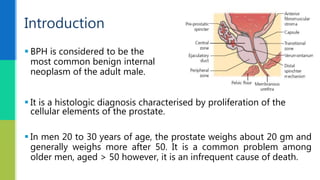
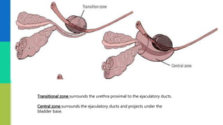
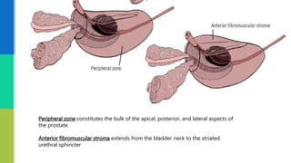
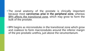
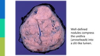
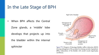
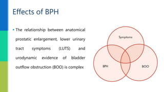
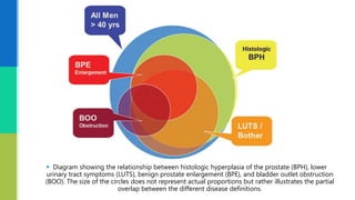
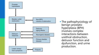
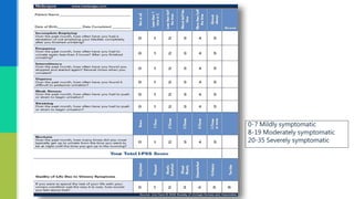
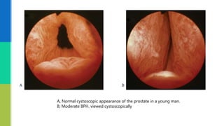
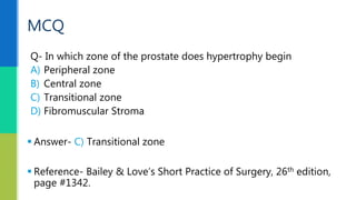
Pathology & Investigation
Benign Prostatic Hyperplasia
Presented by Dr. Ankur Agarwal
Surgical Resident
 Introduction
 Etiology
 Pathology
 Clinical findings
 Investigation
 MCQ
 References
Scheme of Presentation
 It is a histologic diagnosis characterised by proliferation of the
cellular elements of the prostate.
 In men 20 to 30 years of age, the prostate weighs about 20 gm and
generally weighs more after 50. It is a common problem among
older men, aged > 50 however, it is an infrequent cause of death.
 BPH is considered to be the
most common benign internal
neoplasm of the adult male.
Introduction
 The prevalence of histologically diagnosed prostatic hyperplasia
increases from
 8 percent in men aged 31 to 40,
 40 to 50 percent in men aged 51 to 60,
 >80 percent in men older than age 80
 BPH is considered a normal part ageing process in men, serum testosterone
level decreases slowly but the level of oestrogenic steroids is not decreased
equally.
 According to theory, this increased levels of oestrogenic steroids  cause for
prostate enlargement.
 The secretion of intermediate peptide growth factors also plays a part in the
development of BPH.
Etiology
 Androgens are necessary for both normal and abnormal
development of the prostate.
 Testosterone is converted to a more potent androgen,
dihydrotestosterone (DHT), by the enzyme 5-α reductase type 2.
The type 1 form of the enzyme is present in liver and skin.
 Men who congenitally lack 5-α reductase type 2 enzyme have
normal serum testosterone levels but lack dihydrotestosterone.
 Those with this disorder have a rudimentary prostate
throughout life but rarely experience bladder outlet obstruction
secondary to BPH.
 These findings suggest that the active androgen, DHT, is
important in promoting growth of prostate that would
eventually lead to symptomatic BPH.
 Prostatic hyperplasia increases urethral resistance causing
compensatory changes in bladder function.
 Obstruction induced changes in detrusor function, compounded
by age related changes in both bladder and nervous system
function lead to
 Urinary frequency
 Urgency
 Nocturia
Pathophysiology
 McNeal demonstrated that BPH first
develops in the peri-urethral
transition zone of the prostate.
 BPH affects both glandular
epithelium and connective
tissue stroma.
Pathology
Transitional zone surrounds the urethra proximal to the ejaculatory ducts.
Central zone surrounds the ejaculatory ducts and projects under the
bladder base.
Peripheral zone constitutes the bulk of the apical, posterior, and lateral aspects of
the prostate.
Anterior fibromuscular stroma extends from the bladder neck to the striated
urethral sphincter
 The zonal anatomy of the prostate is clinically important
because most carcinomas arise in the peripheral zone, whereas
BPH affects the transitional zone, which may grow to form the
bulk of the prostate.
 BPH begins as micronodules in the transitional zone which grow
and coalesce to form macronodules around the inferior margin
of the pre-prostatic urethra, just above the verumontanum.
 Macronodules in turn compress the surrounding normal tissue
of the peripheral zone postero-inferiorly, creating a ‘false
capsule' around the hyperplastic tissue which coincidentally
provides a plane of cleavage for its surgical enucleation.
Well-defined
nodules compress
the urethra
(arrowheads) into
a slit-like lumen.
 As the transitional zone grows, it produces the appearance of
‘lobes' on either side of the urethra. In due course, these lobes
may compress or distort the preprostatic and prostatic parts of
the urethra and produce symptoms.
 The central zone surrounding the ejaculatory ducts is rarely
involved in any disease.
 BPH typically affects the submucous group of glands in the
transitional zone, forming a nodular enlargement.
 This overgrowth compresses the PZ glands into a false capsule
and causes the appearance of the typical ‘lateral’ lobe.
 The unique feature of human prostate is the presence of prostatic
capsule- plays an important role in the development of Lower urinary
tract symptoms.
 The capsule transmits the “pressure” of tissue expansion to the
urethra and leads to an increase in urethral resistance.
In the Late Stage of BPH
• When BPH affects the Central
Zone glands, a ‘middle’ lobe
develops that projects up into
the bladder within the internal
sphincter
 The relationship between anatomical
prostatic enlargement, lower urinary
tract symptoms (LUTS) and
urodynamic evidence of bladder
outflow obstruction (BOO) is complex
Effects of BPH
 Diagram showing the relationship between histologic hyperplasia of the prostate (BPH), lower
urinary tract symptoms (LUTS), benign prostate enlargement (BPE), and bladder outlet obstruction
(BOO). The size of the circles does not represent actual proportions but rather illustrates the partial
overlap between the different disease definitions.
 The pathophysiology of
benign prostatic
hyperplasia (BPH)
involves complex
interactions between
urethral obstruction,
detrusor function and
dysfunction, and urine
production.
 Prostatic hyperplasia increases urethral resistance, resulting in
compensatory changes in bladder function.
 However, the elevated detrusor pressure required to maintain
urinary flow in the presence of increased outflow resistance
occurs at the expense of normal bladder storage function.
 Chronic bladder outlet obstruction (BOO) secondary to BPH
leads to
 Urinary retention
 Renal insufficiency
 Recurrent UTI
 Bladder wall thinning and loss of function due to constant
distension
 Haematuria
 Vesical calculi
 The prostatic urethra is lengthened (twice its normal length), but it is
not narrowed anatomically.
 The normal posterior curve may be so exaggerated- requires a
curved catheter to negotiate it.
 When only one lateral lobe is enlarged, distortion of the prostatic
urethra occurs.
Effects of BPH on the Urethra
 If BPH causes BOO, the musculature
of the bladder hypertrophies to
overcome the obstruction and
appears trabeculated .
 BPH is associated with increased
blood flow, and the resultant veins
at the base of the bladder causes
haematuria.
Effects of BPH on the Urinary Bladder
 Not all symptoms of disturbed voiding in ageing men should
therefore be attributed to BPH causing BOO.
 Pathophysiologically, BOO may be caused in part by increased
smooth muscle tone, which is under the control of a-adrenergic
agonists
Lower Urinary Tract Symptoms (LUTS)
 The following conditions can coexist with BOO:
oIdiopathic detrusor overactivity.
oNeuropathic bladder dysfunction as a result of diabetes, strokes,
Alzheimer’s disease or Parkinson’s disease.
oDegeneration of bladder smooth muscle giving rise to impaired voiding
and detrusor instability;
oBOO due to BPH.
 Hesitancy (worsened if the bladder is very full)
 Poor flow (unimproved by straining)
 Intermittent stream – stops and starts
 Dribbling (including after micturition)
 Sensation of poor bladder emptying
 Episodes of near retention
Voiding symptoms
 Frequency
 Nocturia
 Urgency
 Urge incontinence
 Nocturnal incontinence (enuresis)
Storage symptoms-Overactive Bladder
Syndrome
 This is a urodynamic concept
based on the combination of
low flow rates in the presence
of high voiding pressures.
 Diagnosed definitively only by
pressure-flow studies.
Bladder Outflow Obstruction
 Flow rates provide a useful guide for everyday clinical
management.
 Urodynamically proven BOO may result from:
 BPH
 Bladder neck stenosis
 Bladder neck hypertrophy
 Prostate cancer
 Urethral strictures
 Functional obstruction due to neuropathic conditions
Effects of BOO on the Bladder
Decrease in urinary flow rates :
For a voided volume > 200 ml, a peak flow rate of > 15 ml/s is normal, 10–15 ml/s is equivocal
and < 10 ml/s is low.
 Increase in voiding pressures
:
 Pressures > 80 cmH2O
are high, pressures
between 60 and 80
cmH2O are equivocal and
pressures < 60 cm H2O
are normal.
 Acute retention of urine
 Chronic retention
 Impaired emptying of bladder
 Haematuria
Complications of BOO
 Other than pain from retention, pain is not a symptom of BOO
 Its presence should prompt the exclusion of acute retention,
urinary infection, stones, carcinoma of the prostate and
carcinoma in situ of the bladder
 It is critical to exclude causes of LUTS other than BPH before medical or
surgical treatment.
 The differential diagnosis of lower urinary tract symptoms in addition to
BPH includes the following:
 Urethral stricture
 Bladder neck contracture
 Carcinoma of the prostate
 Carcinoma of the bladder
 Bladder calculi
 Urinary tract infection and prostatitis
 Neurogenic bladder
 History:
 Symptom assessment:
 The International Prostate Symptom Score (IPSS) is
recommended scoring system for the baseline assessment
of symptom, severity in men presenting with LUTS
Assessment of the Patient with LUTS
 Generally WNL
 Chronic retention- distended bladder
 Loss of the transverse supra pubic skin crease
 Examination of the external urethral meatus is done to exclude
stenosis or a palpable urethral mass
 Epididymides are palpated for signs of inflammation
Abdominal Examination
 The posterior surface of the prostate is
smooth, convex and firm in consistency.
 The rectal mucosa can be made to move
over the prostate.
 Residual urine may be felt as a fluctuating
swelling above the prostate.
 Considerable amount of residual urine if
present, pushes the prostate downwards,
making it appear larger than it is.
Digital Rectal Examination
 To eliminate neurological conditions like diabetes mellitus, tabes
dorsalis, disseminated sclerosis, cervical spondylosis, Parkinson’s
disease may mimic prostatic obstruction.
 Examination of perianal sensation and anal tone is useful in
detection of an S2 to S4 cauda equina lesion.
Nervous System
 A urinalysis should be done either by using a dipstick test or
microscopic examination of the spun sediment to rule out UTI
and haematuria, either of which strongly suggest a non-BPH
pathologic process as a cause of symptoms.
 Urine cytology should always be requested in men with severe
irritable symptoms and dysuria, especially if they have a
smoking history. Carcinoma in-situ of the bladder is a diagnosis
that may have serious consequences if overlooked.
Urinalysis
 Postvoid residual urine volume is the amount of fluid remaining in
the bladder immediately after the completion of micturition.
 Studies indicate that PVR urine volume normally ranges from 0.09 to
2.24 mL, with the mean being 0.53 mL (Hinman and Cox, 1967).
 Seventy-eight percent of normal men have PVR volumes of less than
5 mL, and 100% have volumes of less than 12 mL(DiMare et al, 1963).
USG & Postvoid Residual Urine Volume
 Traditionally, urologists have assumed that increasing amounts
of PVR urine denote BPH progression and are thus an
“indication” for surgery.
 Normally present in Serum but if > 2.5–4 nmol/L, then
transrectal ultrasound scanning (TRUS) plus multiple transrectal
biopsies (10 biopsies) should be considered.
Serum Prostate – Specific Antigen
 Special flow rate clinic.
 Coupled with ultrasound measurement
of post-void residual urine
Flow Rate Measurement
A, Normal cystoscopic appearance of the prostate in a young man.
B, Moderate BPH, viewed cystoscopically
Q- In which zone of the prostate does hypertrophy begin
A) Peripheral zone
B) Central zone
C) Transitional zone
D) Fibromuscular Stroma
MCQ
 Answer- C) Transitional zone
 Reference- Bailey & Love’s Short Practice of Surgery, 26th edition,
page #1342.
 Bailey and Love’s Short Practice of Surgery, 26th Edition
 Sabiston Textbook of Surgery, 19th Edition
 Campbell – Walsh Urology, 10th Edition
 Robbins Basic Pathology, 9th Edition
 Gray’s Anatomy, 40th Edition
References
Thank You

BPH- Pathology & Investigations

  • 1.
    Pathology & Investigation BenignProstatic Hyperplasia Presented by Dr. Ankur Agarwal Surgical Resident
  • 2.
     Introduction  Etiology Pathology  Clinical findings  Investigation  MCQ  References Scheme of Presentation
  • 3.
     It isa histologic diagnosis characterised by proliferation of the cellular elements of the prostate.  In men 20 to 30 years of age, the prostate weighs about 20 gm and generally weighs more after 50. It is a common problem among older men, aged > 50 however, it is an infrequent cause of death.  BPH is considered to be the most common benign internal neoplasm of the adult male. Introduction
  • 4.
     The prevalenceof histologically diagnosed prostatic hyperplasia increases from  8 percent in men aged 31 to 40,  40 to 50 percent in men aged 51 to 60,  >80 percent in men older than age 80
  • 5.
     BPH isconsidered a normal part ageing process in men, serum testosterone level decreases slowly but the level of oestrogenic steroids is not decreased equally.  According to theory, this increased levels of oestrogenic steroids  cause for prostate enlargement.  The secretion of intermediate peptide growth factors also plays a part in the development of BPH. Etiology
  • 6.
     Androgens arenecessary for both normal and abnormal development of the prostate.  Testosterone is converted to a more potent androgen, dihydrotestosterone (DHT), by the enzyme 5-α reductase type 2. The type 1 form of the enzyme is present in liver and skin.
  • 7.
     Men whocongenitally lack 5-α reductase type 2 enzyme have normal serum testosterone levels but lack dihydrotestosterone.  Those with this disorder have a rudimentary prostate throughout life but rarely experience bladder outlet obstruction secondary to BPH.  These findings suggest that the active androgen, DHT, is important in promoting growth of prostate that would eventually lead to symptomatic BPH.
  • 8.
     Prostatic hyperplasiaincreases urethral resistance causing compensatory changes in bladder function.  Obstruction induced changes in detrusor function, compounded by age related changes in both bladder and nervous system function lead to  Urinary frequency  Urgency  Nocturia Pathophysiology
  • 9.
     McNeal demonstratedthat BPH first develops in the peri-urethral transition zone of the prostate.  BPH affects both glandular epithelium and connective tissue stroma. Pathology
  • 11.
    Transitional zone surroundsthe urethra proximal to the ejaculatory ducts. Central zone surrounds the ejaculatory ducts and projects under the bladder base.
  • 12.
    Peripheral zone constitutesthe bulk of the apical, posterior, and lateral aspects of the prostate. Anterior fibromuscular stroma extends from the bladder neck to the striated urethral sphincter
  • 13.
     The zonalanatomy of the prostate is clinically important because most carcinomas arise in the peripheral zone, whereas BPH affects the transitional zone, which may grow to form the bulk of the prostate.  BPH begins as micronodules in the transitional zone which grow and coalesce to form macronodules around the inferior margin of the pre-prostatic urethra, just above the verumontanum.
  • 14.
     Macronodules inturn compress the surrounding normal tissue of the peripheral zone postero-inferiorly, creating a ‘false capsule' around the hyperplastic tissue which coincidentally provides a plane of cleavage for its surgical enucleation.
  • 15.
  • 16.
     As thetransitional zone grows, it produces the appearance of ‘lobes' on either side of the urethra. In due course, these lobes may compress or distort the preprostatic and prostatic parts of the urethra and produce symptoms.  The central zone surrounding the ejaculatory ducts is rarely involved in any disease.
  • 17.
     BPH typicallyaffects the submucous group of glands in the transitional zone, forming a nodular enlargement.  This overgrowth compresses the PZ glands into a false capsule and causes the appearance of the typical ‘lateral’ lobe.
  • 18.
     The uniquefeature of human prostate is the presence of prostatic capsule- plays an important role in the development of Lower urinary tract symptoms.  The capsule transmits the “pressure” of tissue expansion to the urethra and leads to an increase in urethral resistance.
  • 20.
    In the LateStage of BPH • When BPH affects the Central Zone glands, a ‘middle’ lobe develops that projects up into the bladder within the internal sphincter
  • 21.
     The relationshipbetween anatomical prostatic enlargement, lower urinary tract symptoms (LUTS) and urodynamic evidence of bladder outflow obstruction (BOO) is complex Effects of BPH
  • 22.
     Diagram showingthe relationship between histologic hyperplasia of the prostate (BPH), lower urinary tract symptoms (LUTS), benign prostate enlargement (BPE), and bladder outlet obstruction (BOO). The size of the circles does not represent actual proportions but rather illustrates the partial overlap between the different disease definitions.
  • 23.
     The pathophysiologyof benign prostatic hyperplasia (BPH) involves complex interactions between urethral obstruction, detrusor function and dysfunction, and urine production.
  • 24.
     Prostatic hyperplasiaincreases urethral resistance, resulting in compensatory changes in bladder function.  However, the elevated detrusor pressure required to maintain urinary flow in the presence of increased outflow resistance occurs at the expense of normal bladder storage function.
  • 25.
     Chronic bladderoutlet obstruction (BOO) secondary to BPH leads to  Urinary retention  Renal insufficiency  Recurrent UTI  Bladder wall thinning and loss of function due to constant distension  Haematuria  Vesical calculi
  • 26.
     The prostaticurethra is lengthened (twice its normal length), but it is not narrowed anatomically.  The normal posterior curve may be so exaggerated- requires a curved catheter to negotiate it.  When only one lateral lobe is enlarged, distortion of the prostatic urethra occurs. Effects of BPH on the Urethra
  • 27.
     If BPHcauses BOO, the musculature of the bladder hypertrophies to overcome the obstruction and appears trabeculated .  BPH is associated with increased blood flow, and the resultant veins at the base of the bladder causes haematuria. Effects of BPH on the Urinary Bladder
  • 28.
     Not allsymptoms of disturbed voiding in ageing men should therefore be attributed to BPH causing BOO.  Pathophysiologically, BOO may be caused in part by increased smooth muscle tone, which is under the control of a-adrenergic agonists Lower Urinary Tract Symptoms (LUTS)
  • 29.
     The followingconditions can coexist with BOO: oIdiopathic detrusor overactivity. oNeuropathic bladder dysfunction as a result of diabetes, strokes, Alzheimer’s disease or Parkinson’s disease. oDegeneration of bladder smooth muscle giving rise to impaired voiding and detrusor instability; oBOO due to BPH.
  • 30.
     Hesitancy (worsenedif the bladder is very full)  Poor flow (unimproved by straining)  Intermittent stream – stops and starts  Dribbling (including after micturition)  Sensation of poor bladder emptying  Episodes of near retention Voiding symptoms
  • 31.
     Frequency  Nocturia Urgency  Urge incontinence  Nocturnal incontinence (enuresis) Storage symptoms-Overactive Bladder Syndrome
  • 32.
     This isa urodynamic concept based on the combination of low flow rates in the presence of high voiding pressures.  Diagnosed definitively only by pressure-flow studies. Bladder Outflow Obstruction
  • 33.
     Flow ratesprovide a useful guide for everyday clinical management.  Urodynamically proven BOO may result from:  BPH  Bladder neck stenosis  Bladder neck hypertrophy  Prostate cancer  Urethral strictures  Functional obstruction due to neuropathic conditions
  • 34.
    Effects of BOOon the Bladder Decrease in urinary flow rates : For a voided volume > 200 ml, a peak flow rate of > 15 ml/s is normal, 10–15 ml/s is equivocal and < 10 ml/s is low.
  • 35.
     Increase invoiding pressures :  Pressures > 80 cmH2O are high, pressures between 60 and 80 cmH2O are equivocal and pressures < 60 cm H2O are normal.
  • 36.
     Acute retentionof urine  Chronic retention  Impaired emptying of bladder  Haematuria Complications of BOO
  • 37.
     Other thanpain from retention, pain is not a symptom of BOO  Its presence should prompt the exclusion of acute retention, urinary infection, stones, carcinoma of the prostate and carcinoma in situ of the bladder
  • 38.
     It iscritical to exclude causes of LUTS other than BPH before medical or surgical treatment.  The differential diagnosis of lower urinary tract symptoms in addition to BPH includes the following:  Urethral stricture  Bladder neck contracture  Carcinoma of the prostate  Carcinoma of the bladder  Bladder calculi  Urinary tract infection and prostatitis  Neurogenic bladder
  • 39.
     History:  Symptomassessment:  The International Prostate Symptom Score (IPSS) is recommended scoring system for the baseline assessment of symptom, severity in men presenting with LUTS Assessment of the Patient with LUTS
  • 41.
     Generally WNL Chronic retention- distended bladder  Loss of the transverse supra pubic skin crease  Examination of the external urethral meatus is done to exclude stenosis or a palpable urethral mass  Epididymides are palpated for signs of inflammation Abdominal Examination
  • 42.
     The posteriorsurface of the prostate is smooth, convex and firm in consistency.  The rectal mucosa can be made to move over the prostate.  Residual urine may be felt as a fluctuating swelling above the prostate.  Considerable amount of residual urine if present, pushes the prostate downwards, making it appear larger than it is. Digital Rectal Examination
  • 43.
     To eliminateneurological conditions like diabetes mellitus, tabes dorsalis, disseminated sclerosis, cervical spondylosis, Parkinson’s disease may mimic prostatic obstruction.  Examination of perianal sensation and anal tone is useful in detection of an S2 to S4 cauda equina lesion. Nervous System
  • 44.
     A urinalysisshould be done either by using a dipstick test or microscopic examination of the spun sediment to rule out UTI and haematuria, either of which strongly suggest a non-BPH pathologic process as a cause of symptoms.  Urine cytology should always be requested in men with severe irritable symptoms and dysuria, especially if they have a smoking history. Carcinoma in-situ of the bladder is a diagnosis that may have serious consequences if overlooked. Urinalysis
  • 45.
     Postvoid residualurine volume is the amount of fluid remaining in the bladder immediately after the completion of micturition.  Studies indicate that PVR urine volume normally ranges from 0.09 to 2.24 mL, with the mean being 0.53 mL (Hinman and Cox, 1967).  Seventy-eight percent of normal men have PVR volumes of less than 5 mL, and 100% have volumes of less than 12 mL(DiMare et al, 1963). USG & Postvoid Residual Urine Volume
  • 46.
     Traditionally, urologistshave assumed that increasing amounts of PVR urine denote BPH progression and are thus an “indication” for surgery.
  • 48.
     Normally presentin Serum but if > 2.5–4 nmol/L, then transrectal ultrasound scanning (TRUS) plus multiple transrectal biopsies (10 biopsies) should be considered. Serum Prostate – Specific Antigen
  • 49.
     Special flowrate clinic.  Coupled with ultrasound measurement of post-void residual urine Flow Rate Measurement
  • 51.
    A, Normal cystoscopicappearance of the prostate in a young man. B, Moderate BPH, viewed cystoscopically
  • 52.
    Q- In whichzone of the prostate does hypertrophy begin A) Peripheral zone B) Central zone C) Transitional zone D) Fibromuscular Stroma MCQ  Answer- C) Transitional zone  Reference- Bailey & Love’s Short Practice of Surgery, 26th edition, page #1342.
  • 53.
     Bailey andLove’s Short Practice of Surgery, 26th Edition  Sabiston Textbook of Surgery, 19th Edition  Campbell – Walsh Urology, 10th Edition  Robbins Basic Pathology, 9th Edition  Gray’s Anatomy, 40th Edition References
  • 54.