INTRODUCTION
 ACUTE UROLOGICAL CONDITIONS; ONE
OF THE MOST COMMON EMERGENCIES
FACED BY US IN THE CASUALTY
DEPARTMENT.
 INCLUDES DIFFERENT CONDITIONS
RANGING FROM URETHRITIS TO
TESTICULAR TORSION.
CONDITIONS INCLUDED
 NON TRAUMATIC
1) URINARY RETENTION
2) HAEMATURIA
3) RENAL/URETERIC COLIC
4) ACUTE SCROTAL CONDITIONS
5) INFECTIONS
 TRAUMATIC
ACUTE URINARY RETENTION
 Painful inability to void, with relief of pain
following drainage of the bladder by
catheterization.
 Pathophysiology:
 Increased urethral resistance, i.e., bladder outlet
obstruction (BOO)
 Low bladder pressure, i.e., impaired bladder
contractility
 Interruption of sensory or motor innervations of
the bladder
ACUTE URINARY RETENTION
 Causes :
 Men:
 Bladder outlet obstruction (the commonest cause, most
commonly due to BEP)
 Urethral stricture
 Acute urethritis or Prostatitis
 Phimosis
 Women
 Pelvic prolapse (cystocoele, rectocoele, uterine)
 Post surgery for ‘stress’ incontinence
 pelvic masses (e.g., ovarian masses)
 Bladder neck obstruction (rare)
ACUTE URINARY RETENTION
 Both Sex
 Blood clot
 Urethral calculus
 Rupture of the urethra
 Neurogenic (injury or disease of the spinal cord)
 Smooth muscle dysfunction associated with ageing
 Faecal impaction
 Anal pain (haemorrhoidectomy)
 Intensive postoperative analgesic treatment
 Some drugs
 Spinal anaesthesia
ACUTE URINARY RETENTION
 Initial Management :
 Urethral catheterization
 Suprapubic catheter ( SPC)
 Late Management:
 Treating the underlying cause
ACUTE FLANK PAIN—URETERIC OR RENAL COLIC
 The commonest urologic emergency.
 One of the commonest causes of the “Acute
Abdomen”.
 Sudden onset of severe pain in the flank
 Most often due to the passage of a stone
formed in the kidney, down through the
ureter.
RENAL COLIC/ URETERIC COLIC
 When caused by acute obstruction of the renal
pelvis, is typically fixed deep in the loin and
‘bursting’ in character.
 When caused by acute ureteric obstruction (usually
by a stone), is colicky with sharp exacerbations
against a constant background.
 Is liable to be referred to the groin, scrotum or
labium as the calculus obstruction moves distally.
RENAL COLIC/ URETERIC COLIC
Differential diagnoses
 Pyelonephritis
 Abdominal aortic aneurysm
 Appendicitis
 Biliary colic (gallstones)
 Peritonitis
 Diverticulitis
 Salpingitis
 Torted ovarian cyst
 Ectopic pregnancy
 Shingles
RENAL COLIC/ URETERIC COLIC
 Work Up :
 History
 Examination: patient want to move around, in an
attempt to find a comfortable position.
 +/- Fever
 Pregnancy test
 Mid Stream Urine examination
 Immediate Investigations:
 Abdominal Ultra sound
 X-ray KUB Region
RENAL COLIC/ URETERIC COLIC
 Acute Management of Ureteric Stones:
 Pain relief
 NSAIDs
 Intramuscular or intravenous injection, by mouth, or
per rectum
 +/- Opiate analgesics (pethidine or morphine).
 ? Hyper hydration
 ‘watchful waiting’ with analgesic
supplements
 95% of stones measuring 5mm or less pass
spontaneously
RENAL COLIC/ URETERIC COLIC
 Indications for Intervention to Relieve
Obstruction and/or Remove the Stone:
 Pain that fails to respond to analgesics.
 Associated fever.
 Renal function is impaired because of the stone
(solitary kidney obstructed by a stone, bilateral
ureteric stones, or preexisting renal impairment )
 Obstruction unrelieved for >4 weeks
HEMATURIA
 Blood in the urine
 Is always abnormal whether microscopic or
macroscopic.
 May be caused by a lesion anywhere in the
urinary tract.
HEMATURIA
 Causes
 Nephrological (medical) or urological (surgical)
 Medical causes:
 glomerular and nonglomerular
 blood dyscrasias, interstitial nephritis, and renovascular
disease
 Surgical/urological nonglomerular causes:
 renal tumours, urothelial tumours (bladder, ureteric, renal
collecting system), prostate cancer, bleeding from vascular
benign prostatic enlargement, trauma, renal or ureteric
stones, and UTI.
 Haematuria in these situations is usually characterised by
circular erythrocytes and absence of proteinuria and casts.
HEMATURIA
 Work Up :
 History
 Examination
 Investigation :
 Urine culture and cytology
 Renal US
 Flexible cystoscopy,
 IVU or computed tomography (CT) scan in selected
groups.
 Treat the cause
ACUTE SCROTAL CONDITIONS
 Emergency situation requiring prompt
evaluation, differential diagnosis, and
potentially immediate surgical exploration
ACUTE SCROTAL CONDITIONS
1. Testicular Torsion
 Most serious.
2. Torsion of the Testicular and Epididymal
Appendages.
3. Epididymitis or Epididymo-orchitis
 Most common
TESTICULAR TORSION
TESTICULAR TORSION
 True surgical emergency of the highest order
 Irreversible ischemic injury to the testicular
parenchyma may begin as soon as 4 hours
 Testicular salvage ↓
as duration of torsion↑
TESTICULAR TORSION
Presentation
 Acute onset of scrotal pain.
 Majority with history of prior episodes of
severe, self-limited scrotal pain and swelling.
 Nausea or vomiting
 Referred to the ipsi-lateral lower quadrant of the
abdomen.
 Dysuria and other bladder symptoms are
usually absent.
TESTICULAR TORSION
Physical examination:
 The affected testis is high-riding Transverse
orientation.
 Acute hydrocoele or massive scrotal edema
 Cremasteric reflex is absent.
 Tender larger than other side.
 Prehn’s sign Positive.
Manual detortion.
TESTICULAR TORSION
 To aid in differential diagnosis of the acute
scrotum.
 To confirm the absence of torsion of the cord.
 Doppler examination of the cord and testis
 High false-positive and false-negative results
TESTICULAR TORSION
 Color Doppler ultrasound:
 Assessment of anatomy and determining the
presence or absence of blood flow.
 Sensitivity: 88.9% specificity of 98.8%
 Operator dependent.
TESTICULAR TORSION
EPID.ORCHITIS
 Presentation:
 Indolent process.
 Scrotal swelling, erythema, and pain.
 Dysuria and fever is more common
 Examination:
 localized epididymal tenderness, a swollen and tender epididymis, or
a massively swollen hemiscrotum with absence of landmarks.
 Cremasteric reflex should be present
 Urine:
 pyuria, bacteriuria, or a positive urine culture(Gram-negative
bacteria) .
EPID.ORCHITIS
 Management:
 Bed rest for 1 to 3 days then relative restriction .
 Scrotal elevation, the use of an athletic supporter
 parenteral antibiotic therapy should be instituted
when UTI is documented or suspected.
 Urethral instrumentation should be avoided
PRIAPISM
 Persistent erection of the penis for more than
4 hours that is not related or accompanied by
sexual desire.
 2 Types:
 ischemic (veno-occlusive, low flow (most
common)
-nonischaemic (arterial, high flow).
PRIAPISM
 Causes:
 Primary (Idiopathic) : 30%- 50%
 Secondary:
 Drugs
 Trauma
 Neurological
 Hematological disease
 Tumors
 Miscellaneous
PRIAPISM
 Treatment:
 Depends on the type of priapism.
 Conservative treatment should first be tried
 Medical treatment
 Surgical treatment.
 Treatment of underlying cause
INFECTIONS
 Acute pyelonephritis
 Cystitis
 Urethritis
 Acute prostatitis
 Acute glomerulonephritis
 Perinephric abscess
TRAUMATIC UROLOGICAL EMERGENCIES
 RENAL INJURIES
 BLADDER INJURIES
 URETHRAL INJURIES
 URETERIC INJURIES
 TESTICULAR INJURIES
 PENILE INJURIES
 PENILE FRACTURE
RENAL INJURIES
 The kidneys relatively protected from
traumatic injuries.
 Considerable degree of force is usually
required to injure a kidney.
RENAL INJURIES
 Mechanisms and cause:
 Blunt
 direct blow or acceleration/ deceleration (road traffic
accidents, falls from a height, fall onto flank)
 Penetrating
 knives, gunshots, iatrogenic, e.g., percutaneous
nephrolithotomy (PCNL)
RENAL INJURIES
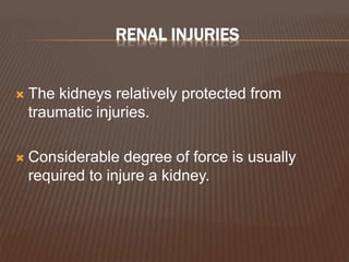
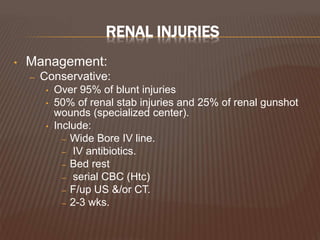
• Indications for renal imaging:
– Macroscopic hematuria
– Penetrating chest, flank, and abdominal wounds
– Microscopic [>5 red blood cells (RBCs) per high
powered field] or dipstick hematuria a
hypotensive patient (SBP <90mmHg )
– A history of a rapid acceleration or deceleration
– Any child with microscopic or dipstick hematuria
who has sustained trauma.
RENAL INJURIES
 What Imaging Study?
 IVU:
 replaced by the contrast-enhanced CT scan
 On-table IVU if patient is transferred immediately to
the operating theatre without having had a CT scan
and a retroperitoneal hematoma is found.
 Spiral CT: does not allow accurate staging
RENAL INJURIES
– Renal US:
• Advantages:
– can certainly establish the presence of two kidneys
– the presence of a retroperitoneal hematoma
– power Doppler can identify the presence of blood flow in the
renal vessels.
• Disadvantages:
– cannot accurately identify parenchymal tears, collecting system
injuries, or extravasations of urine until a later stage when a
urine collection has had time to accumulate.
– Contrast-enhanced CT:
• the imaging study of choice
• accurate, rapid, images other intra-abdominal structures
RENAL INJURIES
 Staging (Grading)
 American Association for the Surgery of
Trauma Organ Injury Severity Scale
RENAL INJURIES
• Management:
– Conservative:
• Over 95% of blunt injuries
• 50% of renal stab injuries and 25% of renal gunshot
wounds (specialized center).
• Include:
– Wide Bore IV line.
– IV antibiotics.
– Bed rest
– serial CBC (Htc)
– F/up US &/or CT.
– 2-3 wks.
RENAL INJURIES
 Surgical exploration:
 Persistent bleeding (persistent tachycardia
and/or hypotension failing to respond to
appropriate fluid and blood replacement
 Expanding perirenal hematoma (again the
patient will show signs of continued bleeding)
 Pulsatile perirenal hematoma
RENAL INJURIES
BLADDER INJURIES
• Causes:
– Iatrogenic injury
• Transurethral resection of bladder tumor (TURBT)
• Cystoscopic bladder biopsy
• Transurethral resection of prostate (TURP)
• Cystolitholapaxy
• Caesarean section, especially as an emergency
• Total hip replacement (very rare)
– Penetrating trauma to the lower abdomen or back
– Blunt pelvic trauma—in association with pelvic fracture or
‘minor’ trauma in the inebriated patient
– Rapid deceleration injury—seat belt injury with full bladder in
the absence of a pelvic fracture
– Spontaneous rupture after bladder augmentation
BLADDER INJURIES
Types of Perforation
 A-intraperitoneal perforation
 B- extra peritoneal perforation
BLADDER INJURIES
 Presentation:
 Recognized intraoperatively
 The classic triad of symptoms and signs that are
suggestive of a bladder rupture
 suprapubic pain and tenderness, difficulty or inability
in passing urine, and haematuria
BLADDER INJURIES
• Management:
– Extra peritoneal
• Bladder drainage +++++
• Open repair +++
– Intra peritoneal :
• open repair…why?
– Unlikely to heal spontaneously.
– Usually large defects.
– Leakage causes peritonitis
– Associated other organ injury.
URETHRAL INJURIES
• ANTERIOR URETHRAL INJURIES
• POSTERIOR URETHRAL INJURIES
ANTERIOR URETHRAL INJURIES
• Rare
• Mechanism:
– The majority a result of a straddle injury in boys
or men.
– Direct injuries to the penis
– Penile fractures
– Inflating a catheter balloon in the anterior urethra
– Penetrating injuries by gunshot wounds.
ANTERIOR URETHRAL INJURIES
 Symptoms and signs:
 Blood at the end of the penis
 Difficulty in passing urine
 Frank hematuria
 Hematoma may around the site of the rupture
 Penile swelling
 Diagnosis:
 Retrograde urethrography
 Contusion: no extravasation of contrast:
 Partial rupture : extravasation of contrast, with contrast also
present in the bladder:.
 Complete disruption: no filling of the posterior urethra or bladder
ANTERIOR URETHRAL INJURIES
• Management
– Contusion
• A small-gauge urethral catheter for one week
– Partial Rupture of Anterior Urethra
• No urethral catheterization
• Majority can be managed by suprapubic urinary diversion for one
week
• Penetrating partial disruption (e.g., knife, gunshot wound), primary
(immediate) repair.
– Complete Rupture of Anterior Urethra
• patient is unstable a suprapubic catheter.
• patient is stable, the urethra may either be immediately repaired or a
suprapubic catheter
– Penetrating Anterior Urethral Injuries
• generally managed by surgical debridement and repair
POSTERIOR URETHRAL INJURIES
 Great majority of posterior urethral injuries
occur in association with pelvic fractures
 Approximately 20% have an associated
bladder rupture.
 Signs
 Blood at the meatus, gross hematuria, and
perineal or scrotal bruising.
 High-riding prostate
POSTERIOR URETHRAL INJURIES
• Classification of posterior urethral injuries
– type I:(rare )
• stretch injury with intact urethra
– type II : (25%)
• partial tear but some continuity remains)
– type III:(75%)
• complete tear with no evidence of continuity
– In women, partial rupture at the anterior position
is the most common urethral injury associated
with pelvic fracture.
POSTERIOR URETHRAL INJURIES
 Management:
 Stretch injury (type I) and incomplete urethral
tears (type II) are best treated by stenting with a
urethral catheter.
 Type III
 Patient is at varying risk of urethral stricture, urinary
incontinence, and erectile dysfunction (ED)
 Initial management with suprapubic cystotomy and
attempting primary repair at 7 to 10 days after injury.
FRACTURE PENIS
 Disruption of the tunica albuginea with rupture
of corpus cavernosum.
 Most commonly with sexual intercourse, but
also reported with masturbation, rolling over or
falling on erect penis.
 Diagnosis straightforward by history and
examination.
 Eggplant deformity.
 Should be promptly explored and surgically
repaired.
CONCLUSION
 Acute Urological Conditions are the most
common systemic conditions which one has
to face in an emergency room.
 With prompt diagnosis and proper
management the morbidity and the mortality
associated with these conditions can be
significantly reduced.
THANK YOU…!

Acute urological conditions

  • 1.
    INTRODUCTION  ACUTE UROLOGICALCONDITIONS; ONE OF THE MOST COMMON EMERGENCIES FACED BY US IN THE CASUALTY DEPARTMENT.  INCLUDES DIFFERENT CONDITIONS RANGING FROM URETHRITIS TO TESTICULAR TORSION.
  • 2.
    CONDITIONS INCLUDED  NONTRAUMATIC 1) URINARY RETENTION 2) HAEMATURIA 3) RENAL/URETERIC COLIC 4) ACUTE SCROTAL CONDITIONS 5) INFECTIONS  TRAUMATIC
  • 3.
    ACUTE URINARY RETENTION Painful inability to void, with relief of pain following drainage of the bladder by catheterization.  Pathophysiology:  Increased urethral resistance, i.e., bladder outlet obstruction (BOO)  Low bladder pressure, i.e., impaired bladder contractility  Interruption of sensory or motor innervations of the bladder
  • 4.
    ACUTE URINARY RETENTION Causes :  Men:  Bladder outlet obstruction (the commonest cause, most commonly due to BEP)  Urethral stricture  Acute urethritis or Prostatitis  Phimosis  Women  Pelvic prolapse (cystocoele, rectocoele, uterine)  Post surgery for ‘stress’ incontinence  pelvic masses (e.g., ovarian masses)  Bladder neck obstruction (rare)
  • 5.
    ACUTE URINARY RETENTION Both Sex  Blood clot  Urethral calculus  Rupture of the urethra  Neurogenic (injury or disease of the spinal cord)  Smooth muscle dysfunction associated with ageing  Faecal impaction  Anal pain (haemorrhoidectomy)  Intensive postoperative analgesic treatment  Some drugs  Spinal anaesthesia
  • 6.
    ACUTE URINARY RETENTION Initial Management :  Urethral catheterization  Suprapubic catheter ( SPC)  Late Management:  Treating the underlying cause
  • 8.
    ACUTE FLANK PAIN—URETERICOR RENAL COLIC  The commonest urologic emergency.  One of the commonest causes of the “Acute Abdomen”.  Sudden onset of severe pain in the flank  Most often due to the passage of a stone formed in the kidney, down through the ureter.
  • 9.
    RENAL COLIC/ URETERICCOLIC  When caused by acute obstruction of the renal pelvis, is typically fixed deep in the loin and ‘bursting’ in character.  When caused by acute ureteric obstruction (usually by a stone), is colicky with sharp exacerbations against a constant background.  Is liable to be referred to the groin, scrotum or labium as the calculus obstruction moves distally.
  • 10.
    RENAL COLIC/ URETERICCOLIC Differential diagnoses  Pyelonephritis  Abdominal aortic aneurysm  Appendicitis  Biliary colic (gallstones)  Peritonitis  Diverticulitis  Salpingitis  Torted ovarian cyst  Ectopic pregnancy  Shingles
  • 11.
    RENAL COLIC/ URETERICCOLIC  Work Up :  History  Examination: patient want to move around, in an attempt to find a comfortable position.  +/- Fever  Pregnancy test  Mid Stream Urine examination  Immediate Investigations:  Abdominal Ultra sound  X-ray KUB Region
  • 12.
    RENAL COLIC/ URETERICCOLIC  Acute Management of Ureteric Stones:  Pain relief  NSAIDs  Intramuscular or intravenous injection, by mouth, or per rectum  +/- Opiate analgesics (pethidine or morphine).  ? Hyper hydration  ‘watchful waiting’ with analgesic supplements  95% of stones measuring 5mm or less pass spontaneously
  • 13.
    RENAL COLIC/ URETERICCOLIC  Indications for Intervention to Relieve Obstruction and/or Remove the Stone:  Pain that fails to respond to analgesics.  Associated fever.  Renal function is impaired because of the stone (solitary kidney obstructed by a stone, bilateral ureteric stones, or preexisting renal impairment )  Obstruction unrelieved for >4 weeks
  • 14.
    HEMATURIA  Blood inthe urine  Is always abnormal whether microscopic or macroscopic.  May be caused by a lesion anywhere in the urinary tract.
  • 15.
    HEMATURIA  Causes  Nephrological(medical) or urological (surgical)  Medical causes:  glomerular and nonglomerular  blood dyscrasias, interstitial nephritis, and renovascular disease  Surgical/urological nonglomerular causes:  renal tumours, urothelial tumours (bladder, ureteric, renal collecting system), prostate cancer, bleeding from vascular benign prostatic enlargement, trauma, renal or ureteric stones, and UTI.  Haematuria in these situations is usually characterised by circular erythrocytes and absence of proteinuria and casts.
  • 16.
    HEMATURIA  Work Up:  History  Examination  Investigation :  Urine culture and cytology  Renal US  Flexible cystoscopy,  IVU or computed tomography (CT) scan in selected groups.  Treat the cause
  • 17.
    ACUTE SCROTAL CONDITIONS Emergency situation requiring prompt evaluation, differential diagnosis, and potentially immediate surgical exploration
  • 19.
    ACUTE SCROTAL CONDITIONS 1.Testicular Torsion  Most serious. 2. Torsion of the Testicular and Epididymal Appendages. 3. Epididymitis or Epididymo-orchitis  Most common
  • 20.
  • 21.
    TESTICULAR TORSION  Truesurgical emergency of the highest order  Irreversible ischemic injury to the testicular parenchyma may begin as soon as 4 hours  Testicular salvage ↓ as duration of torsion↑
  • 22.
    TESTICULAR TORSION Presentation  Acuteonset of scrotal pain.  Majority with history of prior episodes of severe, self-limited scrotal pain and swelling.  Nausea or vomiting  Referred to the ipsi-lateral lower quadrant of the abdomen.  Dysuria and other bladder symptoms are usually absent.
  • 23.
    TESTICULAR TORSION Physical examination: The affected testis is high-riding Transverse orientation.  Acute hydrocoele or massive scrotal edema  Cremasteric reflex is absent.  Tender larger than other side.  Prehn’s sign Positive. Manual detortion.
  • 24.
    TESTICULAR TORSION  Toaid in differential diagnosis of the acute scrotum.  To confirm the absence of torsion of the cord.  Doppler examination of the cord and testis  High false-positive and false-negative results
  • 25.
    TESTICULAR TORSION  ColorDoppler ultrasound:  Assessment of anatomy and determining the presence or absence of blood flow.  Sensitivity: 88.9% specificity of 98.8%  Operator dependent.
  • 26.
  • 27.
    EPID.ORCHITIS  Presentation:  Indolentprocess.  Scrotal swelling, erythema, and pain.  Dysuria and fever is more common  Examination:  localized epididymal tenderness, a swollen and tender epididymis, or a massively swollen hemiscrotum with absence of landmarks.  Cremasteric reflex should be present  Urine:  pyuria, bacteriuria, or a positive urine culture(Gram-negative bacteria) .
  • 28.
    EPID.ORCHITIS  Management:  Bedrest for 1 to 3 days then relative restriction .  Scrotal elevation, the use of an athletic supporter  parenteral antibiotic therapy should be instituted when UTI is documented or suspected.  Urethral instrumentation should be avoided
  • 29.
    PRIAPISM  Persistent erectionof the penis for more than 4 hours that is not related or accompanied by sexual desire.  2 Types:  ischemic (veno-occlusive, low flow (most common) -nonischaemic (arterial, high flow).
  • 30.
    PRIAPISM  Causes:  Primary(Idiopathic) : 30%- 50%  Secondary:  Drugs  Trauma  Neurological  Hematological disease  Tumors  Miscellaneous
  • 31.
    PRIAPISM  Treatment:  Dependson the type of priapism.  Conservative treatment should first be tried  Medical treatment  Surgical treatment.  Treatment of underlying cause
  • 32.
    INFECTIONS  Acute pyelonephritis Cystitis  Urethritis  Acute prostatitis  Acute glomerulonephritis  Perinephric abscess
  • 33.
    TRAUMATIC UROLOGICAL EMERGENCIES RENAL INJURIES  BLADDER INJURIES  URETHRAL INJURIES  URETERIC INJURIES  TESTICULAR INJURIES  PENILE INJURIES  PENILE FRACTURE
  • 34.
    RENAL INJURIES  Thekidneys relatively protected from traumatic injuries.  Considerable degree of force is usually required to injure a kidney.
  • 35.
    RENAL INJURIES  Mechanismsand cause:  Blunt  direct blow or acceleration/ deceleration (road traffic accidents, falls from a height, fall onto flank)  Penetrating  knives, gunshots, iatrogenic, e.g., percutaneous nephrolithotomy (PCNL)
  • 36.
    RENAL INJURIES • Indicationsfor renal imaging: – Macroscopic hematuria – Penetrating chest, flank, and abdominal wounds – Microscopic [>5 red blood cells (RBCs) per high powered field] or dipstick hematuria a hypotensive patient (SBP <90mmHg ) – A history of a rapid acceleration or deceleration – Any child with microscopic or dipstick hematuria who has sustained trauma.
  • 37.
    RENAL INJURIES  WhatImaging Study?  IVU:  replaced by the contrast-enhanced CT scan  On-table IVU if patient is transferred immediately to the operating theatre without having had a CT scan and a retroperitoneal hematoma is found.  Spiral CT: does not allow accurate staging
  • 38.
    RENAL INJURIES – RenalUS: • Advantages: – can certainly establish the presence of two kidneys – the presence of a retroperitoneal hematoma – power Doppler can identify the presence of blood flow in the renal vessels. • Disadvantages: – cannot accurately identify parenchymal tears, collecting system injuries, or extravasations of urine until a later stage when a urine collection has had time to accumulate. – Contrast-enhanced CT: • the imaging study of choice • accurate, rapid, images other intra-abdominal structures
  • 39.
    RENAL INJURIES  Staging(Grading)  American Association for the Surgery of Trauma Organ Injury Severity Scale
  • 40.
    RENAL INJURIES • Management: –Conservative: • Over 95% of blunt injuries • 50% of renal stab injuries and 25% of renal gunshot wounds (specialized center). • Include: – Wide Bore IV line. – IV antibiotics. – Bed rest – serial CBC (Htc) – F/up US &/or CT. – 2-3 wks.
  • 41.
    RENAL INJURIES  Surgicalexploration:  Persistent bleeding (persistent tachycardia and/or hypotension failing to respond to appropriate fluid and blood replacement  Expanding perirenal hematoma (again the patient will show signs of continued bleeding)  Pulsatile perirenal hematoma
  • 42.
  • 43.
    BLADDER INJURIES • Causes: –Iatrogenic injury • Transurethral resection of bladder tumor (TURBT) • Cystoscopic bladder biopsy • Transurethral resection of prostate (TURP) • Cystolitholapaxy • Caesarean section, especially as an emergency • Total hip replacement (very rare) – Penetrating trauma to the lower abdomen or back – Blunt pelvic trauma—in association with pelvic fracture or ‘minor’ trauma in the inebriated patient – Rapid deceleration injury—seat belt injury with full bladder in the absence of a pelvic fracture – Spontaneous rupture after bladder augmentation
  • 44.
    BLADDER INJURIES Types ofPerforation  A-intraperitoneal perforation  B- extra peritoneal perforation
  • 45.
    BLADDER INJURIES  Presentation: Recognized intraoperatively  The classic triad of symptoms and signs that are suggestive of a bladder rupture  suprapubic pain and tenderness, difficulty or inability in passing urine, and haematuria
  • 46.
    BLADDER INJURIES • Management: –Extra peritoneal • Bladder drainage +++++ • Open repair +++ – Intra peritoneal : • open repair…why? – Unlikely to heal spontaneously. – Usually large defects. – Leakage causes peritonitis – Associated other organ injury.
  • 47.
    URETHRAL INJURIES • ANTERIORURETHRAL INJURIES • POSTERIOR URETHRAL INJURIES
  • 48.
    ANTERIOR URETHRAL INJURIES •Rare • Mechanism: – The majority a result of a straddle injury in boys or men. – Direct injuries to the penis – Penile fractures – Inflating a catheter balloon in the anterior urethra – Penetrating injuries by gunshot wounds.
  • 49.
    ANTERIOR URETHRAL INJURIES Symptoms and signs:  Blood at the end of the penis  Difficulty in passing urine  Frank hematuria  Hematoma may around the site of the rupture  Penile swelling  Diagnosis:  Retrograde urethrography  Contusion: no extravasation of contrast:  Partial rupture : extravasation of contrast, with contrast also present in the bladder:.  Complete disruption: no filling of the posterior urethra or bladder
  • 50.
    ANTERIOR URETHRAL INJURIES •Management – Contusion • A small-gauge urethral catheter for one week – Partial Rupture of Anterior Urethra • No urethral catheterization • Majority can be managed by suprapubic urinary diversion for one week • Penetrating partial disruption (e.g., knife, gunshot wound), primary (immediate) repair. – Complete Rupture of Anterior Urethra • patient is unstable a suprapubic catheter. • patient is stable, the urethra may either be immediately repaired or a suprapubic catheter – Penetrating Anterior Urethral Injuries • generally managed by surgical debridement and repair
  • 51.
    POSTERIOR URETHRAL INJURIES Great majority of posterior urethral injuries occur in association with pelvic fractures  Approximately 20% have an associated bladder rupture.  Signs  Blood at the meatus, gross hematuria, and perineal or scrotal bruising.  High-riding prostate
  • 52.
    POSTERIOR URETHRAL INJURIES •Classification of posterior urethral injuries – type I:(rare ) • stretch injury with intact urethra – type II : (25%) • partial tear but some continuity remains) – type III:(75%) • complete tear with no evidence of continuity – In women, partial rupture at the anterior position is the most common urethral injury associated with pelvic fracture.
  • 53.
  • 54.
    POSTERIOR URETHRAL INJURIES Management:  Stretch injury (type I) and incomplete urethral tears (type II) are best treated by stenting with a urethral catheter.  Type III  Patient is at varying risk of urethral stricture, urinary incontinence, and erectile dysfunction (ED)  Initial management with suprapubic cystotomy and attempting primary repair at 7 to 10 days after injury.
  • 56.
    FRACTURE PENIS  Disruptionof the tunica albuginea with rupture of corpus cavernosum.  Most commonly with sexual intercourse, but also reported with masturbation, rolling over or falling on erect penis.  Diagnosis straightforward by history and examination.  Eggplant deformity.  Should be promptly explored and surgically repaired.
  • 57.
    CONCLUSION  Acute UrologicalConditions are the most common systemic conditions which one has to face in an emergency room.  With prompt diagnosis and proper management the morbidity and the mortality associated with these conditions can be significantly reduced.
  • 58.