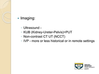
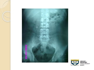
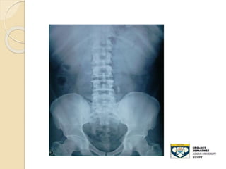
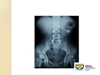
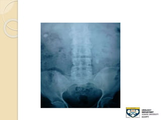
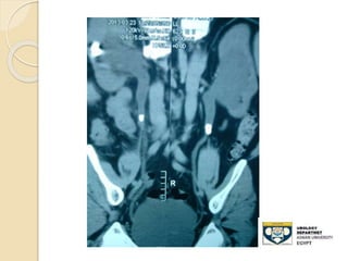
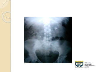
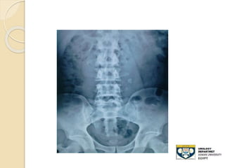
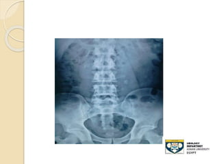
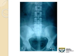
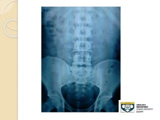
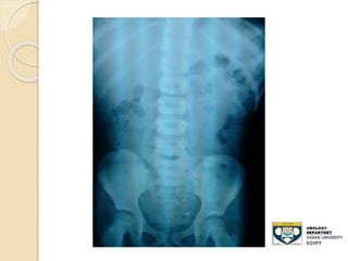
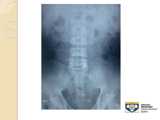
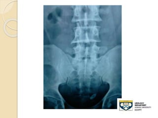
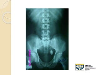
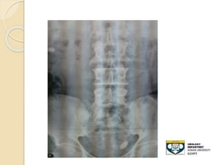
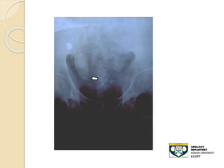
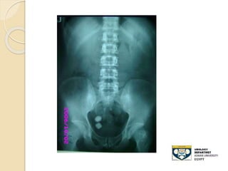
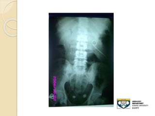
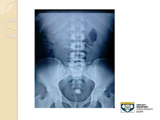
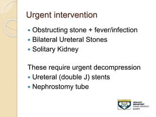
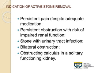
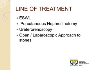
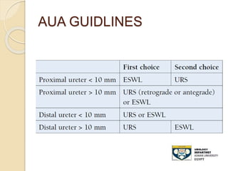
EVALUATION ANDMANAGEMENT
URETERIC STONES
by
Hassaan Ali Gad
Assistant lecturer of urology
Aswan University
hassan.ali@aswu.edu.eg
Learning Objectives
1. List the signs/symptoms and differential
diagnoses of an acute stone episode
2. Describe the imaging studies available
to diagnose ureteral calculi.
3. List the classes of medications effective
for treating the pain of renal colic.
4. Outline the basic treatment options for
ureteral stone
5. Describe the clinical scenarios requiring
urgent decompression of a ureteral
stone.
6. List the basic principles of stone
prevention
URINARY TRACT STONES
The 3rd most common urological
disease preceded only by UTI and
prostate pathology.
OUTLINE
 CLINICAL MANIFESTATIONS
 INVESTIGATION
 MANGMMENT
 PREVENTION
CLINICAL MANIFESTATIONS
 The clinical presentation of patient
with urinary calculi depend mainly on
the site, size, laterality of stone
diseases, and presence or absence of
infection
Presentation maybe
 EMERGENCY
 ELECTIVE
 SILENT OR ASYMPTOMATIC
 COMPLICATIONS
Clinical Manifestations
 Obstructing calculi in the upper urinary
tract cause an extreme crescendo like
pain in the flank that generally radiates
laterally around the abdomen to the
corresponding groin and testicles in
males and labia major in females
Clinical Manifestations
 Colic nature of the pain
◦ Rapid onset
◦ Unable to achieve comfortable position (writhing)
 Radiates from flank to groin
◦ Testis/labia
 Associated nausea/vomiting
 Hematuria
◦ Gross, microscopic (present in 90%; absence doesn’t
r/o)
 Irritative LUTS
◦ May indicate stone near the UVJ/distal ureter
 Associated with fever if UTI
 Anuria
 Tenderness present in the loin or no findings in
abdomen
Urinary Calculus Disease:
Differential Diagnosis
 Bowel:
◦ Inflammatory bowel disease, appendicitis,
diverticulitis
 Gynecologic:
◦ PID, ruptured ovarian cyst, ectopic pregnancy
 Neurologic/Musculoskeletal:
◦ Radicular pain, herpes zoster, muscle
spasm/strain
 Genito-urinary:
◦ Cystitis, pyelonephritis, torsion, UPJ obstruction
Investigations
after careful history taking and physical
examination :
 Labs:
◦ Creatinine& Urea (renal failure)
◦ Urinalysis (microscopy is gold standard to look
for crystals)
◦ CBC&diff (Look for ↑WBC,
◦ Pregnancy Test (HCG) in females
 Imaging:
◦ Ultrasound -
◦ KUB (Kidney-Ureter-Pelvis)=PUT
◦ Non-contrast CT UT (NCCT)
◦ IVP - more or less historical or in remote settings
KUB film
 - Advantages:
◦ 80-90% of stones are radio-opaque
◦ Minimal radiation
 Disadvantages:
◦ Radiolucent stone
◦ Easy to miss mid-ureteral stones over the
sacrum
◦ Bowel gas can obscure its efficacy
◦ Cannot differentiate , Stones,Calcified LN
,
◦ Sensitivity: 50-70%
Stone opacity
 Radiopaque
 Calcium oxalate dihydrate
 Calcium oxalate monohydrate
 Calcium phosphates
 Poor radiopacity
 Magnesium ammonium phosphate
 Cystine
 Radiolucent
 Uric acid
 Ammonium urate
 Xanthine
 Drug-stones
STONE UPPER THIRD URETER
STONE MIDDLE THIRD URETER
STONE LOWER THIRD URETER
Non-contrast CT
 Advantages:
◦ All stone types are visible except indinavir
 Sensitivity - 97%; Specificity - 96%
◦ Rapid,Readily available
◦ Does not require contrast
◦ Other pathologies identified
◦ Information about stone and collecting
system obtained
• Stone density (Stones >500HU are opaque
on KUB)
Non-contrast CT
 Disadvantages:
◦ Increased radiation dose compared with
KUB
◦ Cost
◦ No physiologic information such as
function.
TREATMENT OF STONE
URETER
The overall passage rate of ureteral
stones is:
 Proximal ureteral stones: 25%
 Mid-ureteral stones: 45%
 Distal ureteral stones: 70%
TREATMENT OF STONE
URETER
 Pain Control
 Conservative Treatment
 Active Stone Removal
Pain Control
 NSAID
 OPIOIDS
 IV hydration.
 Anti spasmolytic
 Atypical opioid – tramadol
 Calcium channel blocker
 Alpha - 1 blocker
Nausea and vomiting
 Promethazine ( phenergan )
 Ondansetron ( 5 – HT3 blocker )
Conservative Treatment
 Ideal candidate
 Ureteric stone – especially lower ureteric ( < 8
mm)
 No distal obstruction
 Reasonable pain control
 Adequate kidney function
 Sterile Urine culture
Contraindication of conservative
management
Profession – pilot.
 Obstruction with deteriorating renal
function
 Obstruction with infection
 Persistent pain
Medical Expulsive Therapy
(MET)
 Administration of a drug to facilitate the
passage of the stone
 25 – 30 % more chances of spontaneous
passage
 Decreased time to pass the stone.
 Less surgical intervention.
Should follow-up with urology within 1-2 weeks
Drugs
 Alpha blocker – Tamsulosin
 Calcium channel blocker
 Corticosteroids
 drink >2L of water/day
Urgent intervention
 Obstructing stone + fever/infection
 Bilateral Ureteral Stones
 Solitary Kidney
These require urgent decompression
 Ureteral (double J) stents
 Nephrostomy tube
INDICATION OF ACTIVE STONE REMOVAL
 Persistent pain despite adequate
medication;
 Persistent obstruction with risk of
impaired renal function;
 Stone with urinary tract infection;
 Bilateral obstruction;
 Obstructing calculus in a solitary
functioning kidney.
LINE OF TREATMENT
 ESWL
 Percutaneous Nephrolithotomy
 Ureterorenoscopy
 Open / Laparoscopic Approach to
stones
AUA GUIDLINES
Extracorporeal Shockwave
Lithotripsy (ESWL)
 Least invasive
 Conscious sedation
 Fragments stones that the patient then
passes
 High patient satisfaction
 May require more time to become stone free
 Renal calculi <2cm or ureteral calculi <1cm
 Contra-indications
 Pregnancy
 Bleeding Disorder/anticoagulation (NSAIDS
pre-op)
 Febrile UTI
 Obstruction Distal to the stone being treated
SWL success depends on:
 Stone Size (Better if <1cm)
 Stone Location (Better if renal pelvic,
upper ureter)
 Stone Density/ Composition (Better if
HU<1000)
◦ Hounsfield unit density on NCCT
 Patient Habitus (Better if skin-to-stone
distance <10cm)
 Worse if associated renal anomalies:
◦ UPJ Obstruction
◦ Horseshoe kidney
Ureteroscopic (URS)
 Advantages:
◦ Near 100% stone free rate
◦ Low retreatment rates
◦ Treatment available in most centres
 Disadvantages:
◦ General or spinal anesthesia is usually required
◦ Ureteral stent (DJ) may be left
 Stent symptoms are bothersome to patients
◦ Lower patient satisfaction
 Typically for ureteral calculi and SWL
failures
Stone Prevention:
General Advice
 Increase Hydration to 2-3L per day to achieve
daily urine output of 2.5L
 Diet:
◦ Maintain normal calcium intake (1000-1200mg with
meals)
 Used to advice low calcium diets – Proven to be false
◦ Minimize foods high in oxalate (Spinach, peanut,
rhubarb)
◦ Minimize salt (<2300mg/d) and animal protein
◦ Increase fiber, vegetables and citrus-rich fruits
 Consider urinary alkalinization:
◦ Mainly for uric acid and cystine stones
◦ Potassium citrate - preferred
◦ Sodium citrate - alternative
Conclusions
 Once stone detected – should be removed
 Patients with colic – relief of pain is the priority.
 Obstruction and infection – immediate attention.
 Preventive measures - to avoid recurrence .

Evaluation and management of ureteric stones

  • 1.
    EVALUATION ANDMANAGEMENT URETERIC STONES by HassaanAli Gad Assistant lecturer of urology Aswan University hassan.ali@aswu.edu.eg
  • 2.
    Learning Objectives 1. Listthe signs/symptoms and differential diagnoses of an acute stone episode 2. Describe the imaging studies available to diagnose ureteral calculi. 3. List the classes of medications effective for treating the pain of renal colic. 4. Outline the basic treatment options for ureteral stone 5. Describe the clinical scenarios requiring urgent decompression of a ureteral stone. 6. List the basic principles of stone prevention
  • 3.
    URINARY TRACT STONES The3rd most common urological disease preceded only by UTI and prostate pathology.
  • 4.
    OUTLINE  CLINICAL MANIFESTATIONS INVESTIGATION  MANGMMENT  PREVENTION
  • 5.
    CLINICAL MANIFESTATIONS  Theclinical presentation of patient with urinary calculi depend mainly on the site, size, laterality of stone diseases, and presence or absence of infection Presentation maybe  EMERGENCY  ELECTIVE  SILENT OR ASYMPTOMATIC  COMPLICATIONS
  • 6.
    Clinical Manifestations  Obstructingcalculi in the upper urinary tract cause an extreme crescendo like pain in the flank that generally radiates laterally around the abdomen to the corresponding groin and testicles in males and labia major in females
  • 7.
    Clinical Manifestations  Colicnature of the pain ◦ Rapid onset ◦ Unable to achieve comfortable position (writhing)  Radiates from flank to groin ◦ Testis/labia  Associated nausea/vomiting  Hematuria ◦ Gross, microscopic (present in 90%; absence doesn’t r/o)  Irritative LUTS ◦ May indicate stone near the UVJ/distal ureter  Associated with fever if UTI  Anuria  Tenderness present in the loin or no findings in abdomen
  • 8.
    Urinary Calculus Disease: DifferentialDiagnosis  Bowel: ◦ Inflammatory bowel disease, appendicitis, diverticulitis  Gynecologic: ◦ PID, ruptured ovarian cyst, ectopic pregnancy  Neurologic/Musculoskeletal: ◦ Radicular pain, herpes zoster, muscle spasm/strain  Genito-urinary: ◦ Cystitis, pyelonephritis, torsion, UPJ obstruction
  • 9.
    Investigations after careful historytaking and physical examination :  Labs: ◦ Creatinine& Urea (renal failure) ◦ Urinalysis (microscopy is gold standard to look for crystals) ◦ CBC&diff (Look for ↑WBC, ◦ Pregnancy Test (HCG) in females
  • 10.
     Imaging: ◦ Ultrasound- ◦ KUB (Kidney-Ureter-Pelvis)=PUT ◦ Non-contrast CT UT (NCCT) ◦ IVP - more or less historical or in remote settings
  • 12.
    KUB film  -Advantages: ◦ 80-90% of stones are radio-opaque ◦ Minimal radiation  Disadvantages: ◦ Radiolucent stone ◦ Easy to miss mid-ureteral stones over the sacrum ◦ Bowel gas can obscure its efficacy ◦ Cannot differentiate , Stones,Calcified LN , ◦ Sensitivity: 50-70%
  • 13.
    Stone opacity  Radiopaque Calcium oxalate dihydrate  Calcium oxalate monohydrate  Calcium phosphates  Poor radiopacity  Magnesium ammonium phosphate  Cystine  Radiolucent  Uric acid  Ammonium urate  Xanthine  Drug-stones
  • 14.
  • 23.
  • 26.
  • 38.
    Non-contrast CT  Advantages: ◦All stone types are visible except indinavir  Sensitivity - 97%; Specificity - 96% ◦ Rapid,Readily available ◦ Does not require contrast ◦ Other pathologies identified ◦ Information about stone and collecting system obtained • Stone density (Stones >500HU are opaque on KUB)
  • 39.
    Non-contrast CT  Disadvantages: ◦Increased radiation dose compared with KUB ◦ Cost ◦ No physiologic information such as function.
  • 40.
    TREATMENT OF STONE URETER Theoverall passage rate of ureteral stones is:  Proximal ureteral stones: 25%  Mid-ureteral stones: 45%  Distal ureteral stones: 70%
  • 41.
    TREATMENT OF STONE URETER Pain Control  Conservative Treatment  Active Stone Removal
  • 42.
    Pain Control  NSAID OPIOIDS  IV hydration.  Anti spasmolytic  Atypical opioid – tramadol  Calcium channel blocker  Alpha - 1 blocker Nausea and vomiting  Promethazine ( phenergan )  Ondansetron ( 5 – HT3 blocker )
  • 43.
    Conservative Treatment  Idealcandidate  Ureteric stone – especially lower ureteric ( < 8 mm)  No distal obstruction  Reasonable pain control  Adequate kidney function  Sterile Urine culture
  • 44.
    Contraindication of conservative management Profession– pilot.  Obstruction with deteriorating renal function  Obstruction with infection  Persistent pain
  • 45.
    Medical Expulsive Therapy (MET) Administration of a drug to facilitate the passage of the stone  25 – 30 % more chances of spontaneous passage  Decreased time to pass the stone.  Less surgical intervention. Should follow-up with urology within 1-2 weeks Drugs  Alpha blocker – Tamsulosin  Calcium channel blocker  Corticosteroids  drink >2L of water/day
  • 46.
    Urgent intervention  Obstructingstone + fever/infection  Bilateral Ureteral Stones  Solitary Kidney These require urgent decompression  Ureteral (double J) stents  Nephrostomy tube
  • 47.
    INDICATION OF ACTIVESTONE REMOVAL  Persistent pain despite adequate medication;  Persistent obstruction with risk of impaired renal function;  Stone with urinary tract infection;  Bilateral obstruction;  Obstructing calculus in a solitary functioning kidney.
  • 48.
    LINE OF TREATMENT ESWL  Percutaneous Nephrolithotomy  Ureterorenoscopy  Open / Laparoscopic Approach to stones
  • 49.
  • 50.
    Extracorporeal Shockwave Lithotripsy (ESWL) Least invasive  Conscious sedation  Fragments stones that the patient then passes  High patient satisfaction  May require more time to become stone free  Renal calculi <2cm or ureteral calculi <1cm  Contra-indications  Pregnancy  Bleeding Disorder/anticoagulation (NSAIDS pre-op)  Febrile UTI  Obstruction Distal to the stone being treated
  • 51.
    SWL success dependson:  Stone Size (Better if <1cm)  Stone Location (Better if renal pelvic, upper ureter)  Stone Density/ Composition (Better if HU<1000) ◦ Hounsfield unit density on NCCT  Patient Habitus (Better if skin-to-stone distance <10cm)  Worse if associated renal anomalies: ◦ UPJ Obstruction ◦ Horseshoe kidney
  • 53.
    Ureteroscopic (URS)  Advantages: ◦Near 100% stone free rate ◦ Low retreatment rates ◦ Treatment available in most centres  Disadvantages: ◦ General or spinal anesthesia is usually required ◦ Ureteral stent (DJ) may be left  Stent symptoms are bothersome to patients ◦ Lower patient satisfaction  Typically for ureteral calculi and SWL failures
  • 54.
    Stone Prevention: General Advice Increase Hydration to 2-3L per day to achieve daily urine output of 2.5L  Diet: ◦ Maintain normal calcium intake (1000-1200mg with meals)  Used to advice low calcium diets – Proven to be false ◦ Minimize foods high in oxalate (Spinach, peanut, rhubarb) ◦ Minimize salt (<2300mg/d) and animal protein ◦ Increase fiber, vegetables and citrus-rich fruits  Consider urinary alkalinization: ◦ Mainly for uric acid and cystine stones ◦ Potassium citrate - preferred ◦ Sodium citrate - alternative
  • 55.
    Conclusions  Once stonedetected – should be removed  Patients with colic – relief of pain is the priority.  Obstruction and infection – immediate attention.  Preventive measures - to avoid recurrence .