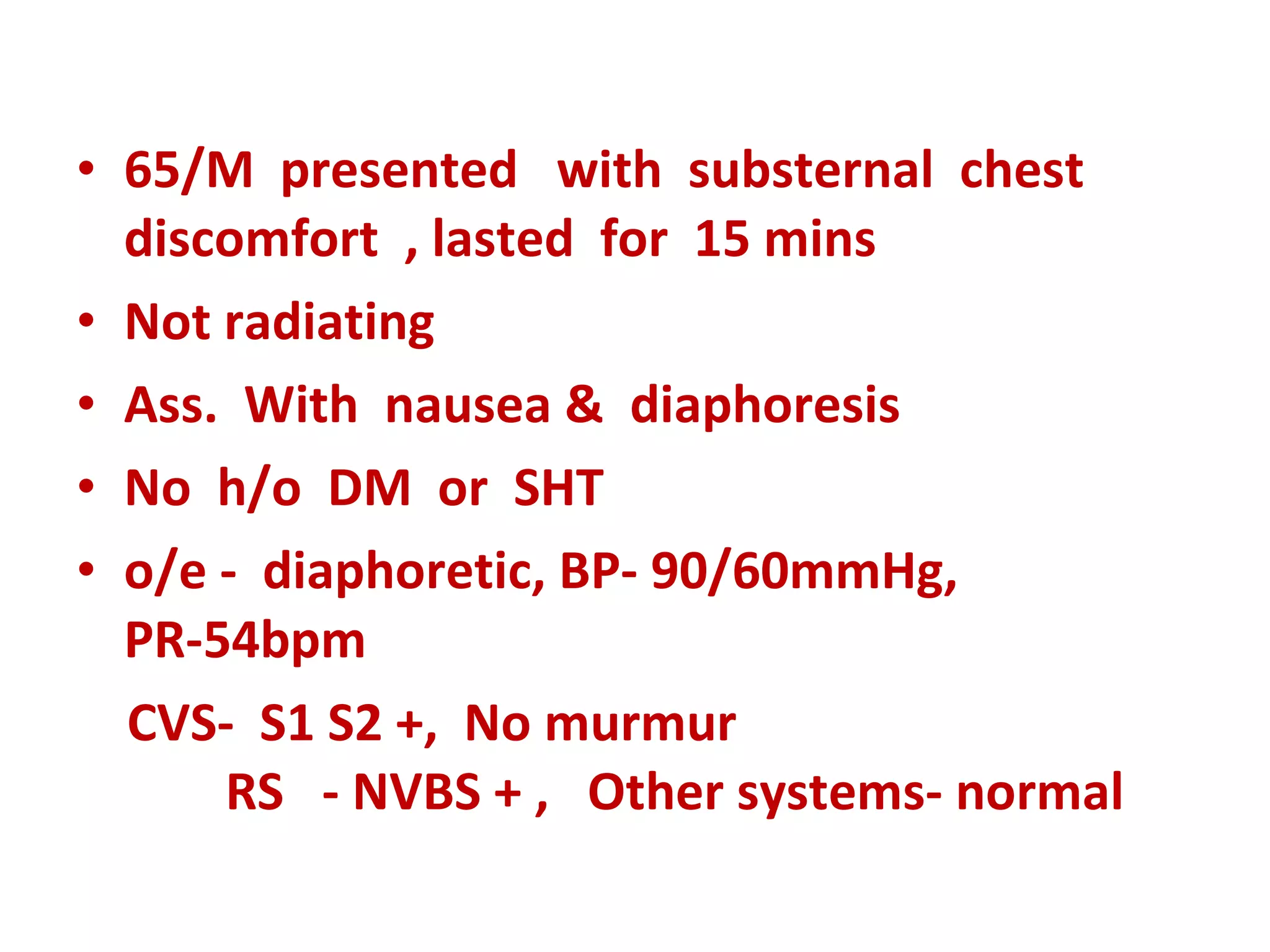
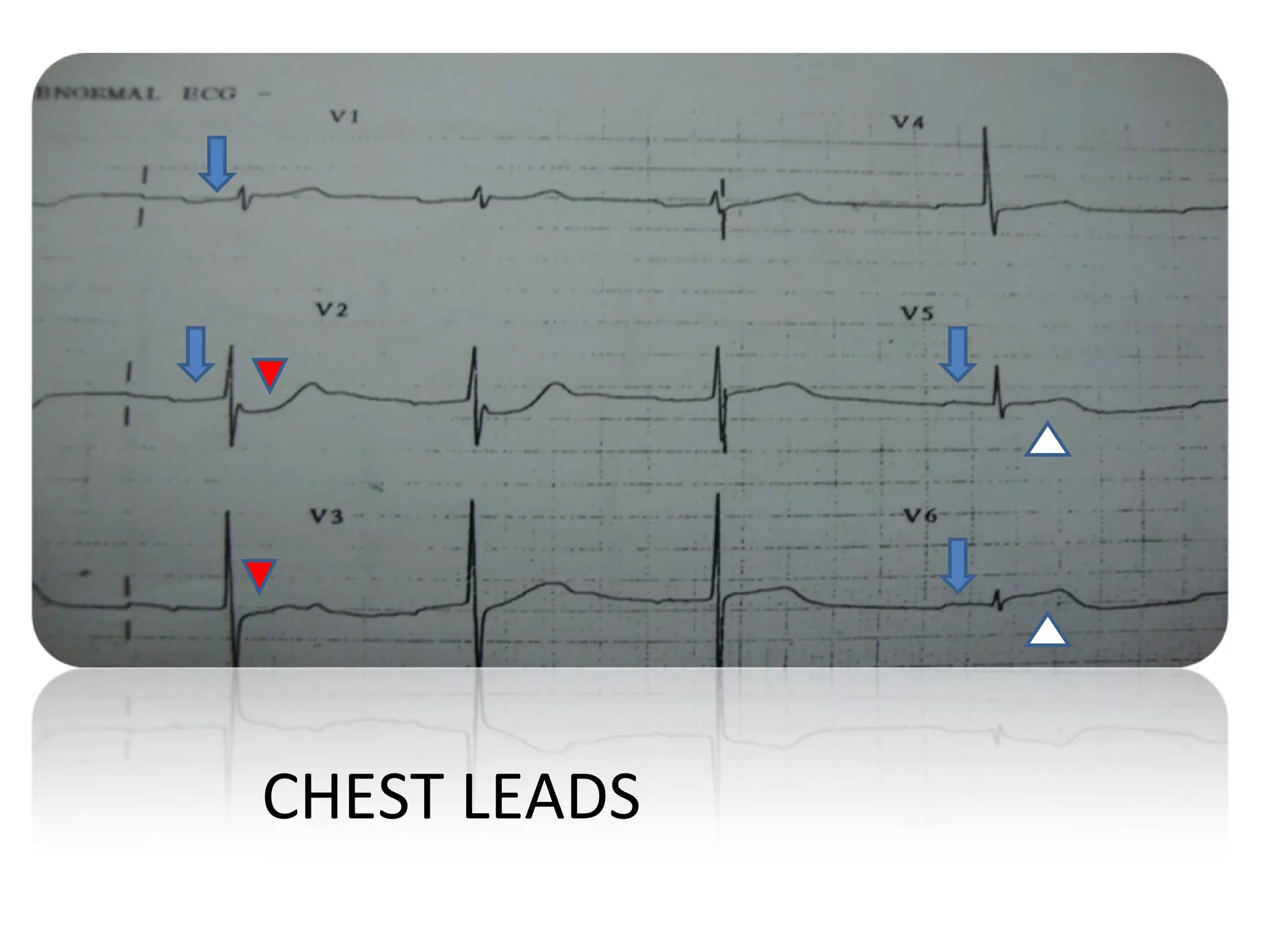
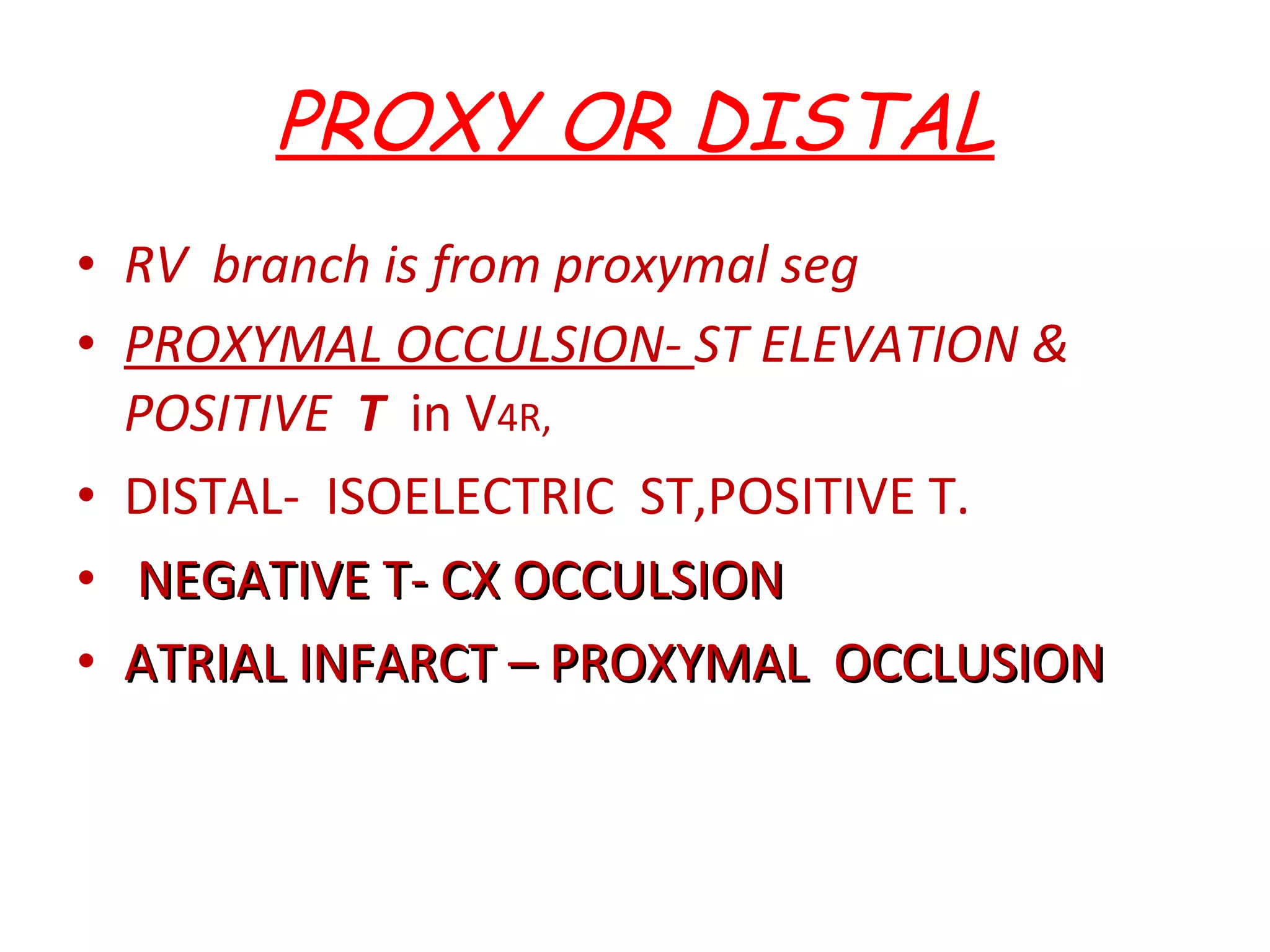
PHYSICIANS MEET PROF.DR.DHANDAPANI’S UNIT AN INTERESTING ECG  DR D SUBBURAJ
65/M  presented  with  substernal  chest discomfort  , lasted  for  15 mins Not radiating  Ass.  With  nausea &  diaphoresis No  h/o  DM  or  SHT o/e -  diaphoretic, BP- 90/60mmHg, PR-54bpm  CVS-  S1 S2 +,  No murmur  RS  - NVBS + ,  Other systems- normal
 
LIMB  LEADS
Chest leads v1
ECG  FINDINGS Sinus rhythm Rate-54 PR-252ms Cardiac  axis  2  degrees QTc-399 ms ST  elevation II,III,a VF,V5,V6.  depression I,a VL,V2,V3 P Ta elevation II,III,a VF,V5 ,V6, depression in V1,V2
Pta SEGMENT ELEVATED IN II,III , aVF.
CHEST LEADS
ATRIAL INFARCT  WITH  INFERO POST LATERAL WALL INFARCT & FIRST DEGREE HEART BLOCK
ATRIAL INFARCTION Seen in upto 10% of patients  with STEMI. Rt atrial  81-98% Biatrial  19-24% Lt atrial  2-19% Often clinically unrecognized because of its subtle ECG changes.
DIAGNOSIS The diagnosis of atrial infarction  is usually made from elevation of P-Ta segment in the clinical setting of MI. The diagnosis may be entertained when the  P-Ta segment is minimally elevated, i.e. in the same direction as the p wave. (Schamroth)
P-Ta segment From  end of P wave to beginning of QRS
DIAGNOSTIC CRITERIA 1. PTa  segment elevation >0.5 mm in  leads v5,v6  with reciprocal PTa segment depression  in leads v1,v2. 2. PTa segment elevation >0.5 mm in  lead I with reciprocal PTa segment depression in leads II,III
PTa segment depression >1.5 mm in  precordial leads . PTa segment depression >1.2 mm inleads I,II,III  and in associaton with any atrial arrhythmias. 5  Abnormal p wave:  flattening of p wave in M pattern,  flattening of p wave in W pattern, irregular or notched p wave  according to lieu et al
COMPLICATIONS Arrhythmias  :61-74% AF, SVT, atrial premature beats Thromboembolism:  84 % systemic, pulmonary Atrial rupture  :4-5% Hemodynamic disturbances
RCA RCA  SUPPLIES  SA node,AV node, RV , posteromedial papillary muscle ,inf part of LV, variabily post&lat segments of LV. RV BRANCH –from proximal seg of RCA RCA OCCLUSION- SA NODE- sinus bradycardia AVNODE-AV nodal block RV-Cardiogenic shock PAPILLARY MUSCLE-MR INFERO POST LATERAL MI
RCA OR CX ? RCA  ST elevation III>II ST depression aVL> I ST dep in I CX  ST elevation in II>III ST isoelectric  LEAD I  avL aVR III II
PROXY OR DISTAL RV  branch is from proxymal seg PROXYMAL OCCULSION-  ST ELEVATION & POSITIVE  T  in V 4R, DISTAL-  ISOELECTRIC  ST,POSITIVE T. NEGATIVE T- CX OCCULSION ATRIAL INFARCT – PROXYMAL  OCCLUSION
ANOTHER  ECG  OF RCA  OCCLUSION I II III aVF aVL aVR V1 V2 V3 V4 V5 V6
REF : HURST 11 th  edition SCHAMROTH  THANK YOU

ECG: Atrial Infarct

  • 1.
    PHYSICIANS MEET PROF.DR.DHANDAPANI’SUNIT AN INTERESTING ECG DR D SUBBURAJ
  • 2.
    65/M presented with substernal chest discomfort , lasted for 15 mins Not radiating Ass. With nausea & diaphoresis No h/o DM or SHT o/e - diaphoretic, BP- 90/60mmHg, PR-54bpm CVS- S1 S2 +, No murmur RS - NVBS + , Other systems- normal
  • 3.
  • 4.
  • 5.
  • 6.
    ECG FINDINGSSinus rhythm Rate-54 PR-252ms Cardiac axis 2 degrees QTc-399 ms ST elevation II,III,a VF,V5,V6. depression I,a VL,V2,V3 P Ta elevation II,III,a VF,V5 ,V6, depression in V1,V2
  • 7.
    Pta SEGMENT ELEVATEDIN II,III , aVF.
  • 8.
  • 9.
    ATRIAL INFARCT WITH INFERO POST LATERAL WALL INFARCT & FIRST DEGREE HEART BLOCK
  • 10.
    ATRIAL INFARCTION Seenin upto 10% of patients with STEMI. Rt atrial 81-98% Biatrial 19-24% Lt atrial 2-19% Often clinically unrecognized because of its subtle ECG changes.
  • 11.
    DIAGNOSIS The diagnosisof atrial infarction is usually made from elevation of P-Ta segment in the clinical setting of MI. The diagnosis may be entertained when the P-Ta segment is minimally elevated, i.e. in the same direction as the p wave. (Schamroth)
  • 12.
    P-Ta segment From end of P wave to beginning of QRS
  • 13.
    DIAGNOSTIC CRITERIA 1.PTa segment elevation >0.5 mm in leads v5,v6 with reciprocal PTa segment depression in leads v1,v2. 2. PTa segment elevation >0.5 mm in lead I with reciprocal PTa segment depression in leads II,III
  • 14.
    PTa segment depression>1.5 mm in precordial leads . PTa segment depression >1.2 mm inleads I,II,III and in associaton with any atrial arrhythmias. 5 Abnormal p wave: flattening of p wave in M pattern, flattening of p wave in W pattern, irregular or notched p wave according to lieu et al
  • 15.
    COMPLICATIONS Arrhythmias :61-74% AF, SVT, atrial premature beats Thromboembolism: 84 % systemic, pulmonary Atrial rupture :4-5% Hemodynamic disturbances
  • 16.
    RCA RCA SUPPLIES SA node,AV node, RV , posteromedial papillary muscle ,inf part of LV, variabily post&lat segments of LV. RV BRANCH –from proximal seg of RCA RCA OCCLUSION- SA NODE- sinus bradycardia AVNODE-AV nodal block RV-Cardiogenic shock PAPILLARY MUSCLE-MR INFERO POST LATERAL MI
  • 17.
    RCA OR CX? RCA ST elevation III>II ST depression aVL> I ST dep in I CX ST elevation in II>III ST isoelectric LEAD I avL aVR III II
  • 18.
    PROXY OR DISTALRV branch is from proxymal seg PROXYMAL OCCULSION- ST ELEVATION & POSITIVE T in V 4R, DISTAL- ISOELECTRIC ST,POSITIVE T. NEGATIVE T- CX OCCULSION ATRIAL INFARCT – PROXYMAL OCCLUSION
  • 19.
    ANOTHER ECG OF RCA OCCLUSION I II III aVF aVL aVR V1 V2 V3 V4 V5 V6
  • 20.
    REF : HURST11 th edition SCHAMROTH THANK YOU