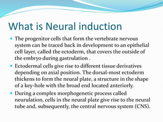
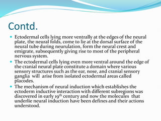
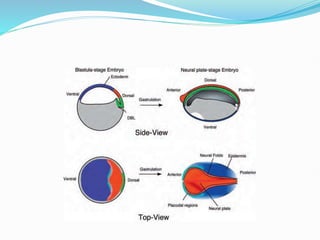
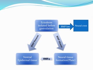
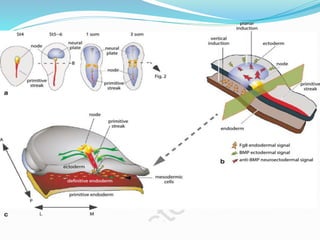
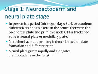
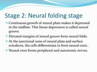
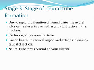
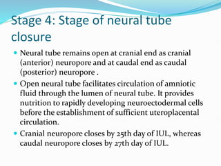
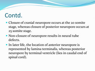
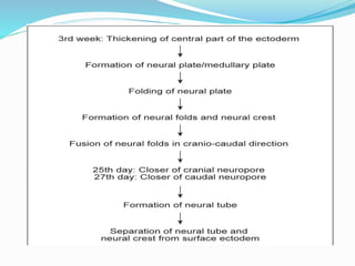
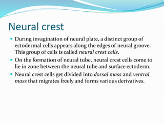
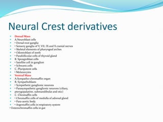
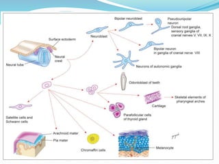
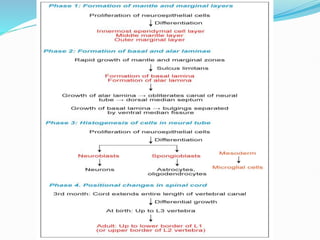
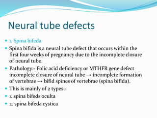
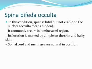
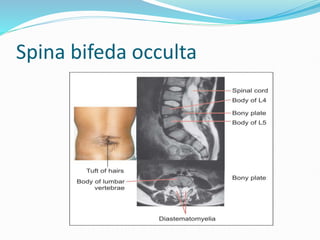
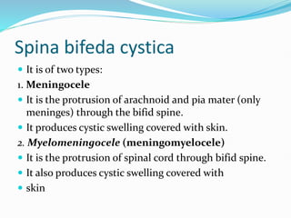
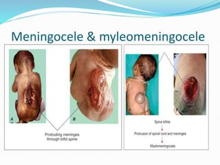
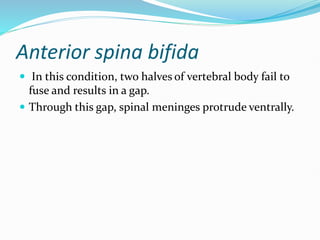
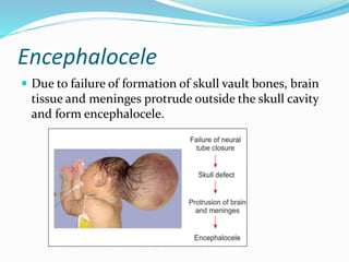
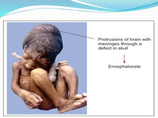
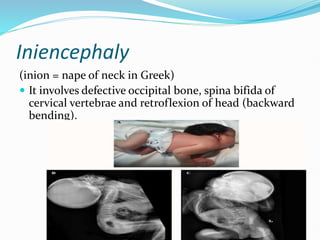
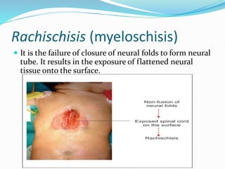
M
Contents
 Introduction
 Discovery and early advancements
 Molecular nature
 Neurulation and its stages
 Differentiation of neural tube
 Neural crest and its derivatives
 Disorders of neural tube and crest.
 Summary
 References
What is Neural induction
 The progenitor cells that form the vertebrate nervous
system can be traced back in development to an epithelial
cell layer, called the ectoderm, that covers the outside of
the embryo during gastrulation .
 Ectodermal cells give rise to different tissue derivatives
depending on axial position. The dorsal-most ectoderm
thickens to form the neural plate, a structure in the shape
of a key-hole with the broad end located anteriorly.
 During a complex morphogenetic process called
neurulation, cells in the neural plate give rise to the neural
tube and, subsequently, the central nervous system (CNS).
Contd.
 Ectodermal cells lying more ventrally at the edges of the neural
plate, the neural folds, come to lie at the dorsal surface of the
neural tube during neurulation, form the neural crest and
emigrate, subsequently giving rise to most of the peripheral
nervous system.
 The ectodermal cells lying even more ventral around the edge of
the cranial neural plate constitute a domain where various
sensory structures such as the ear, nose, and cranial sensory
ganglia will arise from isolated ectodermal areas called
placodes.
 The mechanism of neural induction which establishes the
ectoderm inductive interaction with different subregions was
discovered in early 19th century and now the molecules that
underlie neural induction have been defines and their actions
understood.
Early advancements and discovery
 In the 1930’s, Mangold and Spemann discovered neural
induction during experiments in which they transplanted
small pieces of tissues from one amphibian embryo to
another at pregastrulae stages.
 The key observation was made when they transplanted a
small piece of tissue from a region called the dorsal
blastopore lip (DBL), and the host embryo responded to
the grafted tissue by forming a complete secondary dorsal
axis.
 The secondary dorsal axis contained a complete nervous
system that was derived entirely from the ventral ectoderm
of the host embryo.
Contd.
 The implication of this observation was that the transplanted
tissue can act as a source of inducing signals that can cause
ventral ectoderm to form neural tissue, and that this inductive
interaction normally occurs on the dorsal side of the embryo.
 After Mangold and Spemann’s lead, it was subsequently found
all vertebrate embryos appear to contain a region, called
Spemann’s organizer, which can induce ectoderm to form
neural tissue.
 Tissue in the DBL was later termed the organizer because of its
ability, when transplanted, to reprogram the ventral side of the
embryo to form dorsal tissues, not only in the ectoderm but also
in the internal mesodermal tissues
Contd.
 In these experiments, smaller regions of the DBL were
used, and taken from embryos at different stages.
 The DBL of younger embryos contains the first involuting
tissue and, when transplanted, induces head structures
that contained neural tissue from the anterior portions of
the neuraxis. Conversely, the DBL from older embryos
involutes later and, when transplanted, induces tail
structures that contained neural tissue from just the
posterior portions of the neuraxis.
Molecular nature of Neural
inducers
 It was until the 1990’s, when key biochemical pathways
that mediate cell-cell signaling in animal development
were identified using the tools of molecular genetics
 The core components of this pathway are the BMP ligands,
secreted extracellular proteins that bind and activate a
small family of heterodimeric cell surface receptors, which
in turn transduce a signal by intracellular phosphorylation
events that ultimately lead to changes in the activities of
transcription factors, the SMAD(supressor of mothers
against decapentaplegic) proteins .
Contd.
Role of BMP(Bone morphogenic proteins)
- Fibroblast growth factors (FGF) inhibit BMPs and
transforming growth factor β (TGF-β).
- Notochord produces noggin, chordin, follistatin that
inactivate BMPs and causes neurulation.
Ectoderm
isolated before
gastrulation
Neural tissue
formation(Plate)
BMP 4
Ventral
Epidermal tissue
BMP low Neural crest
Neurulation
 Neural plate gets folded to form neural tube that later
form central nervous system
 The process of formation of neural tube from neural
plate is called neurulation.
 It consists of major 4 phases as described below:-
Stage 1: Neuroectoderm and
neural plate stage
 In presomitic period (16th–19th day): Surface ectoderm
differentiates and thickens in the centre (between the
prechordal plate and primitive node). This thickened
zone is neural plate or medullary plate.
 Notochord acts as a primary inducer for neural plate
formation and differentiation.
 Neural plate grows rapidly and elongates
craniocaudally in the length.
Stage 2: Neural folding stage
 Continuous growth of neural plate makes it depressed
in the midline. This linear depression is called neural
groove.
 Elevated margins of neural groove form neural folds.
 At the junctional zone of neural plate and surface
ectoderm, the cells differentiate to form neural crest..
 Neural crest forms peripheral and autonomic nerves.
Stage 3: Stage of neural tube
formation
 Due to rapid proliferation of neural plate, the neural
folds come closer to each other and start fusion in the
midline.
 On fusion, it forms neural tube.
 Fusion begins in cervical region and extends in cranio-
caudal direction.
 Neural tube forms central nervous system.
Stage 4: Stage of neural tube
closure
 Neural tube remains open at cranial end as cranial
(anterior) neuropore and at caudal end as caudal
(posterior) neuropore .
 Open neural tube facilitates circulation of amniotic
fluid through the lumen of neural tube. It provides
nutrition to rapidly developing neuroectodermal cells
before the establishment of sufficient uteroplacental
circulation.
 Cranial neuropore closes by 25th day of IUL, whereas
caudal neuropore closes by 27th day of IUL.
Contd.
 Closure of cranial neuropore occurs at the 20 somite
stage, whereas closure of posterior neuropore occurs at
25 somite stage.
 Non-closure of neuropore results in neural tube
defects.
 In later life, the location of anterior neuropore is
represented by lamina terminalis, whereas posterior
neuropore by terminal ventricle (lies in caudal end of
spinal cord).
DIFFERENTIATION OF NEURAL
TUBE
 Neural tube has a central cavity (forms ventricles of brain
and central canal of spinal cord) and a peripheral wall
(forms tissue of nervous system).
 Neural tube elongates cranio-caudally.
 Cranial part of the cavity of neural tube dilates to form
brain vesicles, whereas the caudal part of central cavity
remains tubular.
 Dilated cranial part of neural tube forms 3 primary
 brain vesicles :
1. prosencephalon (cranial most)
2. mesencephalon (middle)
3. rhombencephalon (caudal most)
 On further growth, 5 brain vesicles develop as
 follows:
1. Prosencephalon divides into
– Cranial telencephalon—gives rise to two cerebral hemispheres
– Diencephalon—gives rise to optic vesicle,
pineal gland, thalami and hypothalami,
posterior hypophysis.
2. Mesencephalon—gives rise to midbrain
3. Rhombencephalon divides into
– cranial metencephalon—gives rise to pons and cerebellum
– caudal myelencephalon—give rise to medulla oblongata
Neural crest
 During invagination of neural plate, a distinct group of
ectodermal cells appears along the edges of neural groove.
This group of cells is called neural crest cells.
 On the formation of neural tube, neural crest cells come to
lie in zone between the neural tube and surface ectoderm.
 Neural crest cells get divided into dorsal mass and ventral
mass that migrates freely and forms various derivatives.
Neural Crest derivatives
 Dorsal Mass
 A.Neuroblast cells
 • Dorsal root ganglia
 • Sensory ganglia of V, VII, IX and X cranial nerves
 • Skeletal elements of pharyngeal arches
 • Odontoblast of teeth
 • Parafollicular cells of thyroid gland
 B. Spongioblast cells
 • Satellite cell in ganglion
 • Schwann cells
 C. Pluripotent cells
 • Melanocytes
 Ventral Mass
 A.Sympatho-chromaffin organ
 B. Sympathoblasts
 • Sympathetic ganglionic neurons
 • Parasympathetic ganglionic neurons (ciliary,
 pterygopalatine, submandibular and otic)
 C. Chromaffin cells
 • Chromaffin cells of medulla of adrenal gland
 • Para-aortic body
 • Argentaffin cells in respiratory system
• Enterochromaffin cells in gut
Spinal cord development
 Parts of neural tube caudal to the hindbrain
(rhombencephalon) form the spinal cord.
• Formation of spinal cord occurs in 4 phases as follows:
1. Formation of mantle and marginal layers
2. Formation of basal and alar plates
3. Histogenesis of cells and
4. Positional changes of spinal cord.
Neural tube defects
 1. Spina bifeda
 Spina bifida is a neural tube defect that occurs within the
first four weeks of pregnancy due to the incomplete closure
of neural tube.
 Pathology:- Folic acid deficiency or MTHFR gene defect
incomplete closure of neural tube → incomplete formation
of vertebrae → bifid spines of vertebrae (spina bifida).
 This is mainly of 2 types:-
 1. spina bifeds oculta
 2. spina bifeda cystica
Spina bifeda occulta
 In this condition, spine is bifid but not visible on the
surface (occulta means hidden).
– It commonly occurs in lumbosacral region.
– Its location is marked by dimple on the skin and hairy
skin.
– Spinal cord and meninges are normal in position.
Spina bifeda occulta
Spina bifeda cystica
 It is of two types:
1. Meningocele
 It is the protrusion of arachnoid and pia mater (only
meninges) through the bifid spine.
 It produces cystic swelling covered with skin.
2. Myelomeningocele (meningomyelocele)
 It is the protrusion of spinal cord through bifid spine.
 It also produces cystic swelling covered with
 skin
Meningocele & myleomeningocele
Anterior spina bifida
 In this condition, two halves of vertebral body fail to
fuse and results in a gap.
 Through this gap, spinal meninges protrude ventrally.
Encephalocele
 Due to failure of formation of skull vault bones, brain
tissue and meninges protrude outside the skull cavity
and form encephalocele.
Iniencephaly
(inion = nape of neck in Greek)
 It involves defective occipital bone, spina bifida of
cervical vertebrae and retroflexion of head (backward
bending).
Rachischisis (myeloschisis)
 It is the failure of closure of neural folds to form neural
tube. It results in the exposure of flattened neural
tissue onto the surface.
Summary
 Ectodermal cells give rise to different tissue derivatives
depending on position. The dorsal-most ectoderm thickens
to form the neural plate, a structure in the shape of a key-
hole with the broad end located anteriorly.
 Neural plate gets folded to form neural tube that later form
central nervous system.
Refrences
 Testbook of human embryology: Dr yogesh sontake
 Lange basic science, harpers illustration

Neural Induction in human brain: principles and processes

  • 1.
  • 2.
    Contents  Introduction  Discoveryand early advancements  Molecular nature  Neurulation and its stages  Differentiation of neural tube  Neural crest and its derivatives  Disorders of neural tube and crest.  Summary  References
  • 3.
    What is Neuralinduction  The progenitor cells that form the vertebrate nervous system can be traced back in development to an epithelial cell layer, called the ectoderm, that covers the outside of the embryo during gastrulation .  Ectodermal cells give rise to different tissue derivatives depending on axial position. The dorsal-most ectoderm thickens to form the neural plate, a structure in the shape of a key-hole with the broad end located anteriorly.  During a complex morphogenetic process called neurulation, cells in the neural plate give rise to the neural tube and, subsequently, the central nervous system (CNS).
  • 4.
    Contd.  Ectodermal cellslying more ventrally at the edges of the neural plate, the neural folds, come to lie at the dorsal surface of the neural tube during neurulation, form the neural crest and emigrate, subsequently giving rise to most of the peripheral nervous system.  The ectodermal cells lying even more ventral around the edge of the cranial neural plate constitute a domain where various sensory structures such as the ear, nose, and cranial sensory ganglia will arise from isolated ectodermal areas called placodes.  The mechanism of neural induction which establishes the ectoderm inductive interaction with different subregions was discovered in early 19th century and now the molecules that underlie neural induction have been defines and their actions understood.
  • 6.
    Early advancements anddiscovery  In the 1930’s, Mangold and Spemann discovered neural induction during experiments in which they transplanted small pieces of tissues from one amphibian embryo to another at pregastrulae stages.  The key observation was made when they transplanted a small piece of tissue from a region called the dorsal blastopore lip (DBL), and the host embryo responded to the grafted tissue by forming a complete secondary dorsal axis.  The secondary dorsal axis contained a complete nervous system that was derived entirely from the ventral ectoderm of the host embryo.
  • 7.
    Contd.  The implicationof this observation was that the transplanted tissue can act as a source of inducing signals that can cause ventral ectoderm to form neural tissue, and that this inductive interaction normally occurs on the dorsal side of the embryo.  After Mangold and Spemann’s lead, it was subsequently found all vertebrate embryos appear to contain a region, called Spemann’s organizer, which can induce ectoderm to form neural tissue.  Tissue in the DBL was later termed the organizer because of its ability, when transplanted, to reprogram the ventral side of the embryo to form dorsal tissues, not only in the ectoderm but also in the internal mesodermal tissues
  • 8.
    Contd.  In theseexperiments, smaller regions of the DBL were used, and taken from embryos at different stages.  The DBL of younger embryos contains the first involuting tissue and, when transplanted, induces head structures that contained neural tissue from the anterior portions of the neuraxis. Conversely, the DBL from older embryos involutes later and, when transplanted, induces tail structures that contained neural tissue from just the posterior portions of the neuraxis.
  • 9.
    Molecular nature ofNeural inducers  It was until the 1990’s, when key biochemical pathways that mediate cell-cell signaling in animal development were identified using the tools of molecular genetics  The core components of this pathway are the BMP ligands, secreted extracellular proteins that bind and activate a small family of heterodimeric cell surface receptors, which in turn transduce a signal by intracellular phosphorylation events that ultimately lead to changes in the activities of transcription factors, the SMAD(supressor of mothers against decapentaplegic) proteins .
  • 10.
    Contd. Role of BMP(Bonemorphogenic proteins) - Fibroblast growth factors (FGF) inhibit BMPs and transforming growth factor β (TGF-β). - Notochord produces noggin, chordin, follistatin that inactivate BMPs and causes neurulation.
  • 11.
    Ectoderm isolated before gastrulation Neural tissue formation(Plate) BMP4 Ventral Epidermal tissue BMP low Neural crest
  • 12.
    Neurulation  Neural plategets folded to form neural tube that later form central nervous system  The process of formation of neural tube from neural plate is called neurulation.  It consists of major 4 phases as described below:-
  • 14.
    Stage 1: Neuroectodermand neural plate stage  In presomitic period (16th–19th day): Surface ectoderm differentiates and thickens in the centre (between the prechordal plate and primitive node). This thickened zone is neural plate or medullary plate.  Notochord acts as a primary inducer for neural plate formation and differentiation.  Neural plate grows rapidly and elongates craniocaudally in the length.
  • 15.
    Stage 2: Neuralfolding stage  Continuous growth of neural plate makes it depressed in the midline. This linear depression is called neural groove.  Elevated margins of neural groove form neural folds.  At the junctional zone of neural plate and surface ectoderm, the cells differentiate to form neural crest..  Neural crest forms peripheral and autonomic nerves.
  • 16.
    Stage 3: Stageof neural tube formation  Due to rapid proliferation of neural plate, the neural folds come closer to each other and start fusion in the midline.  On fusion, it forms neural tube.  Fusion begins in cervical region and extends in cranio- caudal direction.  Neural tube forms central nervous system.
  • 17.
    Stage 4: Stageof neural tube closure  Neural tube remains open at cranial end as cranial (anterior) neuropore and at caudal end as caudal (posterior) neuropore .  Open neural tube facilitates circulation of amniotic fluid through the lumen of neural tube. It provides nutrition to rapidly developing neuroectodermal cells before the establishment of sufficient uteroplacental circulation.  Cranial neuropore closes by 25th day of IUL, whereas caudal neuropore closes by 27th day of IUL.
  • 18.
    Contd.  Closure ofcranial neuropore occurs at the 20 somite stage, whereas closure of posterior neuropore occurs at 25 somite stage.  Non-closure of neuropore results in neural tube defects.  In later life, the location of anterior neuropore is represented by lamina terminalis, whereas posterior neuropore by terminal ventricle (lies in caudal end of spinal cord).
  • 20.
    DIFFERENTIATION OF NEURAL TUBE Neural tube has a central cavity (forms ventricles of brain and central canal of spinal cord) and a peripheral wall (forms tissue of nervous system).  Neural tube elongates cranio-caudally.  Cranial part of the cavity of neural tube dilates to form brain vesicles, whereas the caudal part of central cavity remains tubular.  Dilated cranial part of neural tube forms 3 primary  brain vesicles : 1. prosencephalon (cranial most) 2. mesencephalon (middle) 3. rhombencephalon (caudal most)
  • 21.
     On furthergrowth, 5 brain vesicles develop as  follows: 1. Prosencephalon divides into – Cranial telencephalon—gives rise to two cerebral hemispheres – Diencephalon—gives rise to optic vesicle, pineal gland, thalami and hypothalami, posterior hypophysis. 2. Mesencephalon—gives rise to midbrain 3. Rhombencephalon divides into – cranial metencephalon—gives rise to pons and cerebellum – caudal myelencephalon—give rise to medulla oblongata
  • 23.
    Neural crest  Duringinvagination of neural plate, a distinct group of ectodermal cells appears along the edges of neural groove. This group of cells is called neural crest cells.  On the formation of neural tube, neural crest cells come to lie in zone between the neural tube and surface ectoderm.  Neural crest cells get divided into dorsal mass and ventral mass that migrates freely and forms various derivatives.
  • 24.
    Neural Crest derivatives Dorsal Mass  A.Neuroblast cells  • Dorsal root ganglia  • Sensory ganglia of V, VII, IX and X cranial nerves  • Skeletal elements of pharyngeal arches  • Odontoblast of teeth  • Parafollicular cells of thyroid gland  B. Spongioblast cells  • Satellite cell in ganglion  • Schwann cells  C. Pluripotent cells  • Melanocytes  Ventral Mass  A.Sympatho-chromaffin organ  B. Sympathoblasts  • Sympathetic ganglionic neurons  • Parasympathetic ganglionic neurons (ciliary,  pterygopalatine, submandibular and otic)  C. Chromaffin cells  • Chromaffin cells of medulla of adrenal gland  • Para-aortic body  • Argentaffin cells in respiratory system • Enterochromaffin cells in gut
  • 26.
    Spinal cord development Parts of neural tube caudal to the hindbrain (rhombencephalon) form the spinal cord. • Formation of spinal cord occurs in 4 phases as follows: 1. Formation of mantle and marginal layers 2. Formation of basal and alar plates 3. Histogenesis of cells and 4. Positional changes of spinal cord.
  • 28.
    Neural tube defects 1. Spina bifeda  Spina bifida is a neural tube defect that occurs within the first four weeks of pregnancy due to the incomplete closure of neural tube.  Pathology:- Folic acid deficiency or MTHFR gene defect incomplete closure of neural tube → incomplete formation of vertebrae → bifid spines of vertebrae (spina bifida).  This is mainly of 2 types:-  1. spina bifeds oculta  2. spina bifeda cystica
  • 29.
    Spina bifeda occulta In this condition, spine is bifid but not visible on the surface (occulta means hidden). – It commonly occurs in lumbosacral region. – Its location is marked by dimple on the skin and hairy skin. – Spinal cord and meninges are normal in position.
  • 30.
  • 31.
    Spina bifeda cystica It is of two types: 1. Meningocele  It is the protrusion of arachnoid and pia mater (only meninges) through the bifid spine.  It produces cystic swelling covered with skin. 2. Myelomeningocele (meningomyelocele)  It is the protrusion of spinal cord through bifid spine.  It also produces cystic swelling covered with  skin
  • 32.
  • 33.
    Anterior spina bifida In this condition, two halves of vertebral body fail to fuse and results in a gap.  Through this gap, spinal meninges protrude ventrally.
  • 34.
    Encephalocele  Due tofailure of formation of skull vault bones, brain tissue and meninges protrude outside the skull cavity and form encephalocele.
  • 36.
    Iniencephaly (inion = napeof neck in Greek)  It involves defective occipital bone, spina bifida of cervical vertebrae and retroflexion of head (backward bending).
  • 37.
    Rachischisis (myeloschisis)  Itis the failure of closure of neural folds to form neural tube. It results in the exposure of flattened neural tissue onto the surface.
  • 38.
    Summary  Ectodermal cellsgive rise to different tissue derivatives depending on position. The dorsal-most ectoderm thickens to form the neural plate, a structure in the shape of a key- hole with the broad end located anteriorly.  Neural plate gets folded to form neural tube that later form central nervous system.
  • 39.
    Refrences  Testbook ofhuman embryology: Dr yogesh sontake  Lange basic science, harpers illustration

Editor's Notes

  • #10 BONE MORPHOGENIC PROTEINS,
  • #11 Fibroblast growth factor, thyrotropic growth factor.
  • #14 FIBROBLAST GROWTH FACTOR