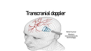
Transcranial doppler ultrasound, principles and procuderes
•Download as PPTX, PDF•
0 likes•38 views

basics principles of transcranial ultrasound procedure in patients

Recommended
More Related Content
Similar to Transcranial doppler ultrasound, principles and procuderes
Similar to Transcranial doppler ultrasound, principles and procuderes (20)
More from Nitish kumar
More from Nitish kumar (8)
Recently uploaded
Recently uploaded (20)
Transcranial doppler ultrasound, principles and procuderes
- 2. Introduced by Rune Aaslid in 1982 for detecting blood flow in the basal intracerebral arteries. Important and established applications now incudes: - Detection of the right to left cardiac shunt. - Cerebral vasomotor reactivity - Monitoring flow velocities for stroke prevention - Continuous monitoring during thrombolysis - Diagnosis of brain death.
- 4. Basic concept of ultrasound physics Ultrasound are the waves of frequency >20KHz and are longitudinal in nature. Types Duty factor: pulse emition/pulse wave interval*100 Pulsed wave USG: transducer receives the signal for most of the time (>90%) Continuous wave ultrasound: requires 2 transducers where one act as emitter and the other as receiver. - Not optimal for diagnostic studies as it provides low spatial resolution.
- 6. Ultrasound transducers Transmitters: converts electric signals into ultrasound Receivers: converts ultrasounds into electric signals Transceivers: can perform both the functions
- 7. • Frequency - Lower frequency and higher wavelength provides better sapling size by increasing penetration through skull bones. - Higher frequency and lower wavelengths probes are used in colour flow and obtains a better structural gray scale imaging.
- 8. • Gate: the range of soft tissue which can be sampled depending upon the depth. It is usually ( +-5% of depth). • Focusing: procedure to shape the ultrasound beam to increase the resolution. It includes:- • 1. External: Changing curvature of the probe by putting lens ahead of it. • 2. internal: changing the curvature of piezoelectric crystals. • 3. Phased array: for phased array transducers in duplex imaging.
- 9. Piezoelectric effect is a property of materials to generate voltage if mechanically deformed and vice versa. • Physical parameters and their determining factors: 1. Sound source: it determines the frequency and period if ultrasound waves. 2. Soft tissues: it determines the propagation speed of the ultrasound waves. -the parameters like wavelength , intensity, amplitude, power is initially determines by the source and as it passes the soft tissues can be altered by them. Propagation speed ∝ stiffness ∝ 1/density
- 10. Hemodynamicprinciplesgoverningdopplerultrasound The factors affecting blood flow across a vessel if determined by several factors:- Blood flow ∝ change in pressure ∝ 1/resistance across the vessel the resistance therefore is determined by – Length of vessel Cross sectional area/ diameter Viscosity of blood
- 11. Winkiesel effect: it is a property of arterial blood vessels to convert energy received from heart to pulsatile energy during systolic ejections & thus converting it into continuous pulsating waves for propagation through the systemic system. ∝ distensibility of arterial walls
- 12. Types of blood flow Laminar flow Turbulant flow Parabolic or pluglike movement of blood in layers Consists of streamline flow as well as eddy currents Minimal energy loss doesn’t takes place due to vessel resistance Energy loss occurs Eg: parabolic flow pattern and pluglike flow pattern. Eg: in stenosis and bifurcation of blood vessels
- 14. Themajoracousticwindowsinpulsed wave ultrasound includes: Transtemporal: can be used to measure velocities in MCA,ACA,PCA& PCOM. Transorbital: ophthalmic artery, Internal carotid artery Suboccipital and transforaminal: allows insonation of vertebral arteries and basilar arteries.
- 16. The Doppler effect It is the change in the observed frequency of a wave when the source and the observer move relative to the medium. Doppler shift is the difference between the emitted and reflected frequency of the wave. - It can be used to determine the velocity and direction of moving objects, such as blood flow, by analyzing the frequency difference between the ultrasound waves. D.S = 2*(blood speed*transducer frequency cosθ)/propagation speed
- 17. For transcranial vessels, major documented parameters 1. Peak systolic velocity: 2. End diastolic velocity 3. Mean flow velocity 4. Pulsatality index 5. Resistivity index:
- 18. • Lindegards ratio: MFV MCA/Unilateral ICA • Soustel ratio: MFV BA/ Unilateral VA
- 20. Normal flow patterns at different levels
- 21. Abnormal findings in transcranial doppler
- 22. Cerebral circulatory arrest (brain death) • Brain death is seen on TCD as variying from high resistance to diastolic flow reversal(reverberating) to absent flow. • TCD is often used as a supplementary test for the confirmation of brain death.
- 23. Methodology 1. Clinical examination followed by NCCT to obtain desired relevant history and side of weakness. 2. The procedure should be started to the contralateral side of the infract, in order to obtain a baseline and normative values for the patient. It also establishes a indication of the affected side. 3. The probe to be placed angulated anterio-superior axis for examining th MCA. 4. Depth and other settings to be adjusted depending upon the patient. 5. For PCA, probe to be tilted medially/posteriorly. 6. USG gel to be used adequately to ensure proper contact b/w the skin and probes.
- 24. Thank you