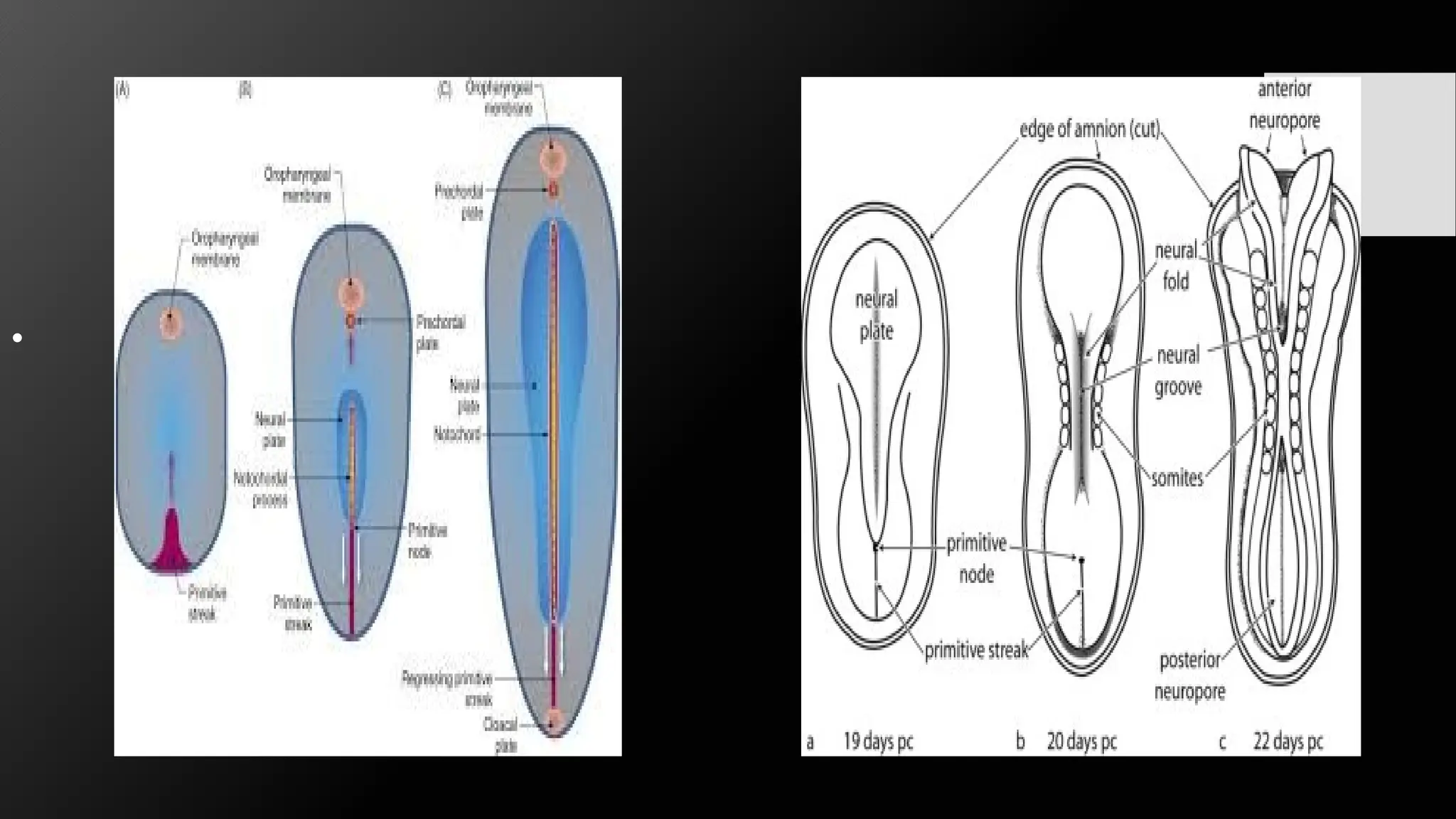
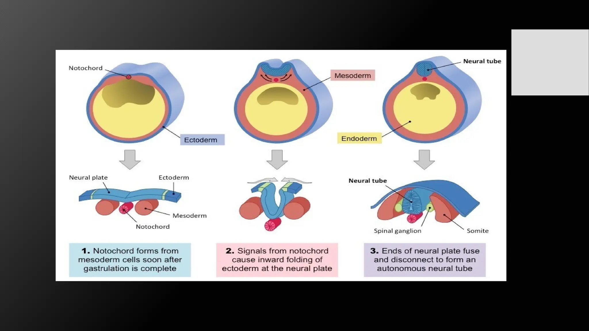
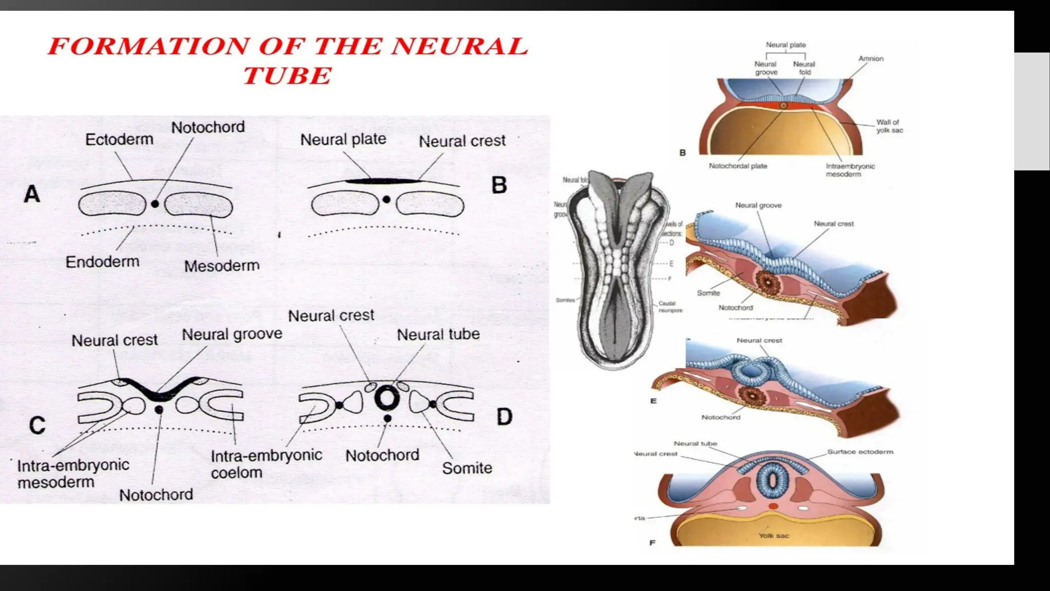
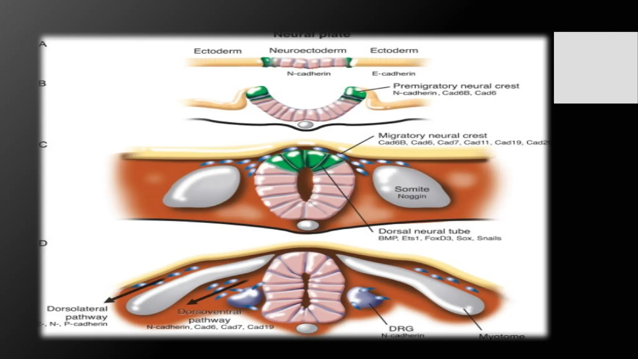
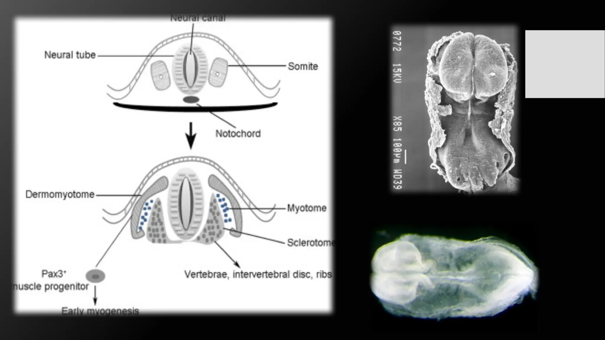
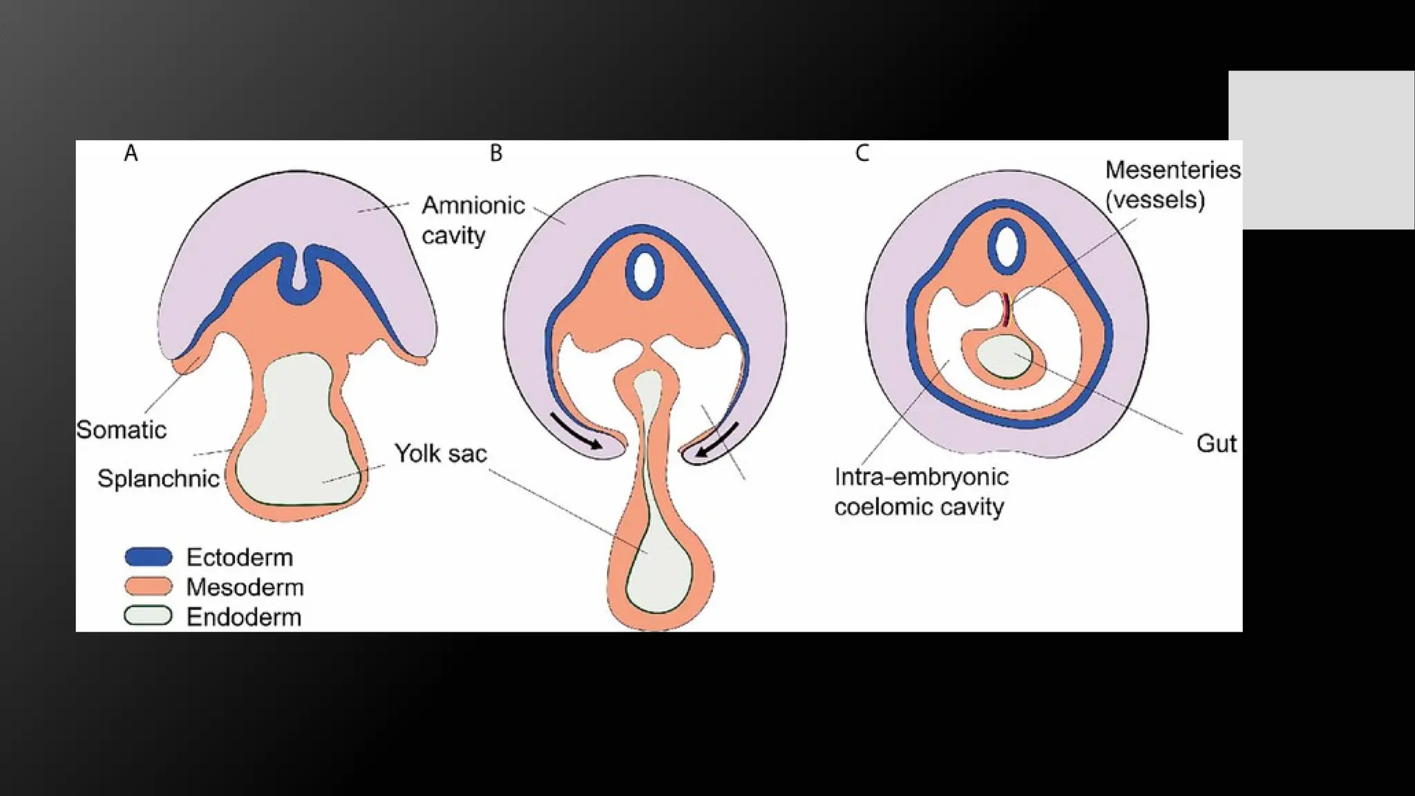
Neurulation
Name: Nolita Turner
Lecture: Dr. D’Aguiar
Course:MD2
Embryology and Histology F24
What is Neurulation?
• Neurulation is the process where a flat sheet of cells called the neural plate
folds inwards and closes to form the neural tube, which will eventually
develop into the central nervous system (brain and spinal cord) of a
vertebrate embryo.
• This process starts in the 3 week post fertilization.
Formation of the Neural Plate
• The neural plate is formed during embryogenesis when the notochord forms
along the midline of the embryo. The notochord releases signaling molecules
to the overlying ectoderm, instructing it to differentiate into neural tissue. The
ectoderm becomes thickened and elongated, forming the neural plate. The
neural plate is a specialized region of the ectoderm, distinct from the
surrounding ectodermal tissue. The cells of the ectoderm under the influence
of signaling molecules change their shape and behavior, leading to the
formation of the neural plate, which will later give rise to the neural tube and
the central nervous system.
•
Formation of neural tube
• The neural plate thickens along its lateral edges, forming neural folds on
either side of a central depression.
• The neural groove, a depression along the midline of the embryo, aligns with
the notochord and is influenced by sonic hedgehog (shh) signals. The neural
folds continue to rise and curl toward the midline, narrowing the groove.
Cellular adhesion molecules, such as N-cadherin and E-cadherin, help
maintain tissue integrity and direct the folding process. The neural groove
deepens further as the neural folds approach each other, preparing for neural
tube closure.
• The neural folds meet and fuse at the midline to form the neural tube. This
fusion occurs from the middle and proceeds both cranially and caudally.
• Closure of the neural tube is complete by the end of the fourth week.
Formation of the neural crest
• The neural crest is a unique embryonic structure in vertebrates
• The neural plate border is established by inductive signals, and neural crest
cells are induced at this border. These progenitors are multipotent and can
differentiate into various cell types.
• Neural crest cells migrate extensively through the embryo, forming
craniofacial cartilage and connective tissue, and forming melanocytes and
contributing to the peripheral nervous system.
Role of Neural Crest in Development
• Neural crest cells are often referred to as the "fourth germ layer" due to their extensive
contribution to multiple systems.
• Peripheral Nervous System: Formation of sensory neurons, autonomic neurons, and Schwann
cells. These structures are essential for neural communication throughout the body.
• Craniofacial Structures: Neural crest cells contribute to the formation of craniofacial bones and
cartilage. They also give rise to connective tissues in the face.
• Melanocytes: These cells migrate into the skin and differentiate into pigment-producing cells.
• Cardiovascular System: They contribute to the formation of parts of the aorticopulmonary
septum and other structures of the heart.
• Endocrine Cells: Neural crest cells form the adrenal medulla and certain hormone-producing
cells.
• Other Structures: Includes teeth (dentine), parts of the thymus, and the inner ear.
Development of Somite's
• Somotogenesis is the process of segmenting the paraxial mesoderm within the
trilaminar embryo body to form pairs of somites, with the first pair appearing at day 20
and increasing at 1 pair/90 minutes.
• Somoete pairs are formed by adding a somite to either side of the notochord, a
process that starts cranially (head end) and extends caudally(tailward). This sequential
process is used to stage the age of various species embryos based on the number of
visible somite pairs. The outer cellular shell of the developing somite is defined by a
mesenchymal to epithelial transition, with the core cells remaining mesenchymal.
• They are precursor populations of cells that give rise to important structures associated
with the vertebrate body plan, such as dermis, skeletal muscle, cartilage, tendons, and
vertebrae. Somites also determine the migratory paths of neural crest cells and spinal
nerve axons.
Development of intraembryonic coelom
• The intraembryonic coelom is a fluid-filled cavity within an embryo's
mesoderm that forms during the third week of embryonic development.
• During the lateral unfolding of the embryo in the 4th week these vacuoles
fuse and form a U-shaped cavity: the intraembryonic coelom, initially a
single horseshoe-shaped cavity
• It splits into a somatic and splanchnic layer.
• Then it eventually divides into pericardial, pleural, and peritoneal body
cavities. These cavities are crucial for the developing organism's major
body cavities.
Neurulation , Harvard University Embryology

Neurulation , Harvard University Embryology

  • 1.
    Neurulation Name: Nolita Turner Lecture:Dr. D’Aguiar Course:MD2 Embryology and Histology F24
  • 2.
    What is Neurulation? •Neurulation is the process where a flat sheet of cells called the neural plate folds inwards and closes to form the neural tube, which will eventually develop into the central nervous system (brain and spinal cord) of a vertebrate embryo. • This process starts in the 3 week post fertilization.
  • 4.
    Formation of theNeural Plate • The neural plate is formed during embryogenesis when the notochord forms along the midline of the embryo. The notochord releases signaling molecules to the overlying ectoderm, instructing it to differentiate into neural tissue. The ectoderm becomes thickened and elongated, forming the neural plate. The neural plate is a specialized region of the ectoderm, distinct from the surrounding ectodermal tissue. The cells of the ectoderm under the influence of signaling molecules change their shape and behavior, leading to the formation of the neural plate, which will later give rise to the neural tube and the central nervous system.
  • 5.
  • 6.
    Formation of neuraltube • The neural plate thickens along its lateral edges, forming neural folds on either side of a central depression. • The neural groove, a depression along the midline of the embryo, aligns with the notochord and is influenced by sonic hedgehog (shh) signals. The neural folds continue to rise and curl toward the midline, narrowing the groove. Cellular adhesion molecules, such as N-cadherin and E-cadherin, help maintain tissue integrity and direct the folding process. The neural groove deepens further as the neural folds approach each other, preparing for neural tube closure. • The neural folds meet and fuse at the midline to form the neural tube. This fusion occurs from the middle and proceeds both cranially and caudally. • Closure of the neural tube is complete by the end of the fourth week.
  • 9.
    Formation of theneural crest • The neural crest is a unique embryonic structure in vertebrates • The neural plate border is established by inductive signals, and neural crest cells are induced at this border. These progenitors are multipotent and can differentiate into various cell types. • Neural crest cells migrate extensively through the embryo, forming craniofacial cartilage and connective tissue, and forming melanocytes and contributing to the peripheral nervous system.
  • 11.
    Role of NeuralCrest in Development • Neural crest cells are often referred to as the "fourth germ layer" due to their extensive contribution to multiple systems. • Peripheral Nervous System: Formation of sensory neurons, autonomic neurons, and Schwann cells. These structures are essential for neural communication throughout the body. • Craniofacial Structures: Neural crest cells contribute to the formation of craniofacial bones and cartilage. They also give rise to connective tissues in the face. • Melanocytes: These cells migrate into the skin and differentiate into pigment-producing cells. • Cardiovascular System: They contribute to the formation of parts of the aorticopulmonary septum and other structures of the heart. • Endocrine Cells: Neural crest cells form the adrenal medulla and certain hormone-producing cells. • Other Structures: Includes teeth (dentine), parts of the thymus, and the inner ear.
  • 13.
    Development of Somite's •Somotogenesis is the process of segmenting the paraxial mesoderm within the trilaminar embryo body to form pairs of somites, with the first pair appearing at day 20 and increasing at 1 pair/90 minutes. • Somoete pairs are formed by adding a somite to either side of the notochord, a process that starts cranially (head end) and extends caudally(tailward). This sequential process is used to stage the age of various species embryos based on the number of visible somite pairs. The outer cellular shell of the developing somite is defined by a mesenchymal to epithelial transition, with the core cells remaining mesenchymal. • They are precursor populations of cells that give rise to important structures associated with the vertebrate body plan, such as dermis, skeletal muscle, cartilage, tendons, and vertebrae. Somites also determine the migratory paths of neural crest cells and spinal nerve axons.
  • 15.
    Development of intraembryoniccoelom • The intraembryonic coelom is a fluid-filled cavity within an embryo's mesoderm that forms during the third week of embryonic development. • During the lateral unfolding of the embryo in the 4th week these vacuoles fuse and form a U-shaped cavity: the intraembryonic coelom, initially a single horseshoe-shaped cavity • It splits into a somatic and splanchnic layer. • Then it eventually divides into pericardial, pleural, and peritoneal body cavities. These cavities are crucial for the developing organism's major body cavities.

Editor's Notes

  • #2 Basically : it's the formation of the neural tube from the neural plate during early embryonic development
  • #4 The neural plate is the thicken of the ectoderm Ectoderm The outermost layer that gives rise to the nervous system, skin, hair, nails, and the lens of the eye. It also forms the epithelia of the nasal cavity, mouth, sinuses, and anal canal. Mesoderm Gives rise to the skeletal and muscular systems, as well as connective tissue, cartilage, and the circulatory system. Endoderm Forms the linings of the digestive and respiratory systems, as well as organs like the liver and pancreas. It also forms the epithelial linings of organ-specific glands. The notochord secretes sonic hedgehog (Shh), a signaling protein that instructs the overlying ectoderm to differentiate into neural tissue
  • #13 and any disruption in the cycle can result in anomalies such as congenital vertebral defects and kidney problems The first set of somites, or the initial pair of somites, form in the anterior part of the trunk, near the notochord, during embryonic development 44 pairs of somites