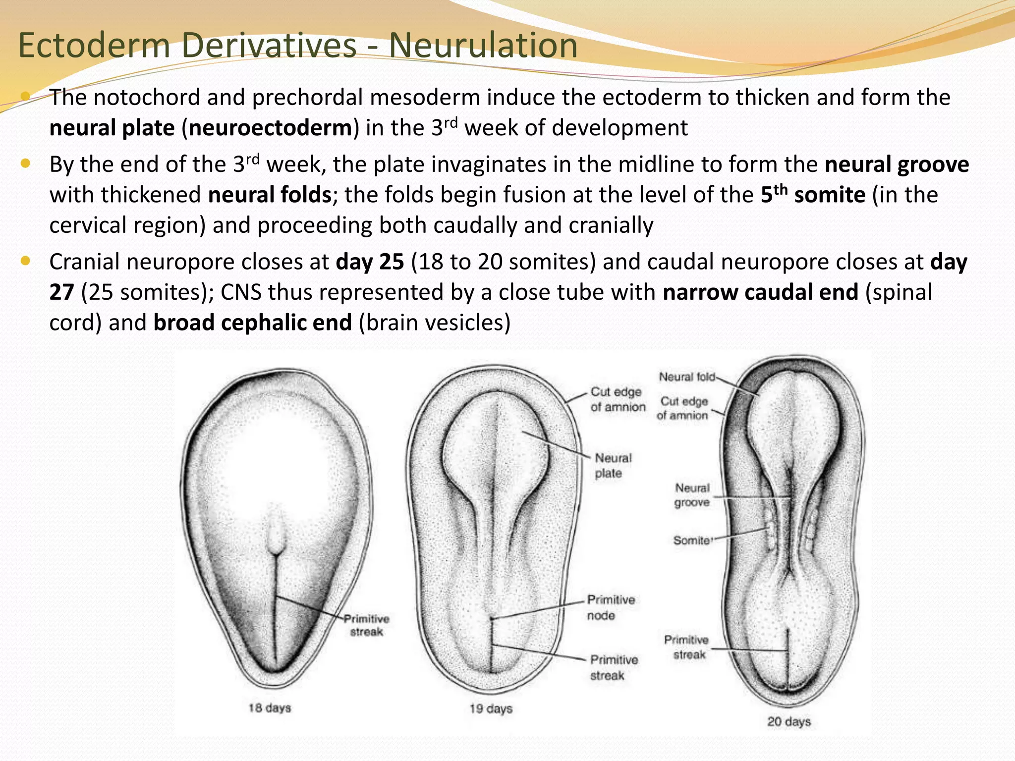
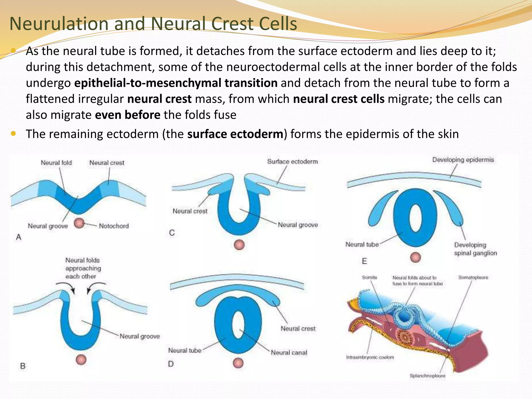
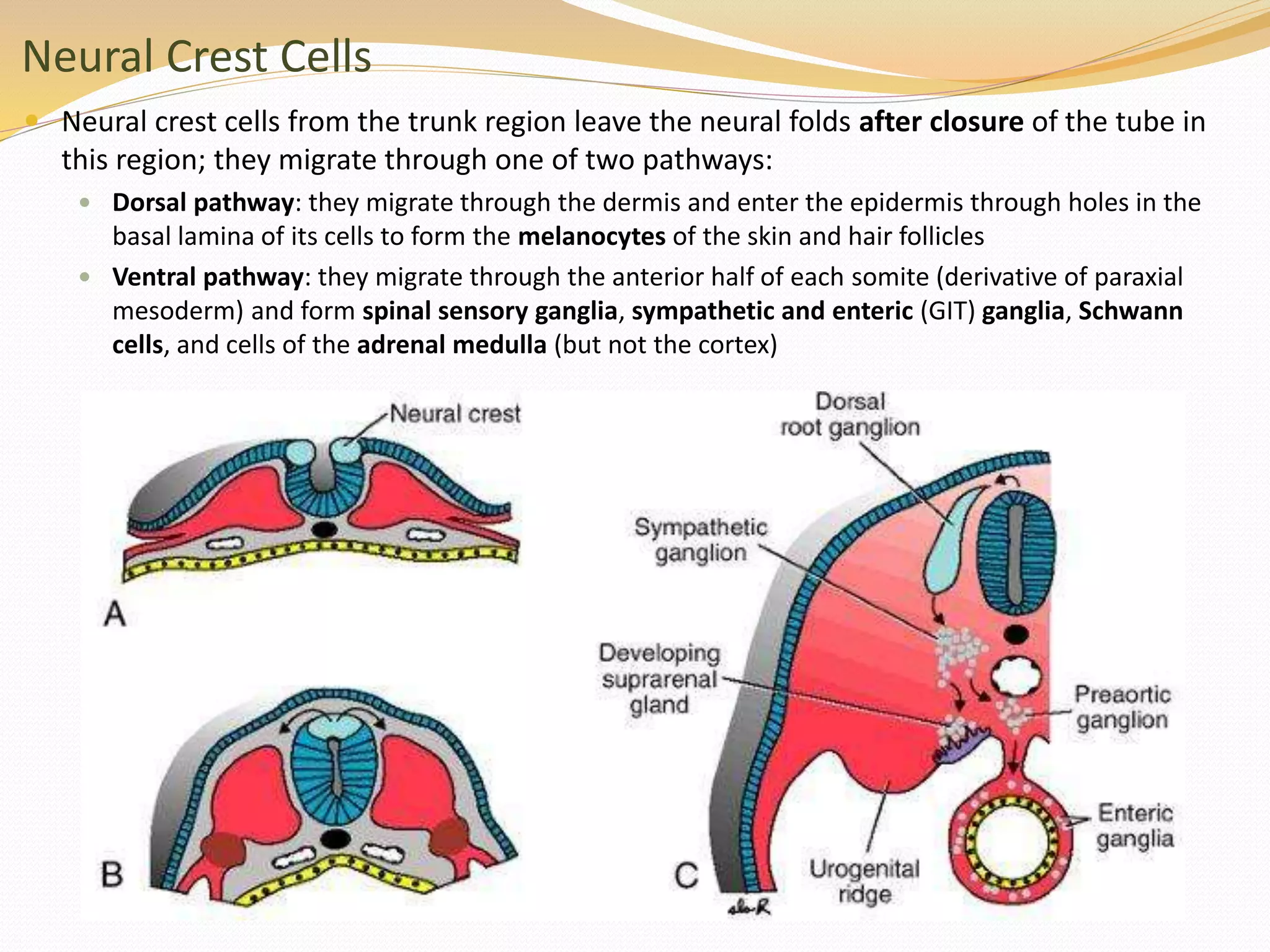
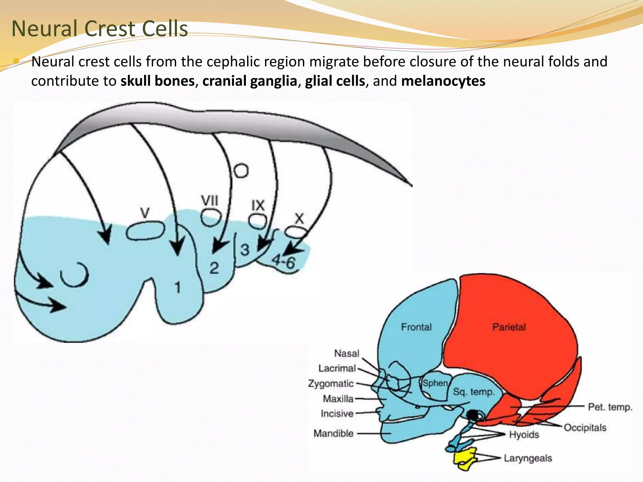
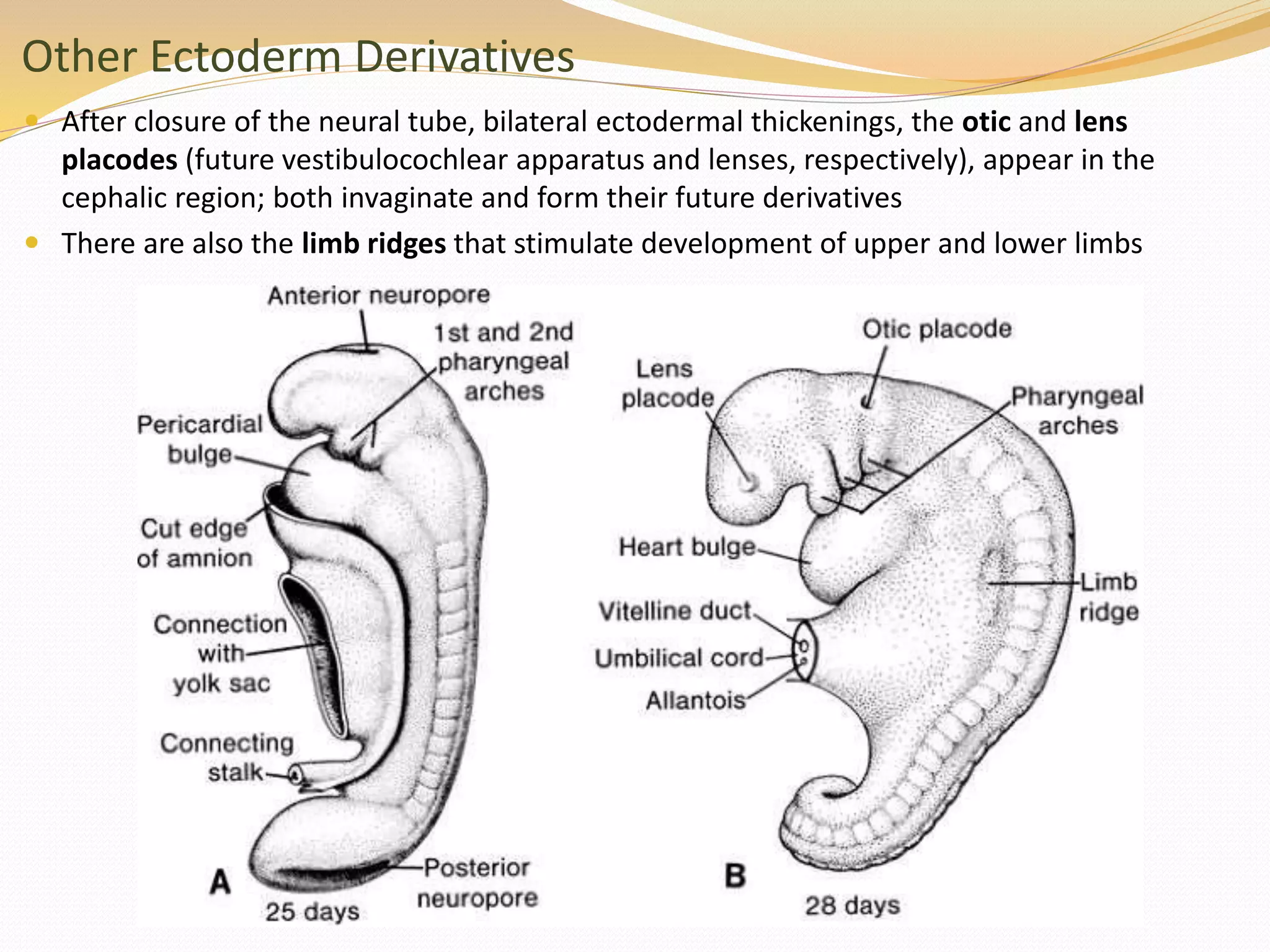
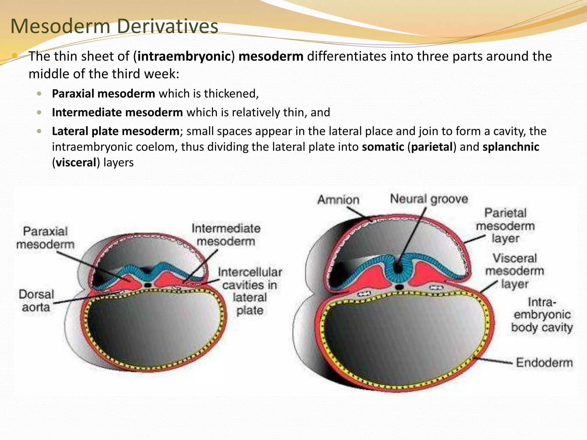
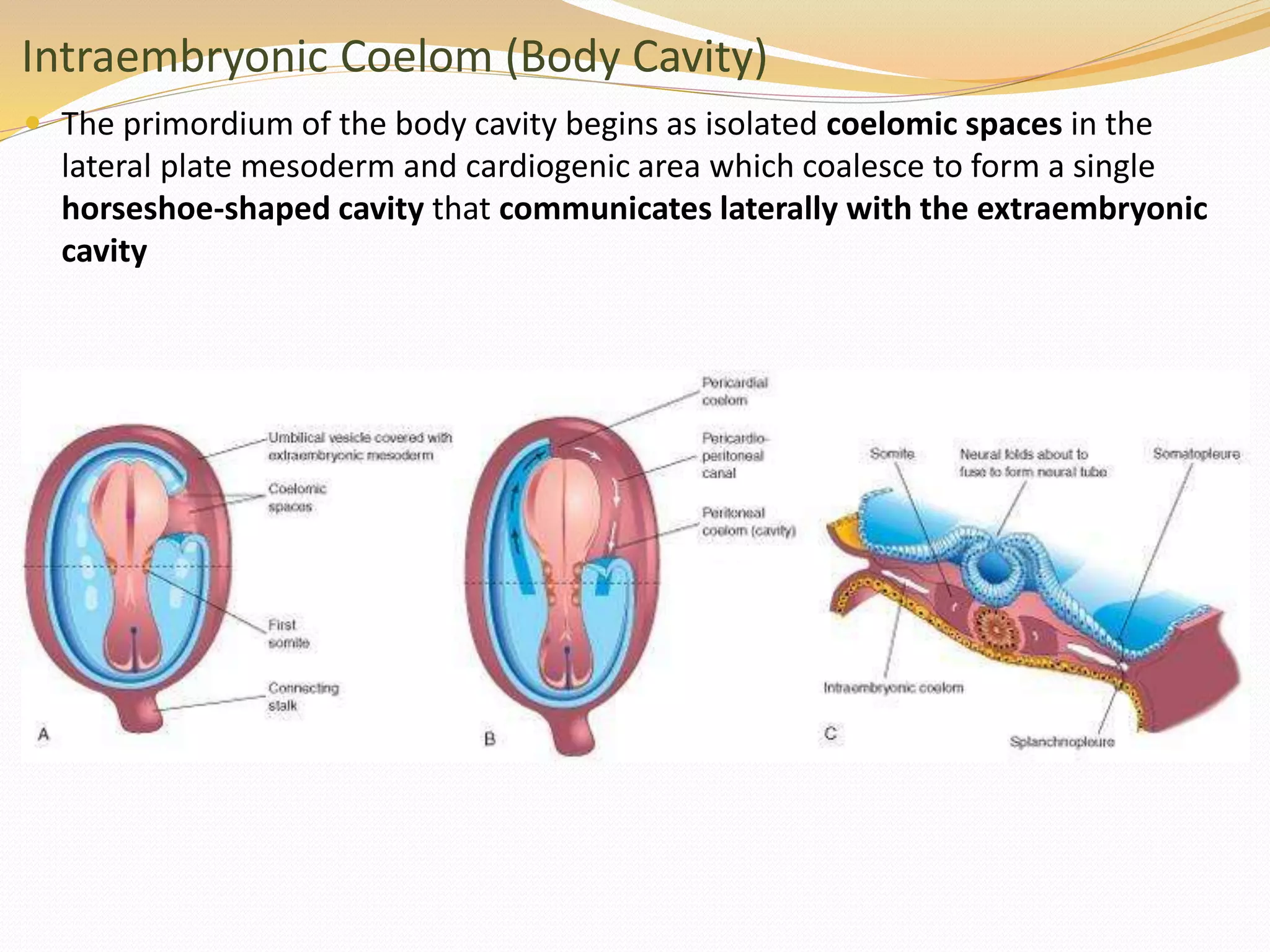
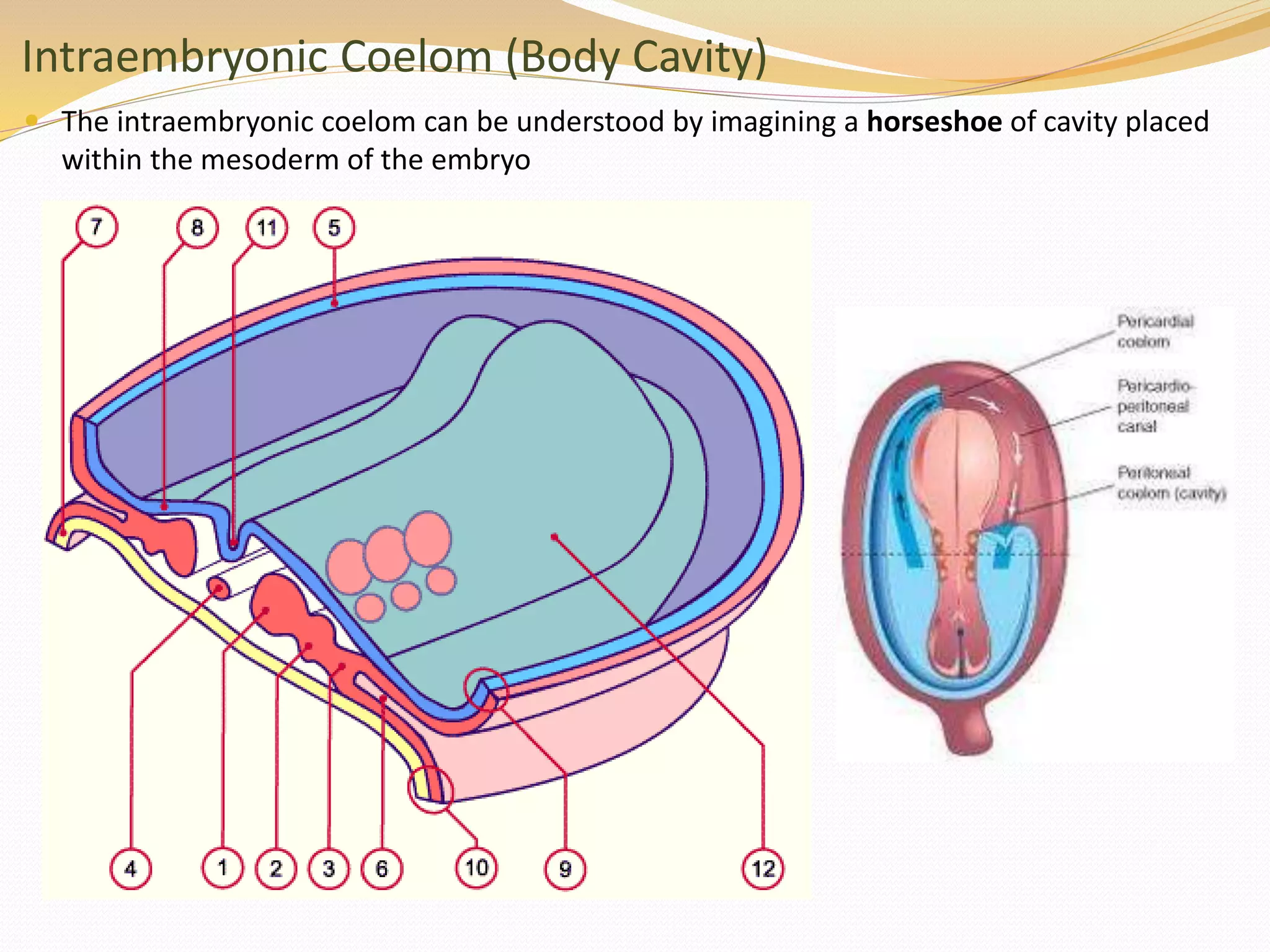
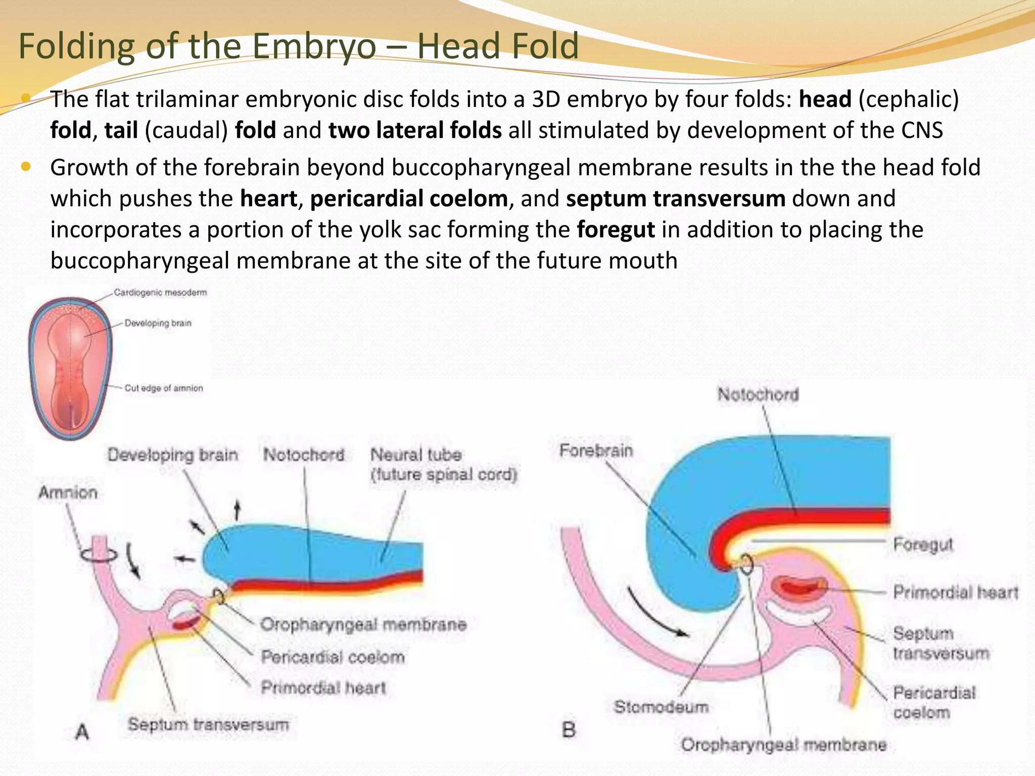
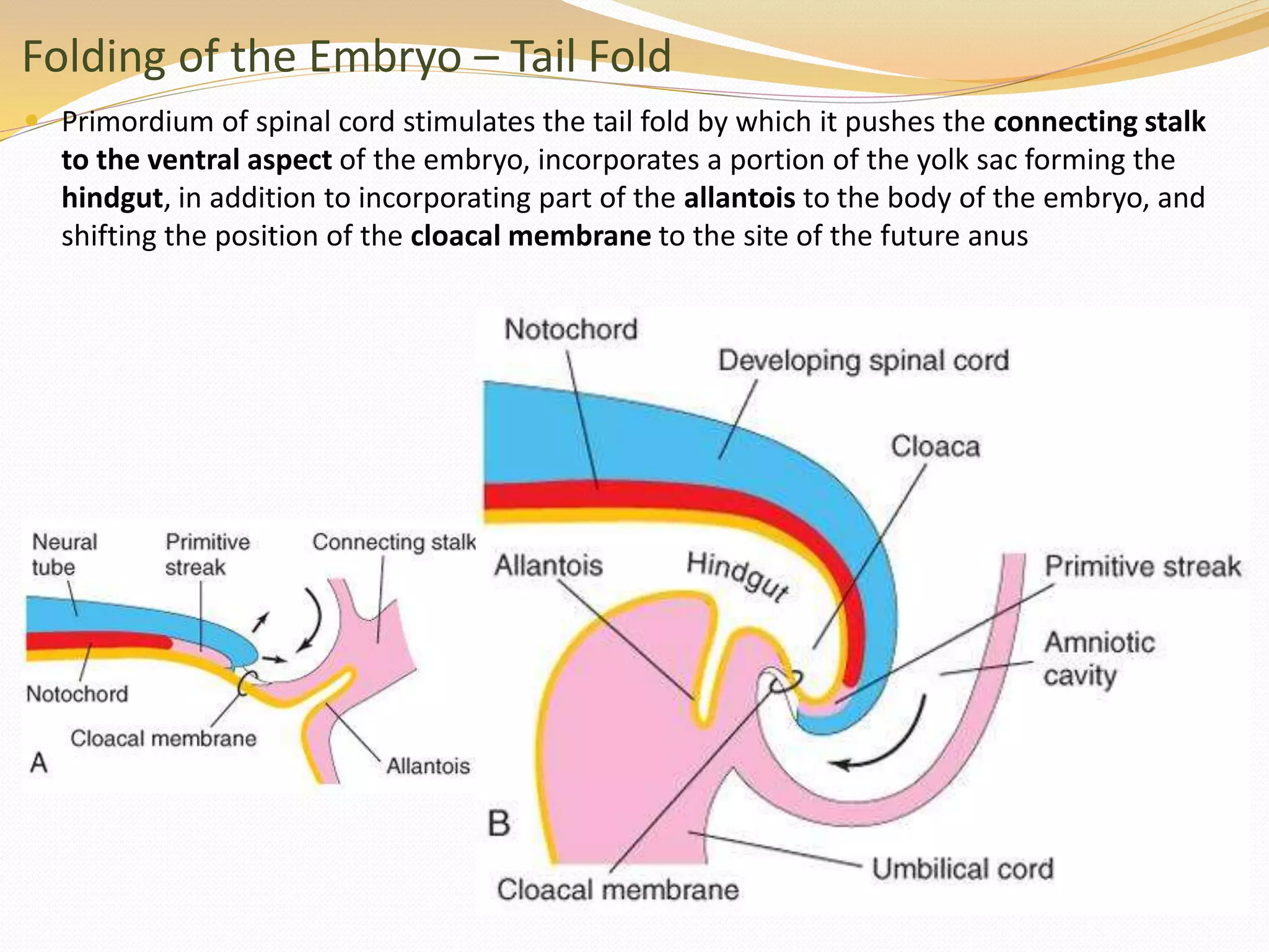
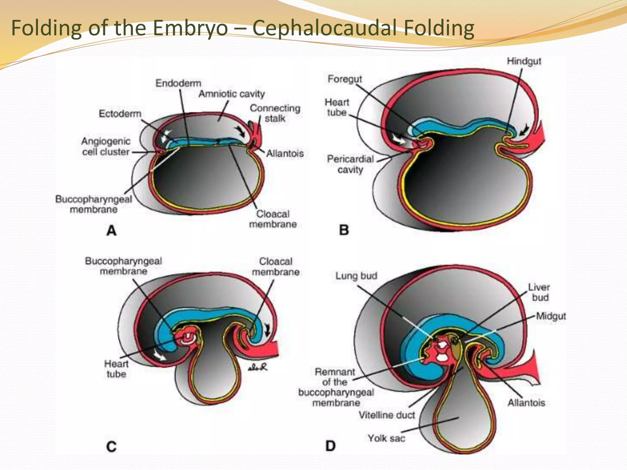
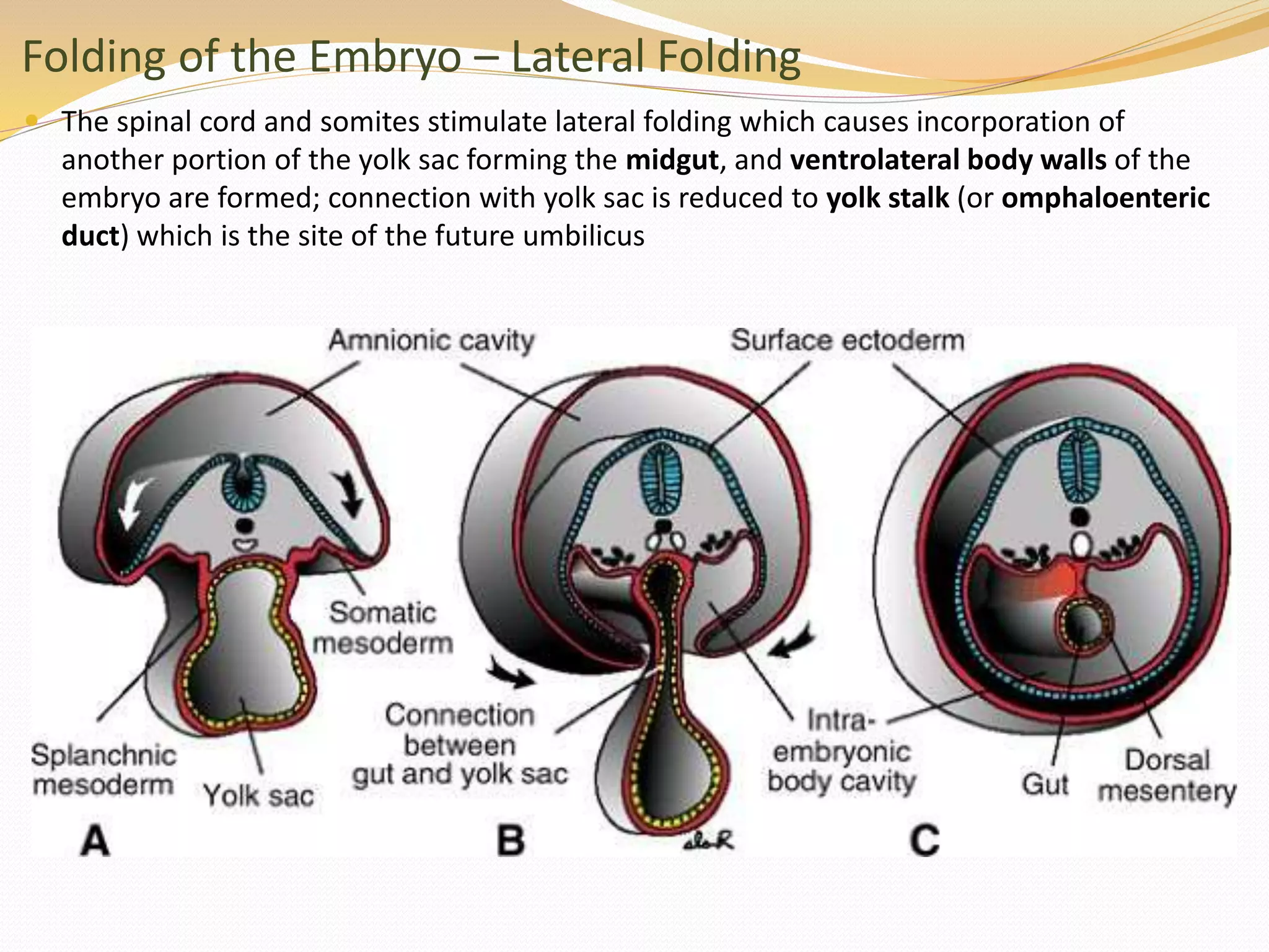
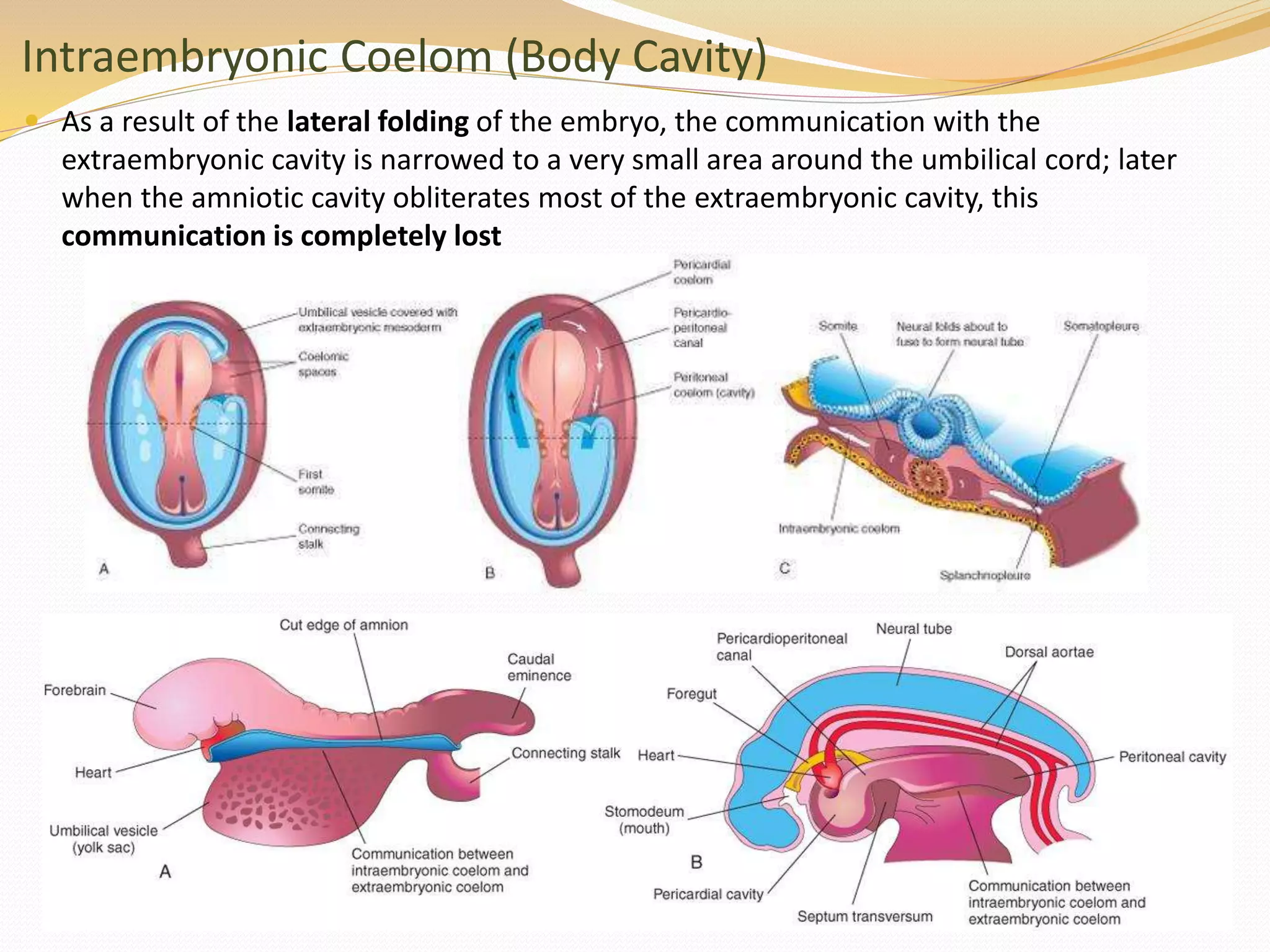
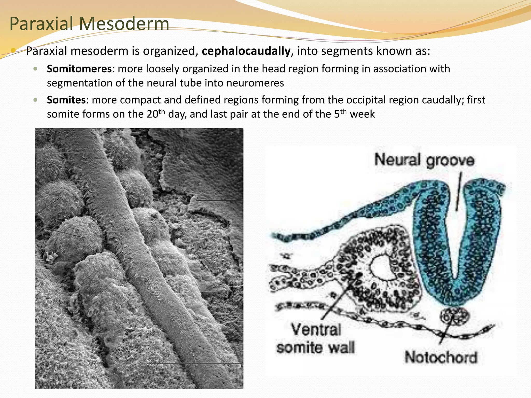
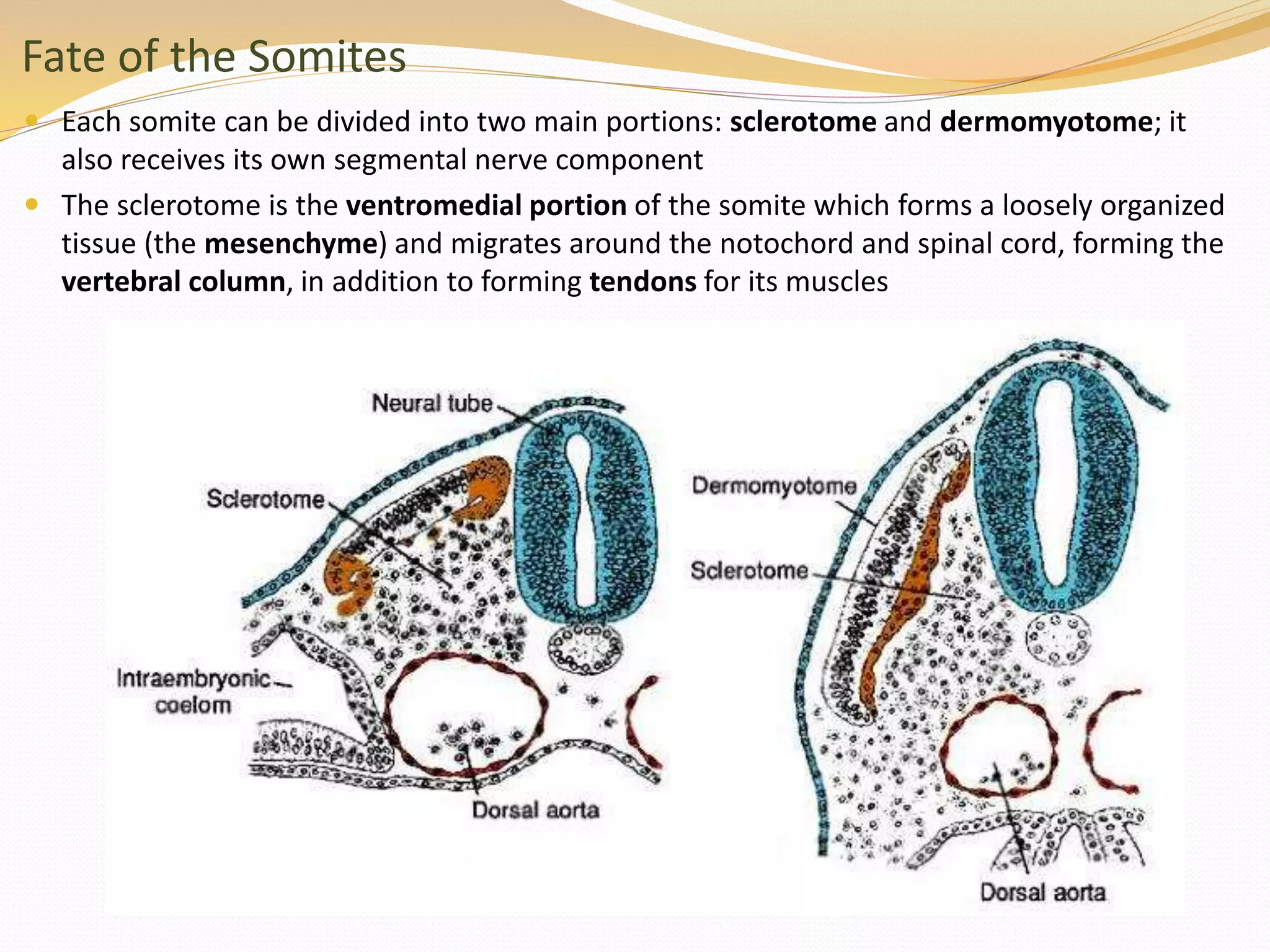
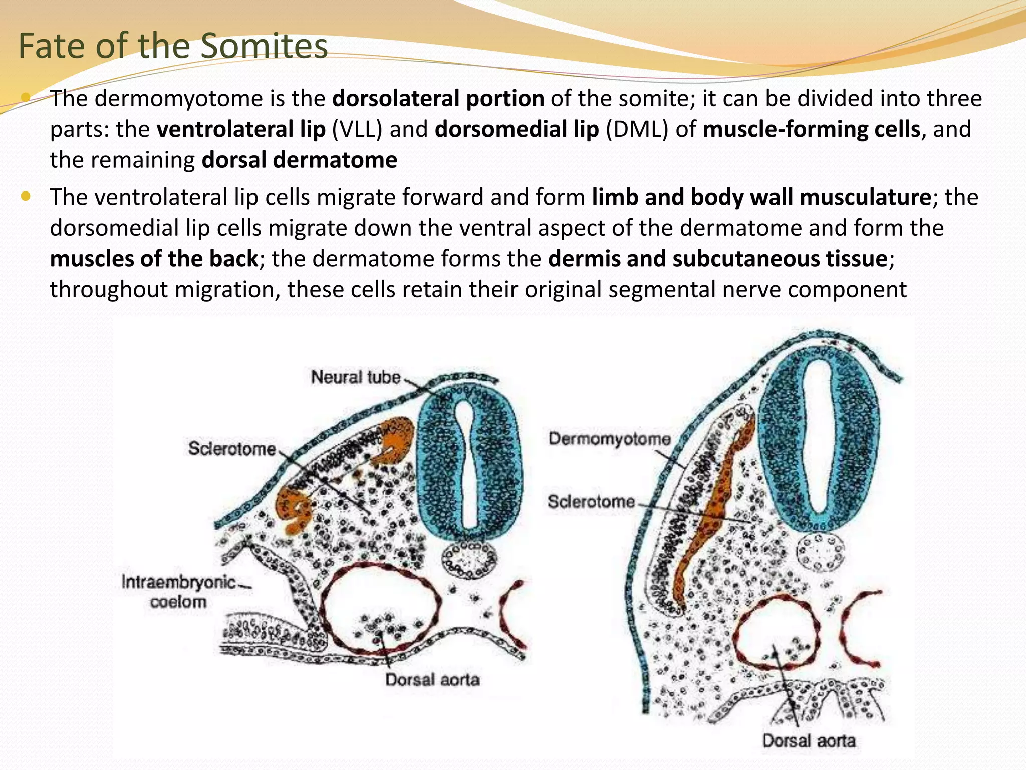
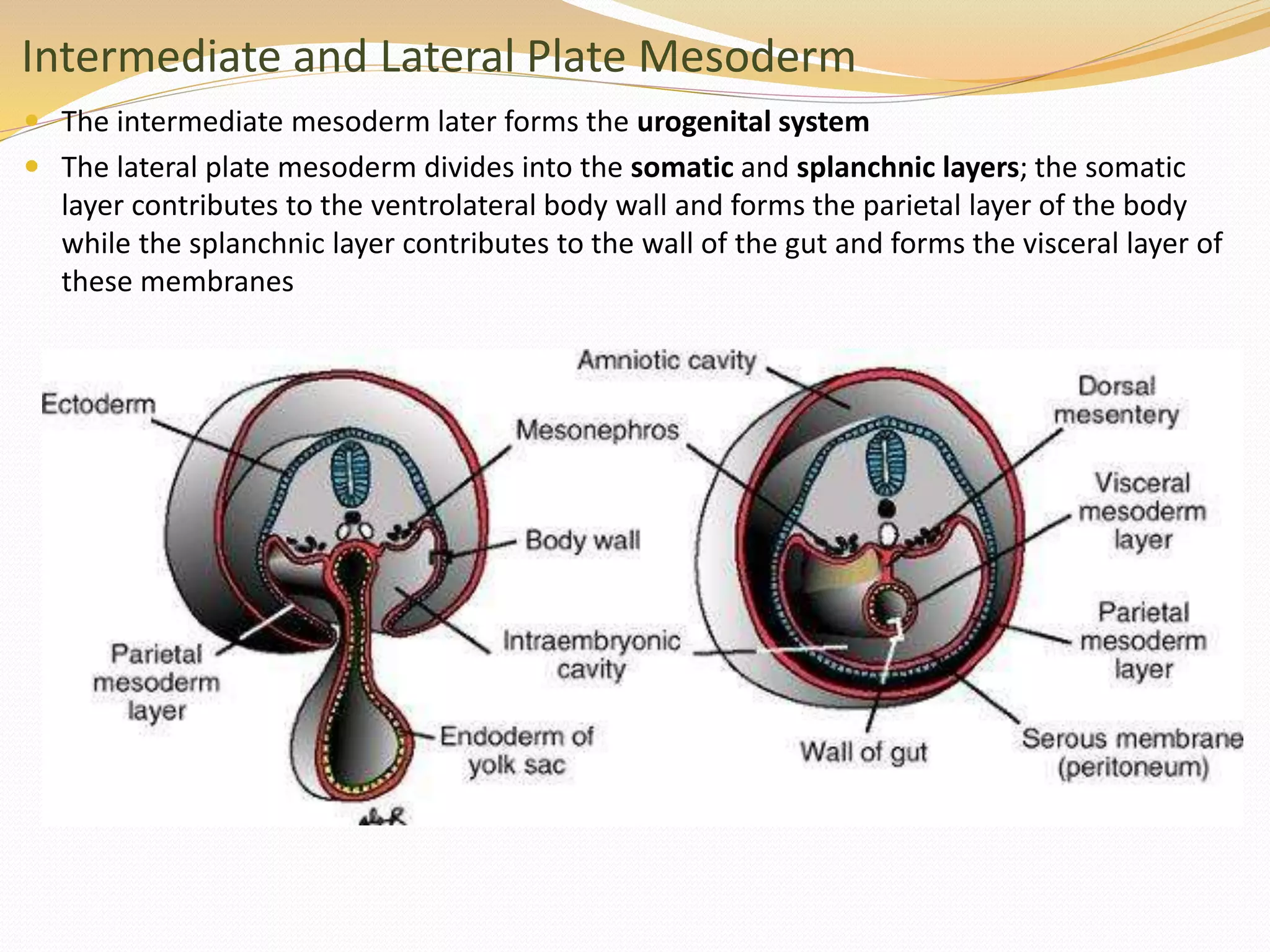
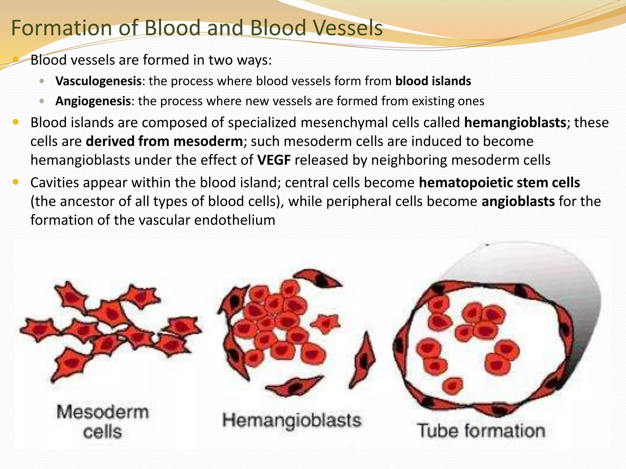
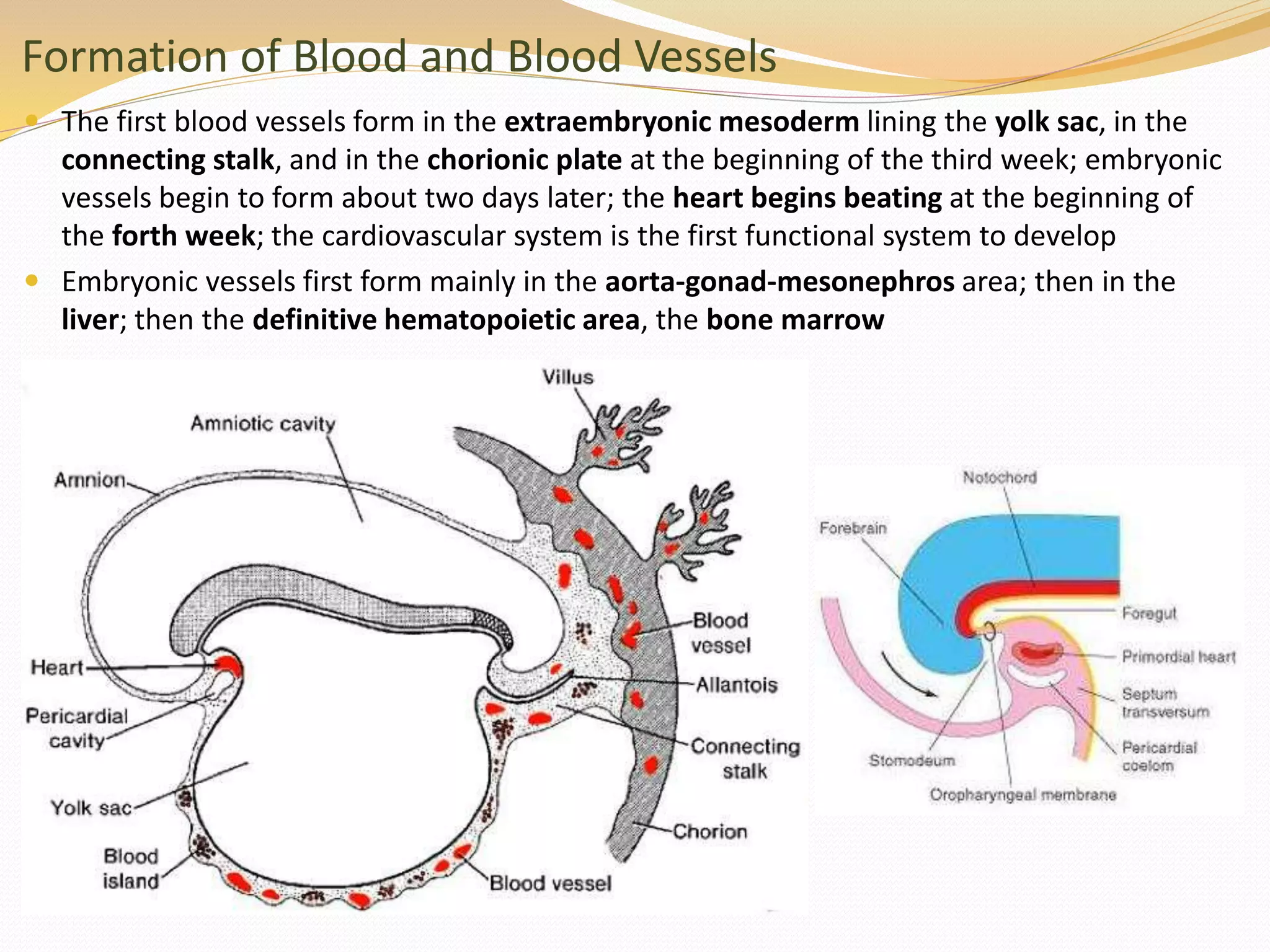
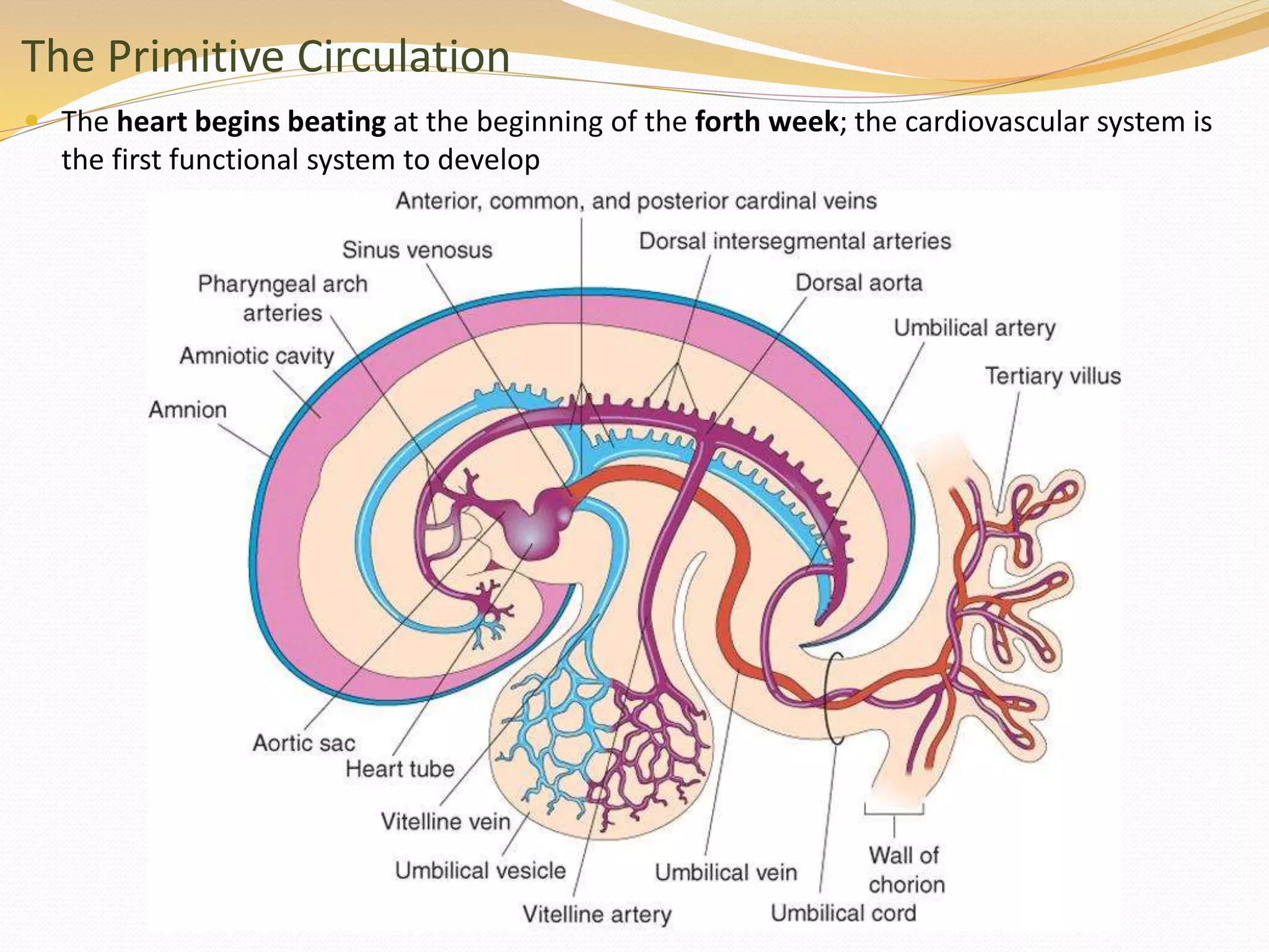
Ectoderm Derivatives - Neurulation
 The notochord and prechordal mesoderm induce the ectoderm to thicken and form the
neural plate (neuroectoderm) in the 3rd week of development
 By the end of the 3rd week, the plate invaginates in the midline to form the neural groove
with thickened neural folds; the folds begin fusion at the level of the 5th somite (in the
cervical region) and proceeding both caudally and cranially
 Cranial neuropore closes at day 25 (18 to 20 somites) and caudal neuropore closes at day
27 (25 somites); CNS thus represented by a close tube with narrow caudal end (spinal
cord) and broad cephalic end (brain vesicles)
Neurulation and Neural Crest Cells
 As the neural tube is formed, it detaches from the surface ectoderm and lies deep to it;
during this detachment, some of the neuroectodermal cells at the inner border of the folds
undergo epithelial-to-mesenchymal transition and detach from the neural tube to form a
flattened irregular neural crest mass, from which neural crest cells migrate; the cells can
also migrate even before the folds fuse
 The remaining ectoderm (the surface ectoderm) forms the epidermis of the skin
Neural Crest Cells
 Neural crest cells from the trunk region leave the neural folds after closure of the tube in
this region; they migrate through one of two pathways:
 Dorsal pathway: they migrate through the dermis and enter the epidermis through holes in the
basal lamina of its cells to form the melanocytes of the skin and hair follicles
 Ventral pathway: they migrate through the anterior half of each somite (derivative of paraxial
mesoderm) and form spinal sensory ganglia, sympathetic and enteric (GIT) ganglia, Schwann
cells, and cells of the adrenal medulla (but not the cortex)
Neural Crest Cells
 Neural crest cells from the cephalic region migrate before closure of the neural folds and
contribute to skull bones, cranial ganglia, glial cells, and melanocytes
Other Ectoderm Derivatives
 After closure of the neural tube, bilateral ectodermal thickenings, the otic and lens
placodes (future vestibulocochlear apparatus and lenses, respectively), appear in the
cephalic region; both invaginate and form their future derivatives
 There are also the limb ridges that stimulate development of upper and lower limbs
Mesoderm Derivatives
 The thin sheet of (intraembryonic) mesoderm differentiates into three parts around the
middle of the third week:
 Paraxial mesoderm which is thickened,
 Intermediate mesoderm which is relatively thin, and
 Lateral plate mesoderm; small spaces appear in the lateral place and join to form a cavity, the
intraembryonic coelom, thus dividing the lateral plate into somatic (parietal) and splanchnic
(visceral) layers
The Mesoderm Layers and the Two Coeloms
Intraembryonic Coelom (Body Cavity)
 The primordium of the body cavity begins as isolated coelomic spaces in the
lateral plate mesoderm and cardiogenic area which coalesce to form a single
horseshoe-shaped cavity that communicates laterally with the extraembryonic
cavity
Intraembryonic Coelom (Body Cavity)
 The intraembryonic coelom can be understood by imagining a horseshoe of cavity placed
within the mesoderm of the embryo
Folding of the Embryo – Head Fold
 The flat trilaminar embryonic disc folds into a 3D embryo by four folds: head (cephalic)
fold, tail (caudal) fold and two lateral folds all stimulated by development of the CNS
 Growth of the forebrain beyond buccopharyngeal membrane results in the the head fold
which pushes the heart, pericardial coelom, and septum transversum down and
incorporates a portion of the yolk sac forming the foregut in addition to placing the
buccopharyngeal membrane at the site of the future mouth
Folding of the Embryo – Tail Fold
 Primordium of spinal cord stimulates the tail fold by which it pushes the connecting stalk
to the ventral aspect of the embryo, incorporates a portion of the yolk sac forming the
hindgut, in addition to incorporating part of the allantois to the body of the embryo, and
shifting the position of the cloacal membrane to the site of the future anus
Folding of the Embryo – Cephalocaudal Folding
Folding of the Embryo – Lateral Folding
 The spinal cord and somites stimulate lateral folding which causes incorporation of
another portion of the yolk sac forming the midgut, and ventrolateral body walls of the
embryo are formed; connection with yolk sac is reduced to yolk stalk (or omphaloenteric
duct) which is the site of the future umbilicus
Intraembryonic Coelom (Body Cavity)
 As a result of the lateral folding of the embryo, the communication with the
extraembryonic cavity is narrowed to a very small area around the umbilical cord; later
when the amniotic cavity obliterates most of the extraembryonic cavity, this
communication is completely lost
Paraxial Mesoderm
 Paraxial mesoderm is organized, cephalocaudally, into segments known as:
 Somitomeres: more loosely organized in the head region forming in association with
segmentation of the neural tube into neuromeres
 Somites: more compact and defined regions forming from the occipital region caudally; first
somite forms on the 20th day, and last pair at the end of the 5th week
Fate of the Somites
 Each somite can be divided into two main portions: sclerotome and dermomyotome; it
also receives its own segmental nerve component
 The sclerotome is the ventromedial portion of the somite which forms a loosely organized
tissue (the mesenchyme) and migrates around the notochord and spinal cord, forming the
vertebral column, in addition to forming tendons for its muscles
Fate of the Somites
 The dermomyotome is the dorsolateral portion of the somite; it can be divided into three
parts: the ventrolateral lip (VLL) and dorsomedial lip (DML) of muscle-forming cells, and
the remaining dorsal dermatome
 The ventrolateral lip cells migrate forward and form limb and body wall musculature; the
dorsomedial lip cells migrate down the ventral aspect of the dermatome and form the
muscles of the back; the dermatome forms the dermis and subcutaneous tissue;
throughout migration, these cells retain their original segmental nerve component
Intermediate and Lateral Plate Mesoderm
 The intermediate mesoderm later forms the urogenital system
 The lateral plate mesoderm divides into the somatic and splanchnic layers; the somatic
layer contributes to the ventrolateral body wall and forms the parietal layer of the body
while the splanchnic layer contributes to the wall of the gut and forms the visceral layer of
these membranes
Formation of Blood and Blood Vessels
 Blood vessels are formed in two ways:
 Vasculogenesis: the process where blood vessels form from blood islands
 Angiogenesis: the process where new vessels are formed from existing ones
 Blood islands are composed of specialized mesenchymal cells called hemangioblasts; these
cells are derived from mesoderm; such mesoderm cells are induced to become
hemangioblasts under the effect of VEGF released by neighboring mesoderm cells
 Cavities appear within the blood island; central cells become hematopoietic stem cells
(the ancestor of all types of blood cells), while peripheral cells become angioblasts for the
formation of the vascular endothelium
Formation of Blood and Blood Vessels
 The first blood vessels form in the extraembryonic mesoderm lining the yolk sac, in the
connecting stalk, and in the chorionic plate at the beginning of the third week; embryonic
vessels begin to form about two days later; the heart begins beating at the beginning of
the forth week; the cardiovascular system is the first functional system to develop
 Embryonic vessels first form mainly in the aorta-gonad-mesonephros area; then in the
liver; then the definitive hematopoietic area, the bone marrow
The Primitive Circulation
 The heart begins beating at the beginning of the forth week; the cardiovascular system is
the first functional system to develop
Left-Out Materials
 When something in the book is not mentioned in the
session, it could be due to one of two reasons:
 It is not a significant concept or structural piece of
information
 It is going to be repeated in greater detail in another
chapter
 Something not discussed in the session will not be
included for the quizzes but may be required at university
Embryology Course III - 3rd to 8th Weeks of Development

Embryology Course III - 3rd to 8th Weeks of Development

  • 2.
    Ectoderm Derivatives -Neurulation  The notochord and prechordal mesoderm induce the ectoderm to thicken and form the neural plate (neuroectoderm) in the 3rd week of development  By the end of the 3rd week, the plate invaginates in the midline to form the neural groove with thickened neural folds; the folds begin fusion at the level of the 5th somite (in the cervical region) and proceeding both caudally and cranially  Cranial neuropore closes at day 25 (18 to 20 somites) and caudal neuropore closes at day 27 (25 somites); CNS thus represented by a close tube with narrow caudal end (spinal cord) and broad cephalic end (brain vesicles)
  • 3.
    Neurulation and NeuralCrest Cells  As the neural tube is formed, it detaches from the surface ectoderm and lies deep to it; during this detachment, some of the neuroectodermal cells at the inner border of the folds undergo epithelial-to-mesenchymal transition and detach from the neural tube to form a flattened irregular neural crest mass, from which neural crest cells migrate; the cells can also migrate even before the folds fuse  The remaining ectoderm (the surface ectoderm) forms the epidermis of the skin
  • 4.
    Neural Crest Cells Neural crest cells from the trunk region leave the neural folds after closure of the tube in this region; they migrate through one of two pathways:  Dorsal pathway: they migrate through the dermis and enter the epidermis through holes in the basal lamina of its cells to form the melanocytes of the skin and hair follicles  Ventral pathway: they migrate through the anterior half of each somite (derivative of paraxial mesoderm) and form spinal sensory ganglia, sympathetic and enteric (GIT) ganglia, Schwann cells, and cells of the adrenal medulla (but not the cortex)
  • 5.
    Neural Crest Cells Neural crest cells from the cephalic region migrate before closure of the neural folds and contribute to skull bones, cranial ganglia, glial cells, and melanocytes
  • 6.
    Other Ectoderm Derivatives After closure of the neural tube, bilateral ectodermal thickenings, the otic and lens placodes (future vestibulocochlear apparatus and lenses, respectively), appear in the cephalic region; both invaginate and form their future derivatives  There are also the limb ridges that stimulate development of upper and lower limbs
  • 7.
    Mesoderm Derivatives  Thethin sheet of (intraembryonic) mesoderm differentiates into three parts around the middle of the third week:  Paraxial mesoderm which is thickened,  Intermediate mesoderm which is relatively thin, and  Lateral plate mesoderm; small spaces appear in the lateral place and join to form a cavity, the intraembryonic coelom, thus dividing the lateral plate into somatic (parietal) and splanchnic (visceral) layers
  • 8.
    The Mesoderm Layersand the Two Coeloms
  • 9.
    Intraembryonic Coelom (BodyCavity)  The primordium of the body cavity begins as isolated coelomic spaces in the lateral plate mesoderm and cardiogenic area which coalesce to form a single horseshoe-shaped cavity that communicates laterally with the extraembryonic cavity
  • 10.
    Intraembryonic Coelom (BodyCavity)  The intraembryonic coelom can be understood by imagining a horseshoe of cavity placed within the mesoderm of the embryo
  • 11.
    Folding of theEmbryo – Head Fold  The flat trilaminar embryonic disc folds into a 3D embryo by four folds: head (cephalic) fold, tail (caudal) fold and two lateral folds all stimulated by development of the CNS  Growth of the forebrain beyond buccopharyngeal membrane results in the the head fold which pushes the heart, pericardial coelom, and septum transversum down and incorporates a portion of the yolk sac forming the foregut in addition to placing the buccopharyngeal membrane at the site of the future mouth
  • 12.
    Folding of theEmbryo – Tail Fold  Primordium of spinal cord stimulates the tail fold by which it pushes the connecting stalk to the ventral aspect of the embryo, incorporates a portion of the yolk sac forming the hindgut, in addition to incorporating part of the allantois to the body of the embryo, and shifting the position of the cloacal membrane to the site of the future anus
  • 13.
    Folding of theEmbryo – Cephalocaudal Folding
  • 14.
    Folding of theEmbryo – Lateral Folding  The spinal cord and somites stimulate lateral folding which causes incorporation of another portion of the yolk sac forming the midgut, and ventrolateral body walls of the embryo are formed; connection with yolk sac is reduced to yolk stalk (or omphaloenteric duct) which is the site of the future umbilicus
  • 15.
    Intraembryonic Coelom (BodyCavity)  As a result of the lateral folding of the embryo, the communication with the extraembryonic cavity is narrowed to a very small area around the umbilical cord; later when the amniotic cavity obliterates most of the extraembryonic cavity, this communication is completely lost
  • 16.
    Paraxial Mesoderm  Paraxialmesoderm is organized, cephalocaudally, into segments known as:  Somitomeres: more loosely organized in the head region forming in association with segmentation of the neural tube into neuromeres  Somites: more compact and defined regions forming from the occipital region caudally; first somite forms on the 20th day, and last pair at the end of the 5th week
  • 17.
    Fate of theSomites  Each somite can be divided into two main portions: sclerotome and dermomyotome; it also receives its own segmental nerve component  The sclerotome is the ventromedial portion of the somite which forms a loosely organized tissue (the mesenchyme) and migrates around the notochord and spinal cord, forming the vertebral column, in addition to forming tendons for its muscles
  • 18.
    Fate of theSomites  The dermomyotome is the dorsolateral portion of the somite; it can be divided into three parts: the ventrolateral lip (VLL) and dorsomedial lip (DML) of muscle-forming cells, and the remaining dorsal dermatome  The ventrolateral lip cells migrate forward and form limb and body wall musculature; the dorsomedial lip cells migrate down the ventral aspect of the dermatome and form the muscles of the back; the dermatome forms the dermis and subcutaneous tissue; throughout migration, these cells retain their original segmental nerve component
  • 19.
    Intermediate and LateralPlate Mesoderm  The intermediate mesoderm later forms the urogenital system  The lateral plate mesoderm divides into the somatic and splanchnic layers; the somatic layer contributes to the ventrolateral body wall and forms the parietal layer of the body while the splanchnic layer contributes to the wall of the gut and forms the visceral layer of these membranes
  • 20.
    Formation of Bloodand Blood Vessels  Blood vessels are formed in two ways:  Vasculogenesis: the process where blood vessels form from blood islands  Angiogenesis: the process where new vessels are formed from existing ones  Blood islands are composed of specialized mesenchymal cells called hemangioblasts; these cells are derived from mesoderm; such mesoderm cells are induced to become hemangioblasts under the effect of VEGF released by neighboring mesoderm cells  Cavities appear within the blood island; central cells become hematopoietic stem cells (the ancestor of all types of blood cells), while peripheral cells become angioblasts for the formation of the vascular endothelium
  • 21.
    Formation of Bloodand Blood Vessels  The first blood vessels form in the extraembryonic mesoderm lining the yolk sac, in the connecting stalk, and in the chorionic plate at the beginning of the third week; embryonic vessels begin to form about two days later; the heart begins beating at the beginning of the forth week; the cardiovascular system is the first functional system to develop  Embryonic vessels first form mainly in the aorta-gonad-mesonephros area; then in the liver; then the definitive hematopoietic area, the bone marrow
  • 22.
    The Primitive Circulation The heart begins beating at the beginning of the forth week; the cardiovascular system is the first functional system to develop
  • 23.
    Left-Out Materials  Whensomething in the book is not mentioned in the session, it could be due to one of two reasons:  It is not a significant concept or structural piece of information  It is going to be repeated in greater detail in another chapter  Something not discussed in the session will not be included for the quizzes but may be required at university