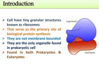
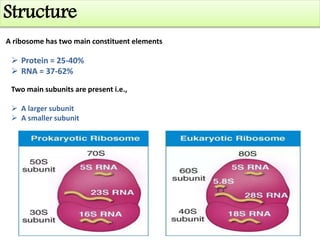
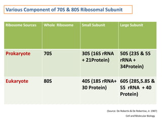
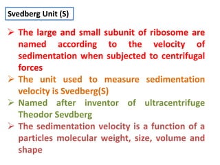
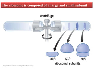
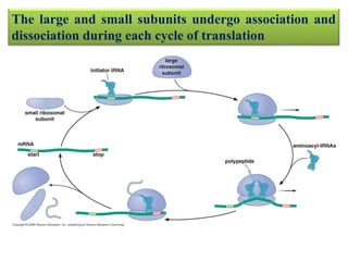
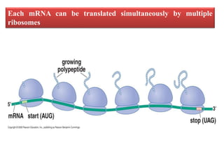
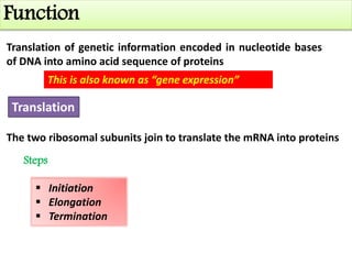
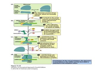
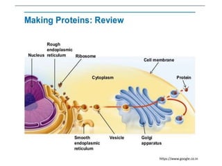
RIBOSOMES
Department of Genetics and Plant Breeding
FACULTY OF AGRICULTURE
BIRSA AGRICULTURAL UNIVERSITY
KANKE, RANCHI – 834 006 (JHARKHAND)
ANJANI KUMAR
Registration No:- A/BAU/5129/2017
Sub:- Molecular Cell Biology (MBB-503)
Introduction
 Cell have tiny granular structures
known as ribosomes
 That serve as the primary site of
biological protein synthesis
 They are not membrane bounded
 They are the only organelle found
in prokaryotic cell
 Found in both Prokaryotes &
Eukaryotes
Discovery
 Ribosomes were first seen in cellular
homogenates by dark-field microscopy in the
late 1930s by Albert Claude who referred to
them as “microsomes.”
 First observed in the mid-1950s by Romanian-
American cell biologist George Emil Palade,
using an electron microscope, as dense
particles or granules
 Ribosomes were discovered by Robinson and
Brown (1953) in plant cells and by Palade
(1955) in animal cells.
 Palade (1955) also coined the term of
ribosome. Source: Ribosomes: Discovery, Occurrence and
Functions- M. Manisha
https://en.wikipedia.org
Number
Quantity of ribosomes vary depend upon
the type of cell e.g.,
 Escherichia coli = 20,000
 Yeast= 200000
Quantity depends upon the physiological
ability of cell to produce proteins
Location
 Dispersed freely in the cytosol
 Attached to the surface of ER
Ribosomes can found either:
Origin
In prokaryotes Completely cytoplasmic
 23S,16S & 5S rRNA are transcribed by
specific segments of nucleoid.
 23S & 5S rRNA associates with cytoplasmic
proteins to form 50S subunit.
 16S rRNA alongwith their proteins
form 30S subunit.
In eukaryotes Partly nucleolar & partly cytoplasmic in origin
 Nucleolar organising region transcribes 45S nucleolar RNA
(precursor of 5.8,28,18S rRNAs)
 5S rRNA is transcribed from the r-DNA lying adjacent to NOR.
On the basis of location ribosomes are
divided into two types:
 Free ribosomes
 Bounded ribosomes
Types
On the basis of sedimentation coefficient,
ribosomes are of two types:
 70S Ribosomes:
 80S Ribosomes:
Found in prokaryotes
Found in cytoplasm of eukaryotes
Free Ribosomes
 These ribosomes are found freely dispersed in the cytosol
 They are involved in the synthesis of proteins that work inside the
cytosol
 They vary in number depending upon the functionality of the cell types
and its need to synthesize proteins
Bounded Ribosomes
 They are found attached to the surface of Endoplasmic reticulum making
them “Rough Endoplasmic Reticulum”
 The proteins assembled in these ribosomes are either transported to the outside of
the cell or are included in the cell membrane
Contd…
Structure
A ribosome has two main constituent elements
 Protein = 25-40%
 RNA = 37-62%
Two main subunits are present i.e.,
 A larger subunit
 A smaller subunit
Various Component of 70S & 80S Ribosomal Subunit
Ribosome Sources Whole Ribosome Small Subunit Large Subunit
Prokaryote 70S 30S (16S rRNA
+ 21Protein)
50S (23S & 5S
rRNA +
34Protein)
Eukaryote 80S 40S (18S rRNA+
30 Protein)
60S (28S,5.8S &
5S rRNA + 40
Protein)
(Source: De Robertis & De Robertise, Jr. 1987)
Cell and Molecular Biology
Svedberg Unit (S)
 The large and small subunit of ribosome are
named according to the velocity of
sedimentation when subjected to centrifugal
forces
 The unit used to measure sedimentation
velocity is Svedberg(S)
 Named after inventor of ultracentrifuge
Theodor Sevdberg
 The sedimentation velocity is a function of a
particles molecular weight, size, volume and
shape
The ribosome is composed of a large and small subunit
The large and small subunits undergo association and
dissociation during each cycle of translation
Views of 3-D structure of the ribosome including three bound tRNAs
Each mRNA can be translated simultaneously by multiple
ribosomes
Domains of Ribosomes
There are two domains of Ribosomes
Translational Domain
 The region responsible for translation is called the
Translational domain
 Both subunits contribute to this domain, located in
the upper half of the small subunit and in the
associated areas of the large subunit
Exit Domain
 The growing peptide chain emerges from
the large subunit at the exit domain
 This is located on the side of the subunit
https://www.google.co.in
The ribosome has three binding sites for tRNA
Function
Translation of genetic information encoded in nucleotide bases
of DNA into amino acid sequence of proteins
This is also known as “gene expression”
Translation
The two ribosomal subunits join to translate the mRNA into proteins
Steps
 Initiation
 Elongation
 Termination
https://www.google.co.in
The Nobel Prize in Chemistry 2009
"for studies of the structure and function of the ribosome"
Venkatraman Ramakrishnan
1/3 of the prize
United Kingdom
Thomas A. Steitz
1/3 of the prize
USA
Ada E. Yonath
1/3 of the prize
Israel
Weizmann
Institute of
Science
Rehovot, Israel
MRC Laboratory of
Molecular Biology
Cambridge, United
Kingdom
Yale University New
Haven, CT, USA; Howard
Hughes Medical
Institute
Ribosome

Ribosome

  • 1.
    RIBOSOMES Department of Geneticsand Plant Breeding FACULTY OF AGRICULTURE BIRSA AGRICULTURAL UNIVERSITY KANKE, RANCHI – 834 006 (JHARKHAND) ANJANI KUMAR Registration No:- A/BAU/5129/2017 Sub:- Molecular Cell Biology (MBB-503)
  • 2.
    Introduction  Cell havetiny granular structures known as ribosomes  That serve as the primary site of biological protein synthesis  They are not membrane bounded  They are the only organelle found in prokaryotic cell  Found in both Prokaryotes & Eukaryotes
  • 3.
    Discovery  Ribosomes werefirst seen in cellular homogenates by dark-field microscopy in the late 1930s by Albert Claude who referred to them as “microsomes.”  First observed in the mid-1950s by Romanian- American cell biologist George Emil Palade, using an electron microscope, as dense particles or granules  Ribosomes were discovered by Robinson and Brown (1953) in plant cells and by Palade (1955) in animal cells.  Palade (1955) also coined the term of ribosome. Source: Ribosomes: Discovery, Occurrence and Functions- M. Manisha https://en.wikipedia.org
  • 4.
    Number Quantity of ribosomesvary depend upon the type of cell e.g.,  Escherichia coli = 20,000  Yeast= 200000 Quantity depends upon the physiological ability of cell to produce proteins
  • 5.
    Location  Dispersed freelyin the cytosol  Attached to the surface of ER Ribosomes can found either:
  • 6.
    Origin In prokaryotes Completelycytoplasmic  23S,16S & 5S rRNA are transcribed by specific segments of nucleoid.  23S & 5S rRNA associates with cytoplasmic proteins to form 50S subunit.  16S rRNA alongwith their proteins form 30S subunit. In eukaryotes Partly nucleolar & partly cytoplasmic in origin  Nucleolar organising region transcribes 45S nucleolar RNA (precursor of 5.8,28,18S rRNAs)  5S rRNA is transcribed from the r-DNA lying adjacent to NOR.
  • 7.
    On the basisof location ribosomes are divided into two types:  Free ribosomes  Bounded ribosomes Types On the basis of sedimentation coefficient, ribosomes are of two types:  70S Ribosomes:  80S Ribosomes: Found in prokaryotes Found in cytoplasm of eukaryotes
  • 8.
    Free Ribosomes  Theseribosomes are found freely dispersed in the cytosol  They are involved in the synthesis of proteins that work inside the cytosol  They vary in number depending upon the functionality of the cell types and its need to synthesize proteins Bounded Ribosomes  They are found attached to the surface of Endoplasmic reticulum making them “Rough Endoplasmic Reticulum”  The proteins assembled in these ribosomes are either transported to the outside of the cell or are included in the cell membrane Contd…
  • 9.
    Structure A ribosome hastwo main constituent elements  Protein = 25-40%  RNA = 37-62% Two main subunits are present i.e.,  A larger subunit  A smaller subunit
  • 10.
    Various Component of70S & 80S Ribosomal Subunit Ribosome Sources Whole Ribosome Small Subunit Large Subunit Prokaryote 70S 30S (16S rRNA + 21Protein) 50S (23S & 5S rRNA + 34Protein) Eukaryote 80S 40S (18S rRNA+ 30 Protein) 60S (28S,5.8S & 5S rRNA + 40 Protein) (Source: De Robertis & De Robertise, Jr. 1987) Cell and Molecular Biology
  • 11.
    Svedberg Unit (S) The large and small subunit of ribosome are named according to the velocity of sedimentation when subjected to centrifugal forces  The unit used to measure sedimentation velocity is Svedberg(S)  Named after inventor of ultracentrifuge Theodor Sevdberg  The sedimentation velocity is a function of a particles molecular weight, size, volume and shape
  • 12.
    The ribosome iscomposed of a large and small subunit
  • 13.
    The large andsmall subunits undergo association and dissociation during each cycle of translation
  • 14.
    Views of 3-Dstructure of the ribosome including three bound tRNAs
  • 15.
    Each mRNA canbe translated simultaneously by multiple ribosomes
  • 16.
    Domains of Ribosomes Thereare two domains of Ribosomes Translational Domain  The region responsible for translation is called the Translational domain  Both subunits contribute to this domain, located in the upper half of the small subunit and in the associated areas of the large subunit Exit Domain  The growing peptide chain emerges from the large subunit at the exit domain  This is located on the side of the subunit https://www.google.co.in
  • 17.
    The ribosome hasthree binding sites for tRNA
  • 18.
    Function Translation of geneticinformation encoded in nucleotide bases of DNA into amino acid sequence of proteins This is also known as “gene expression” Translation The two ribosomal subunits join to translate the mRNA into proteins Steps  Initiation  Elongation  Termination
  • 20.
  • 21.
    The Nobel Prizein Chemistry 2009 "for studies of the structure and function of the ribosome" Venkatraman Ramakrishnan 1/3 of the prize United Kingdom Thomas A. Steitz 1/3 of the prize USA Ada E. Yonath 1/3 of the prize Israel Weizmann Institute of Science Rehovot, Israel MRC Laboratory of Molecular Biology Cambridge, United Kingdom Yale University New Haven, CT, USA; Howard Hughes Medical Institute