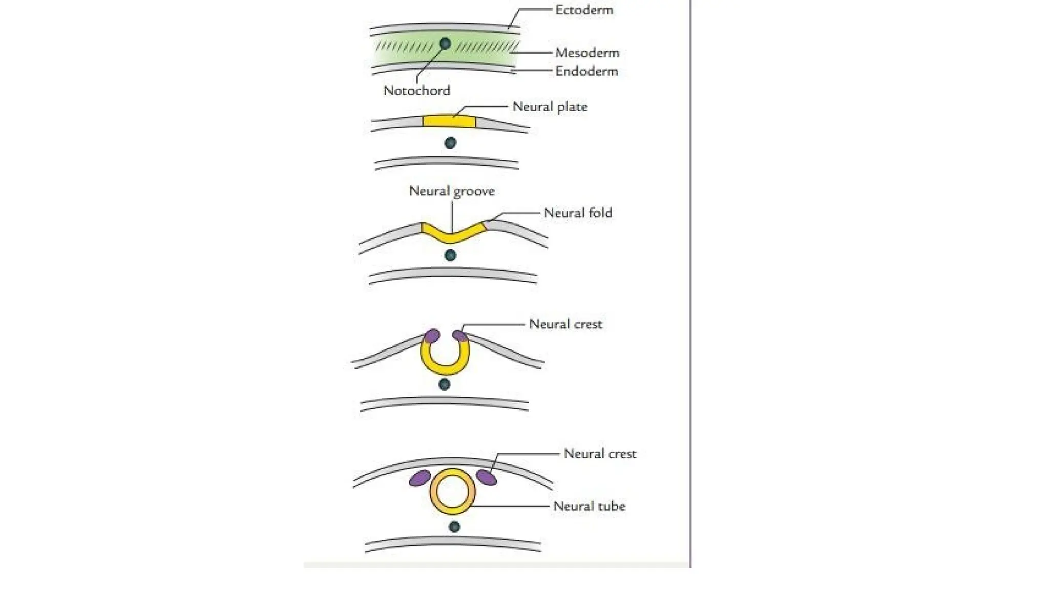
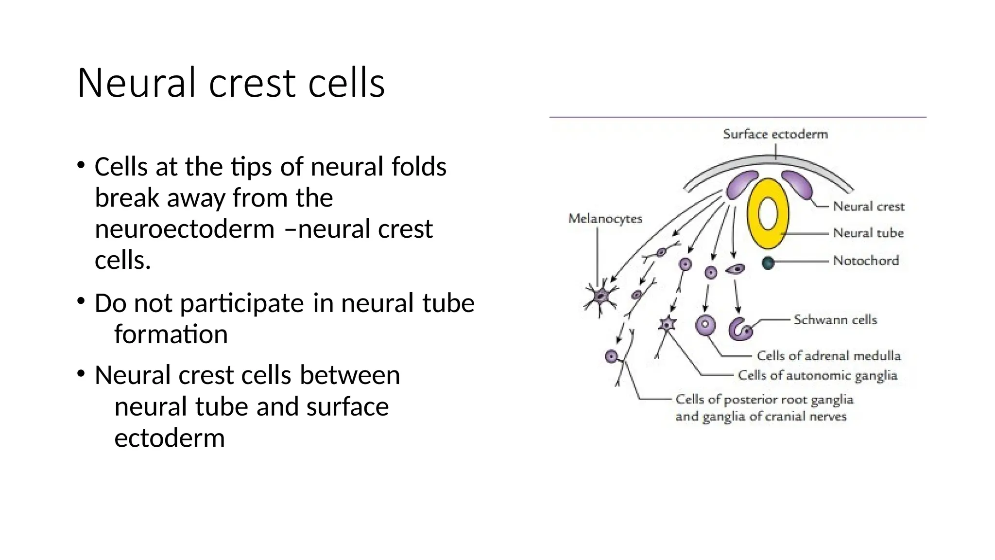
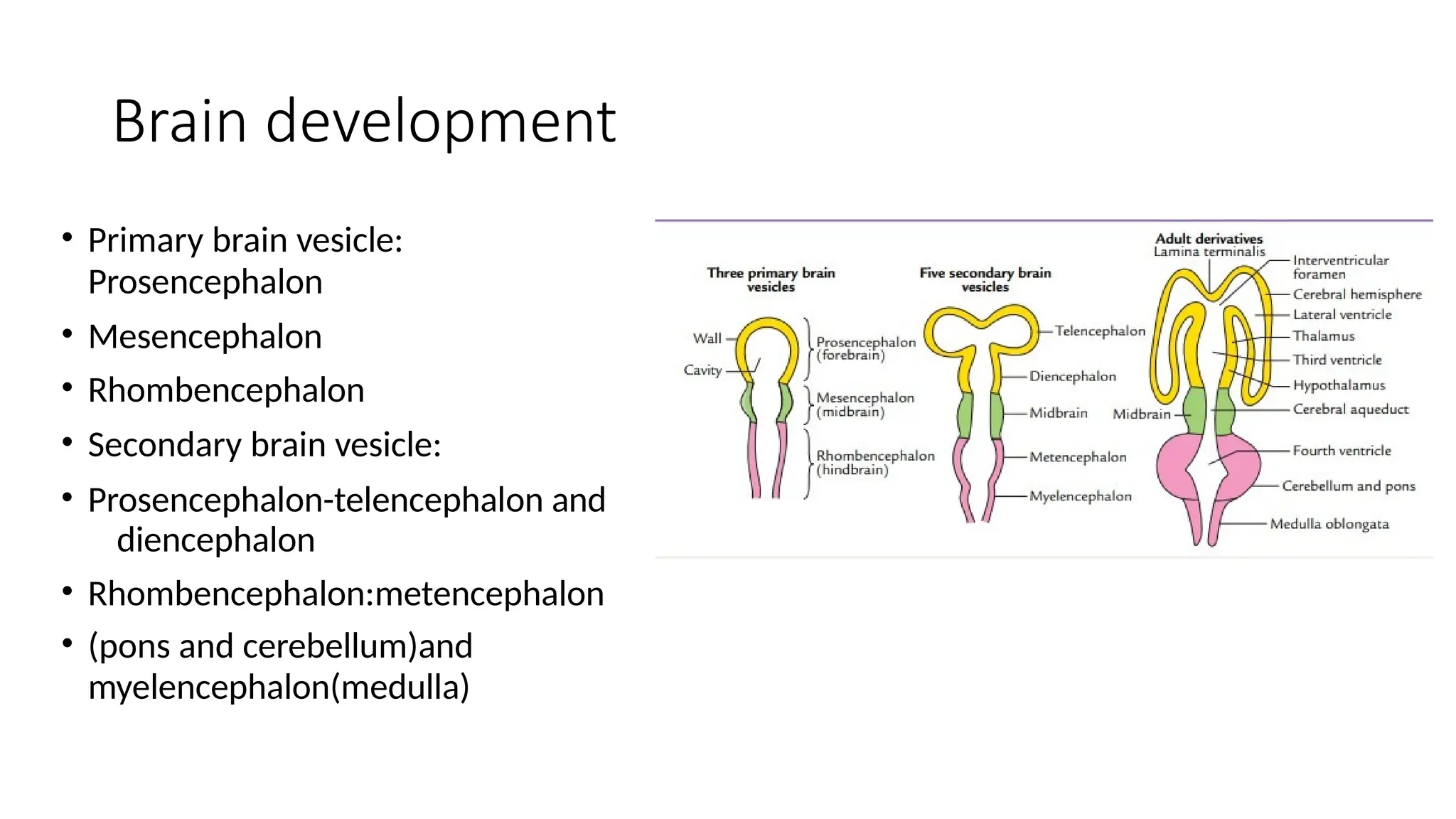
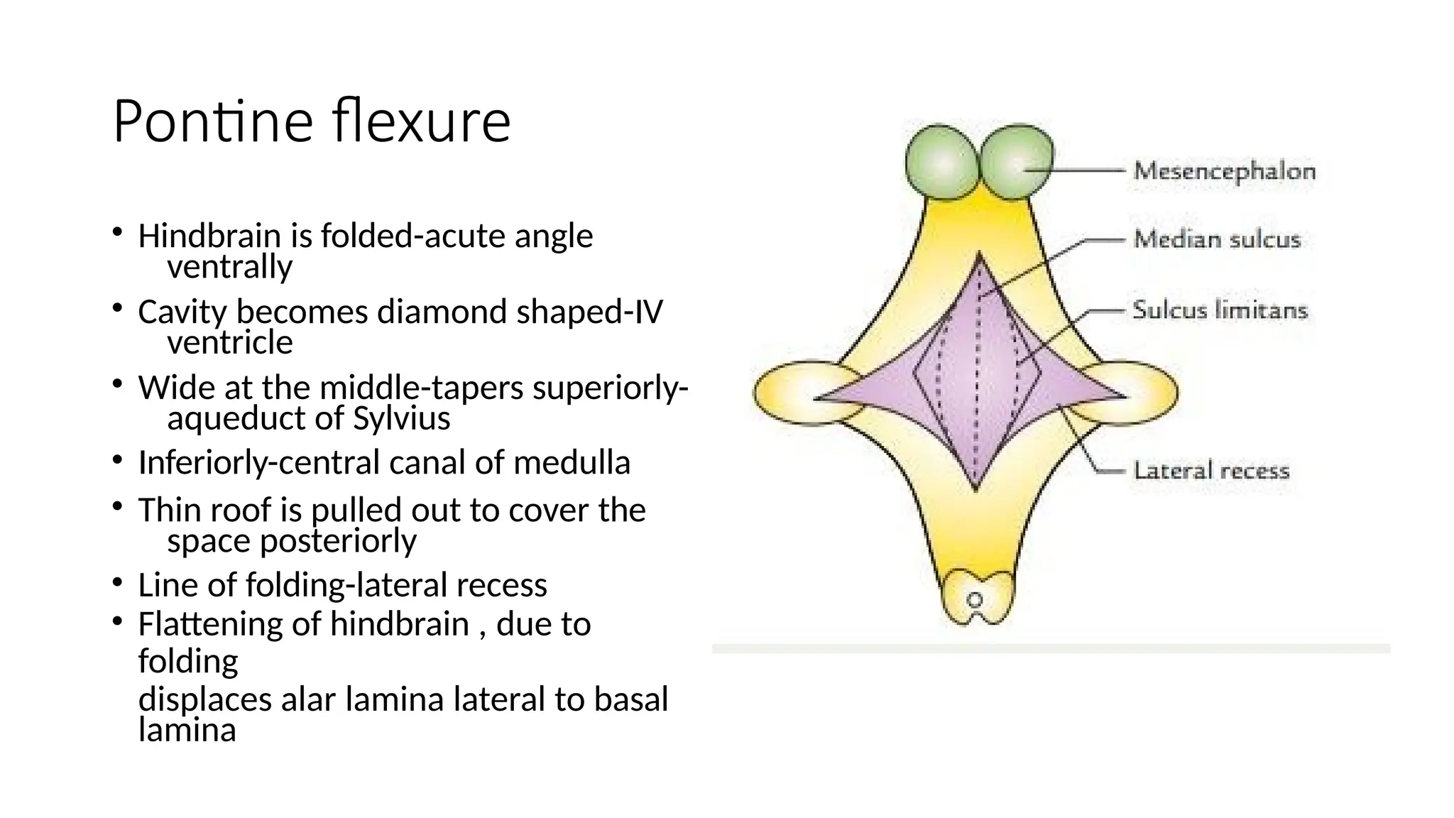
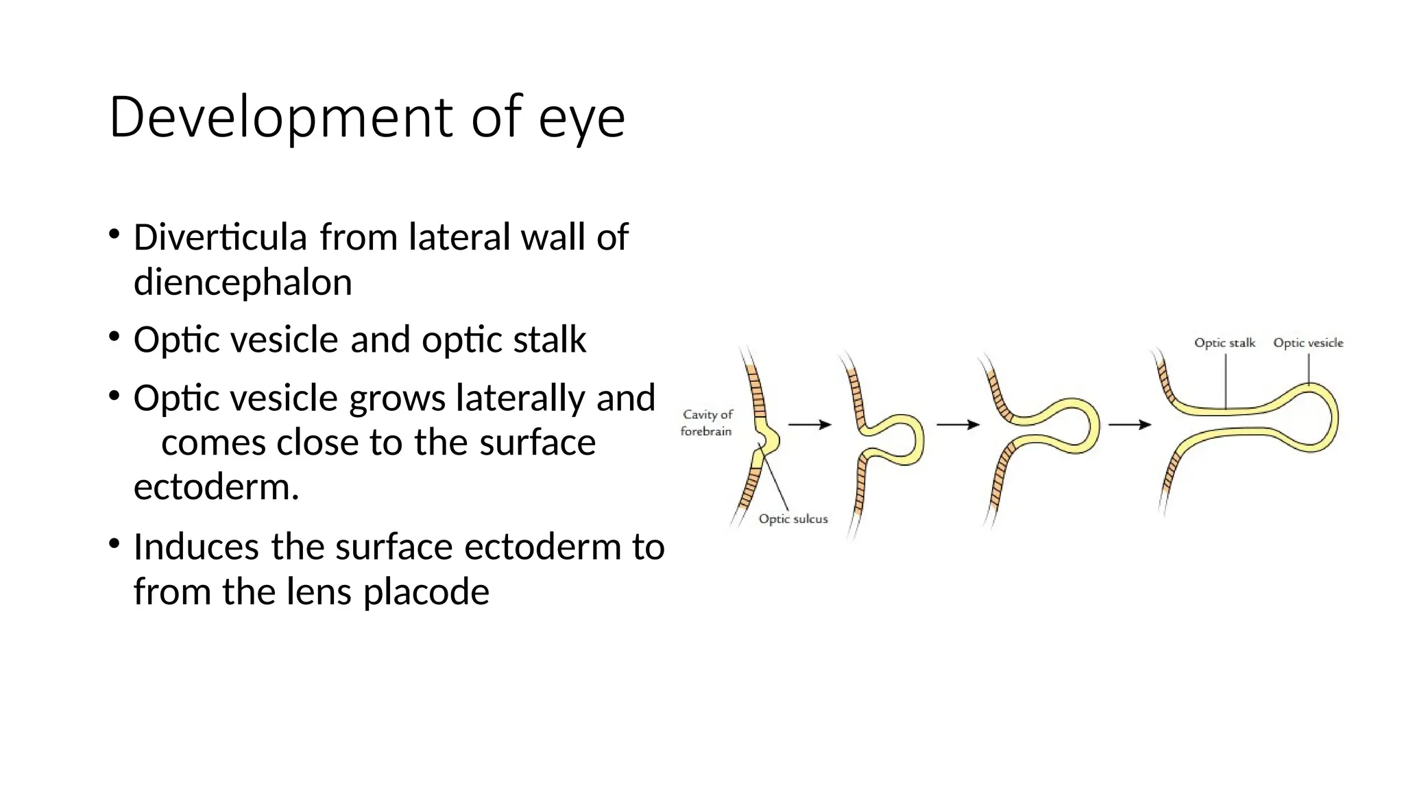
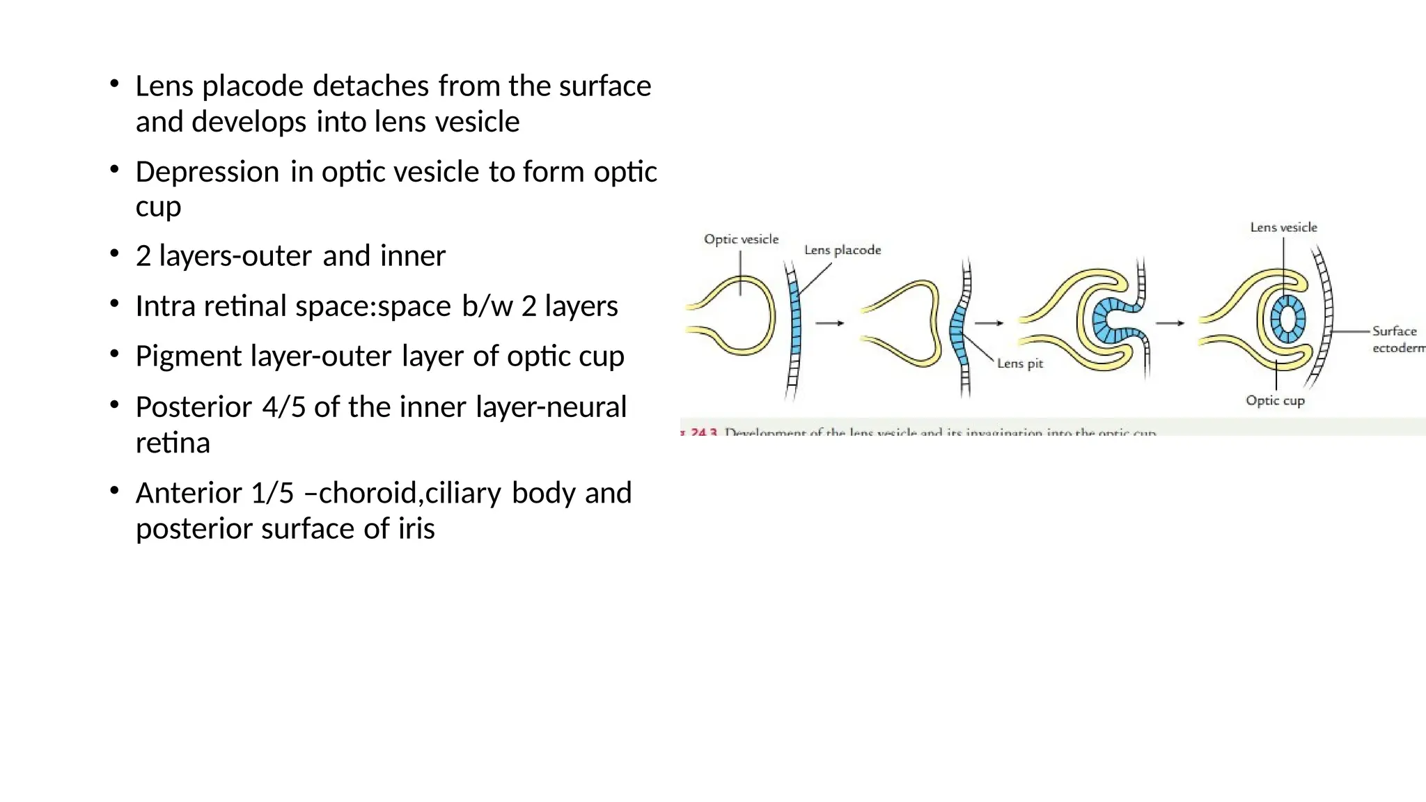
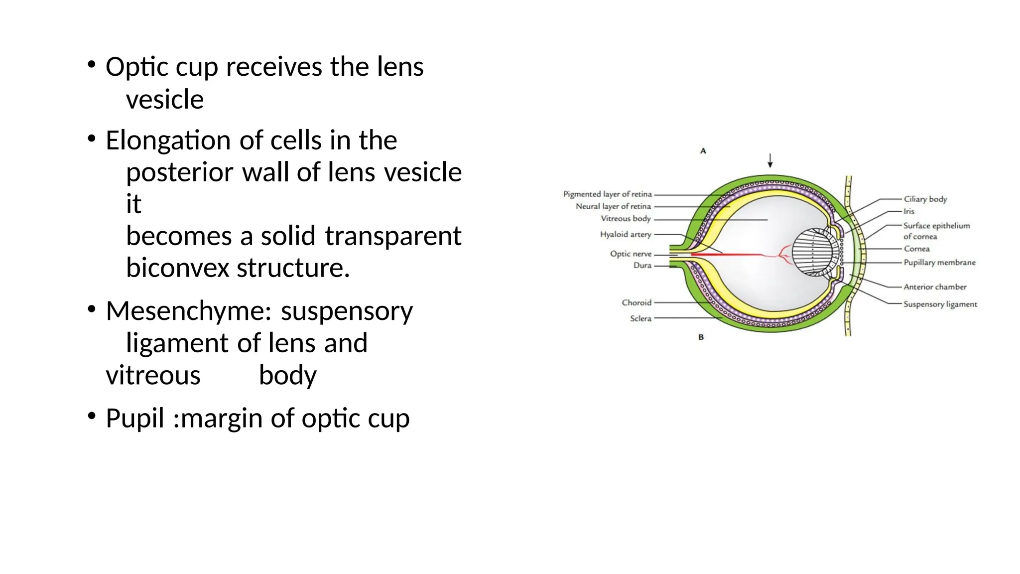
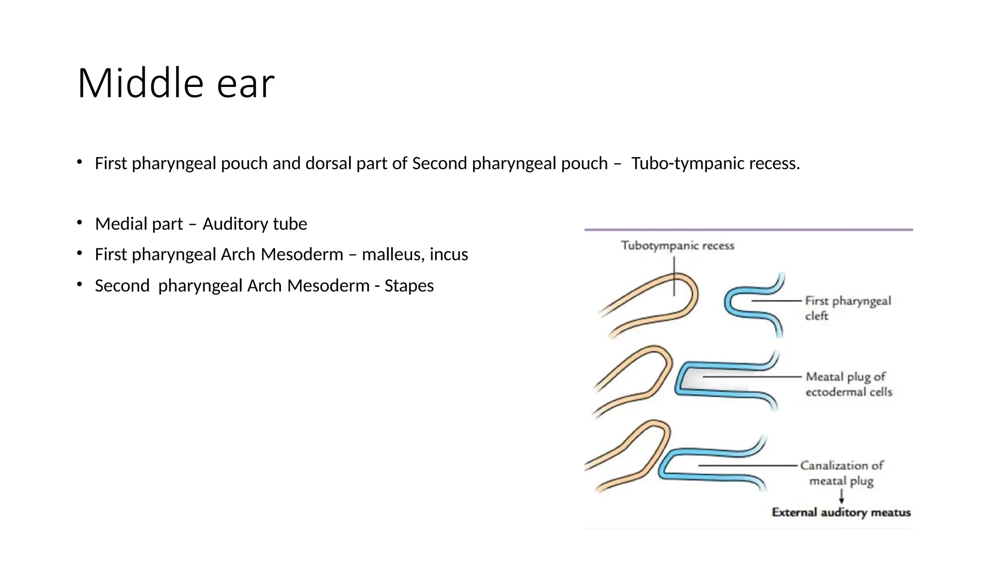
Development of CNS
Neurulation
• Ectoderm overlying the notochord
thickens-neural plate
• Development of mesoderm on either
side of notochord
• Neural fold formation
• Neural groove
• Movement of neural folds towards
each other –neural tube
• Anterior (25th day)and posterior
neuropore(27th day)
• Cranial part and caudal part
Neural crest cells
• Cells at the tips of neural folds
break away from the
neuroectoderm –neural crest
cells.
• Do not participate in neural tube
formation
• Neural crest cells between
neural tube and surface
ectoderm
Formation of ectodermal placodes
• Neural fold –neural crest cells and neuroepithelial cells
• Neuroepithelial cells get incorporated into the surface
ectoderm.- ecodermal placodes
Spinal cord development
• Neural tube increases in
thickness
• 5th week of IUL-T.S,of neural
tube-matrix zone,mantle
zone,marginal zone
• Matrix zone-neuroblasts and
spongioblast-neurons and
neuroglial cells
• Mantle zone-spinal gray
matter
• Marginal zone-white matter
Development of Functional columns
• Neural tube cavity-vertical slit
• Dorsal and ventral walls of NT-roof
and floor plates.
• Thickening of lateral wall-sulcus
limitans –dorsal and ventral walls
• Dorsal lamina/alar lamina-
sensory/afferent
• Ventral lamina/basal lamina-
efferent/motor
• Spinal nerve
Functional components
• Alar lamina
• General somatic afferent: superficial
and deep receptors
• General visceral afferent:
thoracolumbar and sacral regions-
viscera and blood vessels
• Basal lamina:
• General visceral efferent:
thoracolumbar and sacral regions-
viscera and blood vessels
• General somatic efferent:innervate
skeletal muscles
Positional changes of spinal cord
• 8 weeks-equal length
• 24 weeks-S1 vertebra
• Birth-L3 level
• Adults-L1 level
Brain development
• Primary brain vesicle:
Prosencephalon
• Mesencephalon
• Rhombencephalon
• Secondary brain vesicle:
• Prosencephalon-telencephalon and
diencephalon
• Rhombencephalon:metencephalon
• (pons and cerebellum)and
myelencephalon(medulla)
Flexures of the brain
• Pontine flexure:middle
of rhombencephalon
• Cephalic:regions of
midbrain
• Cervical
flexure:rhombencephalon and
spinal cord.
• Cephalic and Cervical flexure
concave ventrally
Pontine flexure
• Hindbrain is folded-acute angle
ventrally
• Cavity becomes diamond shaped-IV
ventricle
• Wide at the middle-tapers superiorly-
aqueduct of Sylvius
• Inferiorly-central canal of medulla
• Thin roof is pulled out to cover the
space posteriorly
• Line of folding-lateral recess
• Flattening of hindbrain , due to
folding
displaces alar lamina lateral to basal
lamina
• Cervical flexure: convex dorsally
and present at the junction
between spinal cord and
hindbrain
• Cephalic flexure: convex dorsally
and appears at midbrain level.
Ventricular system
• Hindbrain-fourth ventricle
• Mesencephalic cavity:cerebral
aqueduct
• Diencephalic cavity:third ventricle
• Telencephalic cavity:lateral ventricle
Hindbrain
• Floor of IV ventricle-myelencephalon and metencephalon
Basal and alar lamina
Development of eye
• Diverticula from lateral wall of
diencephalon
• Optic vesicle and optic stalk
• Optic vesicle grows laterally and
comes close to the surface
ectoderm.
• Induces the surface ectoderm to
from the lens placode
• Lens placode detaches from the surface
and develops into lens vesicle
• Depression in optic vesicle to form optic
cup
• 2 layers-outer and inner
• Intra retinal space:space b/w 2 layers
• Pigment layer-outer layer of optic cup
• Posterior 4/5 of the inner layer-neural
retina
• Anterior 1/5 –choroid,ciliary body and
posterior surface of iris
• Optic cup receives the lens
vesicle
• Elongation of cells in the
posterior wall of lens vesicle
it
becomes a solid transparent
biconvex structure.
• Mesenchyme: suspensory
ligament of lens and
vitreous body
• Pupil :margin of optic cup
• Optic stalk develops a fissure-
choroid fissure
• Choroid fissure carries hyaloid
artery –posterior part of the
lens.
• Optic stalk:optic nerve.
Development of Ear
• External Ear – Pinna or the
auricle is formed by the Six
Cartilaginous masses present on
the either side of EAM.
• EAM- develops from First
Ectodermal cleft.
Middle ear
• First pharyngeal pouch and dorsal part of Second pharyngeal pouch – Tubo-tympanic recess.
• Medial part – Auditory tube
• First pharyngeal Arch Mesoderm – malleus, incus
• Second pharyngeal Arch Mesoderm - Stapes
Tympanic membrane
• External – Ectoderm of first Cleft
• Internal – Endoderm of Tubo-Tympanic Recess
• Middle Mesoderm between First Cleft and
Pouch

Development of CNS.pptx kjhjjnjjjkjkkkkkk

  • 1.
  • 2.
    Neurulation • Ectoderm overlyingthe notochord thickens-neural plate • Development of mesoderm on either side of notochord • Neural fold formation • Neural groove • Movement of neural folds towards each other –neural tube • Anterior (25th day)and posterior neuropore(27th day) • Cranial part and caudal part
  • 4.
    Neural crest cells •Cells at the tips of neural folds break away from the neuroectoderm –neural crest cells. • Do not participate in neural tube formation • Neural crest cells between neural tube and surface ectoderm
  • 5.
    Formation of ectodermalplacodes • Neural fold –neural crest cells and neuroepithelial cells • Neuroepithelial cells get incorporated into the surface ectoderm.- ecodermal placodes
  • 6.
    Spinal cord development •Neural tube increases in thickness • 5th week of IUL-T.S,of neural tube-matrix zone,mantle zone,marginal zone • Matrix zone-neuroblasts and spongioblast-neurons and neuroglial cells • Mantle zone-spinal gray matter • Marginal zone-white matter
  • 7.
    Development of Functionalcolumns • Neural tube cavity-vertical slit • Dorsal and ventral walls of NT-roof and floor plates. • Thickening of lateral wall-sulcus limitans –dorsal and ventral walls • Dorsal lamina/alar lamina- sensory/afferent • Ventral lamina/basal lamina- efferent/motor • Spinal nerve
  • 8.
    Functional components • Alarlamina • General somatic afferent: superficial and deep receptors • General visceral afferent: thoracolumbar and sacral regions- viscera and blood vessels • Basal lamina: • General visceral efferent: thoracolumbar and sacral regions- viscera and blood vessels • General somatic efferent:innervate skeletal muscles
  • 9.
    Positional changes ofspinal cord • 8 weeks-equal length • 24 weeks-S1 vertebra • Birth-L3 level • Adults-L1 level
  • 10.
    Brain development • Primarybrain vesicle: Prosencephalon • Mesencephalon • Rhombencephalon • Secondary brain vesicle: • Prosencephalon-telencephalon and diencephalon • Rhombencephalon:metencephalon • (pons and cerebellum)and myelencephalon(medulla)
  • 12.
    Flexures of thebrain • Pontine flexure:middle of rhombencephalon • Cephalic:regions of midbrain • Cervical flexure:rhombencephalon and spinal cord. • Cephalic and Cervical flexure concave ventrally
  • 13.
    Pontine flexure • Hindbrainis folded-acute angle ventrally • Cavity becomes diamond shaped-IV ventricle • Wide at the middle-tapers superiorly- aqueduct of Sylvius • Inferiorly-central canal of medulla • Thin roof is pulled out to cover the space posteriorly • Line of folding-lateral recess • Flattening of hindbrain , due to folding displaces alar lamina lateral to basal lamina
  • 14.
    • Cervical flexure:convex dorsally and present at the junction between spinal cord and hindbrain • Cephalic flexure: convex dorsally and appears at midbrain level.
  • 15.
    Ventricular system • Hindbrain-fourthventricle • Mesencephalic cavity:cerebral aqueduct • Diencephalic cavity:third ventricle • Telencephalic cavity:lateral ventricle
  • 16.
    Hindbrain • Floor ofIV ventricle-myelencephalon and metencephalon Basal and alar lamina
  • 17.
    Development of eye •Diverticula from lateral wall of diencephalon • Optic vesicle and optic stalk • Optic vesicle grows laterally and comes close to the surface ectoderm. • Induces the surface ectoderm to from the lens placode
  • 18.
    • Lens placodedetaches from the surface and develops into lens vesicle • Depression in optic vesicle to form optic cup • 2 layers-outer and inner • Intra retinal space:space b/w 2 layers • Pigment layer-outer layer of optic cup • Posterior 4/5 of the inner layer-neural retina • Anterior 1/5 –choroid,ciliary body and posterior surface of iris
  • 20.
    • Optic cupreceives the lens vesicle • Elongation of cells in the posterior wall of lens vesicle it becomes a solid transparent biconvex structure. • Mesenchyme: suspensory ligament of lens and vitreous body • Pupil :margin of optic cup
  • 21.
    • Optic stalkdevelops a fissure- choroid fissure • Choroid fissure carries hyaloid artery –posterior part of the lens. • Optic stalk:optic nerve.
  • 22.
    Development of Ear •External Ear – Pinna or the auricle is formed by the Six Cartilaginous masses present on the either side of EAM. • EAM- develops from First Ectodermal cleft.
  • 23.
    Middle ear • Firstpharyngeal pouch and dorsal part of Second pharyngeal pouch – Tubo-tympanic recess. • Medial part – Auditory tube • First pharyngeal Arch Mesoderm – malleus, incus • Second pharyngeal Arch Mesoderm - Stapes
  • 24.
    Tympanic membrane • External– Ectoderm of first Cleft • Internal – Endoderm of Tubo-Tympanic Recess • Middle Mesoderm between First Cleft and Pouch