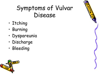
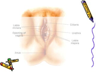
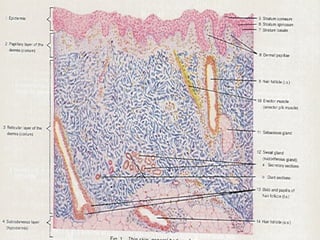
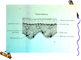
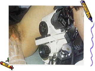
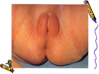
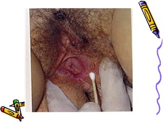
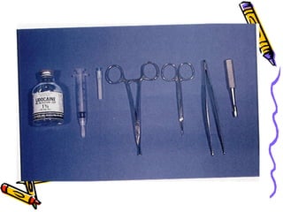
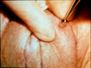
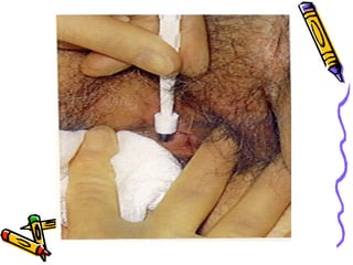
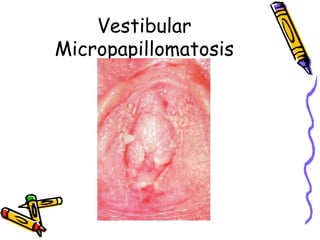
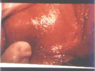
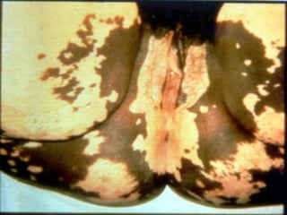
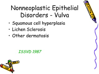
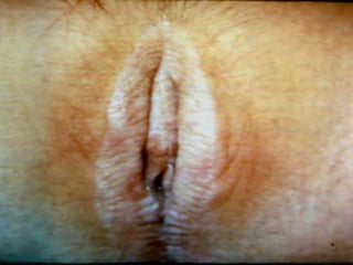
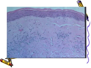
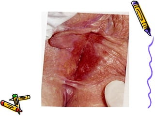
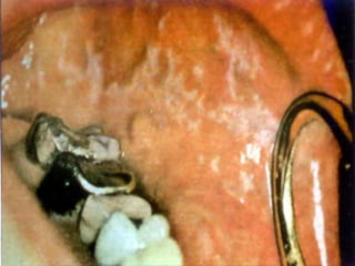
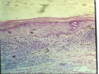
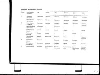
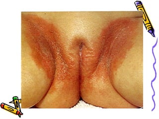
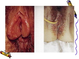
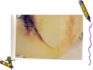
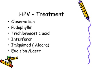
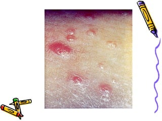
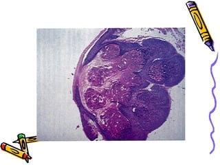
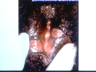
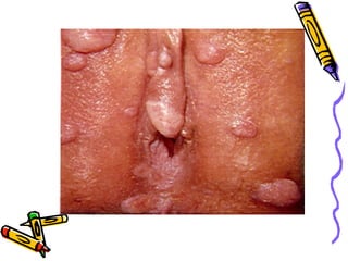
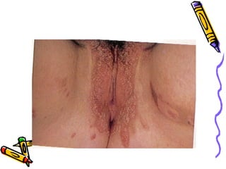
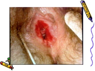
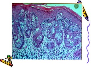
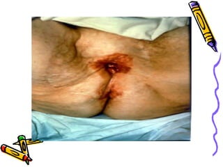
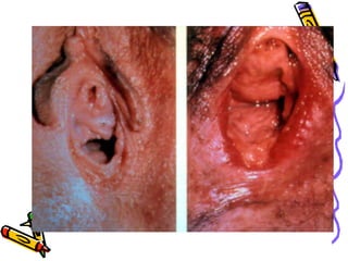
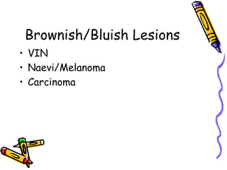
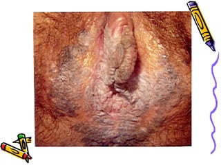
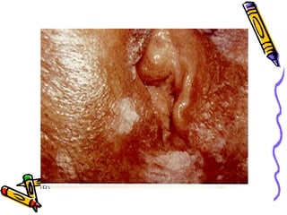
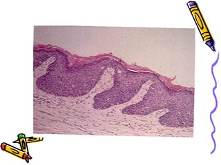
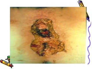
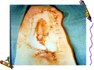
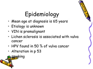
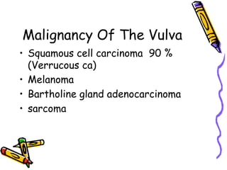
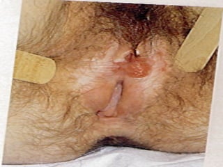
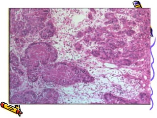
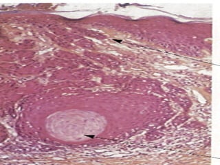
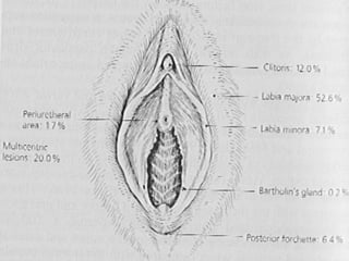
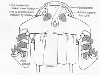
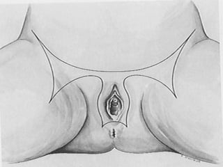
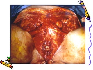
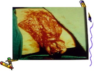
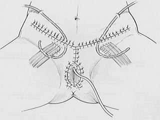
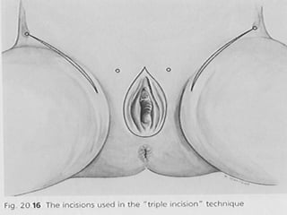
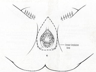
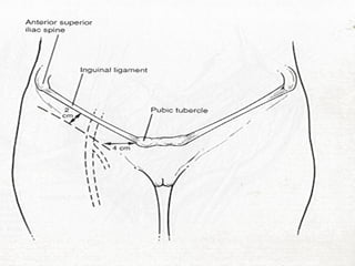
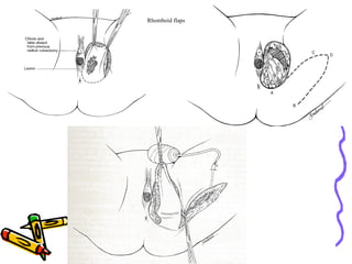
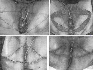
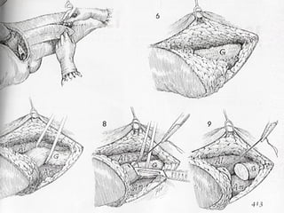
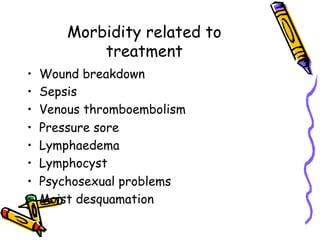
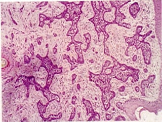
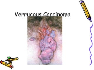
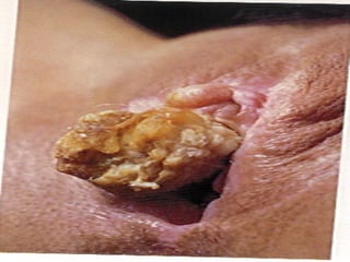
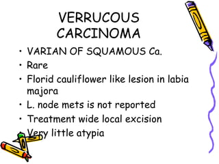
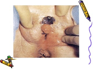
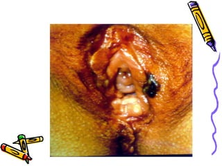
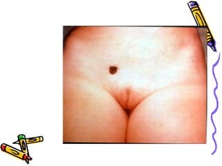
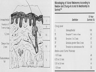
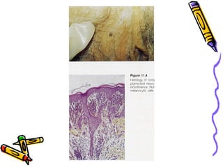
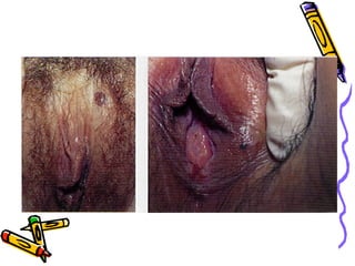
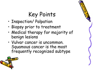
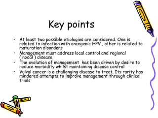
Benign and malignant
disease of the vulva
Khalid Sait ( FRCSC)
Gynecological Oncology
Professor
Faculty of Medicine
King Abdulaziz University
Symptoms of Vulvar
Disease
•  Itching
•  Burning
•  Dyspareunia
•  Discharge
•  Bleeding
General Pathology Term.
•  Epidermis
•  Acanthosis
•  Parakeratosis
•  Hyperkeratosis
Vestibular
Micropapillomatosis
General Guide to
Diagnosis
•  White Lesions
•  Red Lesions
•  Brown/Bluish Lesions
White Lesions
•  Vitilligo
•  Nonneoplastic epithelial disorders
Nonneoplastic Epithelial
Disorders - Vulva
•  Squamous cell hyperplasia
•  Lichen Sclerosis
•  Other dermatosis
ISSVD 1987
Lichen Sclerosis
•  Steroid
•  Testesterone
•  Progesterone
Squamous cell
hyperplasia
•  Steroid
Red Lesions
•  Infective
•  Dermatitis
•  Psoriasis
•  Pagets
•  VIN/Ca
HPV - Treatment
•  Observation
•  Podophyllin
•  Trichloroacetic acid
•  Interferon
•  Imiquimod ( Aldara)
•  Excision /Laser
Brownish/Bluish Lesions
•  VIN
•  Naevi/Melanoma
•  Carcinoma
Malignant Disease Of The
Vulva
Female Genital Tract
Malignancy
•  Uterus
•  Ovary
•  Cervix
•  Vulva
•  Vagina
•  Fallopian Tubes.
Epidemiology
•  Mean age at diagnosis is 65 years
•  Etiology is unknown
•  VIN is premalignant
•  Lichen sclerosis is associated with vulva
cancer
•  HPV found in 50 % of vulva cancer
•  Alteration in p 53
•  smoking
Malignancy Of The Vulva
•  Squamous cell carcinoma 90 %
(Verrucous ca)
•  Melanoma
•  Bartholine gland adenocarcinoma
•  sarcoma
Carcinoma Of The Vulva
•  Preoperative workup
•  History
•  Physical Examination
•  CBC, U/E and LFT
•  CXR a-p/lat
Management
Staging: surgico - pathological
•  Management of the vulva
•  Management of the groin
Microinvasion
•  Lesion less than 2 cm with depth of
less than 1 mm with noLVS inv. and
no clinical positive node
•  Incidence of L.node mets is 0 %
•  Treatment wide local excision down
to colles’ fascia
Lateral T1 lesion
•  Radical local excision and groin unilateral
node dissection
•  Send for frozen if positive do the other
side
•  If negative unilat node only 10 % will have
positive contra lateral node usually in
lesion with risk factors
•  20-30 % of non suspicious node will have
mets
Lateral T2 lesion
•  Unilateral hemivulvectomy and
bilateral node groin dissection
Prevalence of local invasive recurrence after radical
local excision and radical vulvectomy for early T1 lesion
procedure Pts number recurrence Dead of
disease
Radical local
excision
165 12(7.2%) 1(0.6%
Radical
vulvectomy
365 23(6.3%) 2(0.6%
T1 or T2 midline or
bilateral lesion
•  Radical vulvectomy and bilateral groin
node dissection
Post operative adjuvant
therapy
•  Close margin(< 8mm) re-excision
versus RT. to the vulva
•  Poorly diff., large tumor, LVS
involvement and diffuse infiltrative
lesion :
Radiation to the vulva a lone and may
be to the medial groin area
Respectable vulva lesion with clinically positive groin
node
•  Radical vulvectomy with bilateral
groin node dissection followed BY
radiation to groin and pelvis
•  With multiple inguinal node 20 % risk
of pelvic node involvment
•  22 % of clinical positive groin node
will show no histology evidence of
mets
T3 Involvement of distal urethra , vagina or
anus
•  Preoperative radiation and chemotherapy
to allow conservative surgery with
affecting sphencter followed by:
if residual disease radical vulvectomy and
groin lymph node dissection
If no residual disease do multiple vulval
biopsies and node dissection
•  If bulky node remove bulky node initially if
not fixed or ulcerated
T4 Primary lesion with massive bladder and or rectum
involvement
•  Selective Exenteration with radical
vulvectomy and groin node dissection
•  Remove bulky nodes if not fixed or
ulcerated and give radiation to the
groin and pelvic nodes as well as
radiation to vulva with 5 FU followed
by debulking surgery or a modified
exentrative procedure
Chemotherapy
•  5 FU 1gm/m2/d 1-4 days over 2 4
hoyrs infusion 96 ml start 24 hours
before radiation and be given on day
29-32 of radiation
Radical Vulvectomy
• Radical en-bloc dissection
Taussig and Way 1949-1960
• Triple incision technique
Rhomboid flaps
Morbidity related to
treatment
•  Wound breakdown
•  Sepsis
•  Venous thromboembolism
•  Pressure sore
•  Lymphaedema
•  Lymphocyst
•  Psychosexual problems
•  Moist desquamation
Carcinoma Of The Vulva
stage 5 years survival
0 100 %
1 95 %
2 83%
3 36 %
4 29 %
Prognostic Factors
•  Stage
•  Histological
•  Status of l.node an d number involved and
capsule
•  Vascular invasion
•  Tumor thickness
•  Depth of invasion
•  Amount of keratin
•  performance status
•  DNA polidy
Recurrence
•  In vulva: excision with margin if
possible with 5 years survival up to
50 % or RT if not given before
•  In groin: IF RT NO GIVEN
BEFORE---à RT IF RT GIVEN
BEFORE---à excision IF POSSIBLE
•  In pelvis: radiation if not given
before
•  Distal or after radiation:
chemotherapy
Verrucous Carcinoma
VERRUCOUS
CARCINOMA
•  VARIAN OF SQUAMOUS Ca.
•  Rare
•  Florid cauliflower like lesion in labia
majora
•  L. node mets is not reported
•  Treatment wide local excision
•  Very little atypia
MELANOMA
•  2-10 % of vulva malignancy
•  5 % of melanoma of white female occur in
vulva
•  Mean age 63
•  STAGE I
•  STAGE II
•  STAGE III
•  STAGE IV
•  Node resection need to be done for
prognostic or if found to be positive
clinically
Key Points
•  Inspection/ Palpation
•  Biopsy prior to treatment
•  Medical therapy for majority of
benign lesions
•  Vulvar cancer is uncommon.
Squamous cancer is the most
frequently recognized subtype
Key points
•  At least two possible etiologies are considered. One is
related to infection with oncogenic HPV , other is related to
maturation disorders
•  Management must address local control and regional
( nodal ) disease
•  The evolution of management has been driven by desire to
reduce morbidity whilst maintaining disease control
•  Vulval cancer is a challenging disease to treat. Its rarity has
mindered attempts to improve management through clinical
trials

Vulva disease

  • 1.
    Benign and malignant diseaseof the vulva Khalid Sait ( FRCSC) Gynecological Oncology Professor Faculty of Medicine King Abdulaziz University
  • 2.
    Symptoms of Vulvar Disease • Itching •  Burning •  Dyspareunia •  Discharge •  Bleeding
  • 4.
    General Pathology Term. • Epidermis •  Acanthosis •  Parakeratosis •  Hyperkeratosis
  • 17.
  • 19.
    General Guide to Diagnosis • White Lesions •  Red Lesions •  Brown/Bluish Lesions
  • 20.
    White Lesions •  Vitilligo • Nonneoplastic epithelial disorders
  • 22.
    Nonneoplastic Epithelial Disorders -Vulva •  Squamous cell hyperplasia •  Lichen Sclerosis •  Other dermatosis ISSVD 1987
  • 25.
    Lichen Sclerosis •  Steroid • Testesterone •  Progesterone
  • 28.
  • 33.
    Red Lesions •  Infective • Dermatitis •  Psoriasis •  Pagets •  VIN/Ca
  • 41.
    HPV - Treatment • Observation •  Podophyllin •  Trichloroacetic acid •  Interferon •  Imiquimod ( Aldara) •  Excision /Laser
  • 51.
    Brownish/Bluish Lesions •  VIN • Naevi/Melanoma •  Carcinoma
  • 57.
  • 58.
    Female Genital Tract Malignancy • Uterus •  Ovary •  Cervix •  Vulva •  Vagina •  Fallopian Tubes.
  • 59.
    Epidemiology •  Mean ageat diagnosis is 65 years •  Etiology is unknown •  VIN is premalignant •  Lichen sclerosis is associated with vulva cancer •  HPV found in 50 % of vulva cancer •  Alteration in p 53 •  smoking
  • 60.
    Malignancy Of TheVulva •  Squamous cell carcinoma 90 % (Verrucous ca) •  Melanoma •  Bartholine gland adenocarcinoma •  sarcoma
  • 65.
    Carcinoma Of TheVulva •  Preoperative workup •  History •  Physical Examination •  CBC, U/E and LFT •  CXR a-p/lat
  • 66.
    Management Staging: surgico -pathological •  Management of the vulva •  Management of the groin
  • 68.
    Microinvasion •  Lesion lessthan 2 cm with depth of less than 1 mm with noLVS inv. and no clinical positive node •  Incidence of L.node mets is 0 % •  Treatment wide local excision down to colles’ fascia
  • 69.
    Lateral T1 lesion • Radical local excision and groin unilateral node dissection •  Send for frozen if positive do the other side •  If negative unilat node only 10 % will have positive contra lateral node usually in lesion with risk factors •  20-30 % of non suspicious node will have mets
  • 70.
    Lateral T2 lesion • Unilateral hemivulvectomy and bilateral node groin dissection
  • 72.
    Prevalence of localinvasive recurrence after radical local excision and radical vulvectomy for early T1 lesion procedure Pts number recurrence Dead of disease Radical local excision 165 12(7.2%) 1(0.6% Radical vulvectomy 365 23(6.3%) 2(0.6%
  • 73.
    T1 or T2midline or bilateral lesion •  Radical vulvectomy and bilateral groin node dissection
  • 74.
    Post operative adjuvant therapy • Close margin(< 8mm) re-excision versus RT. to the vulva •  Poorly diff., large tumor, LVS involvement and diffuse infiltrative lesion : Radiation to the vulva a lone and may be to the medial groin area
  • 75.
    Respectable vulva lesionwith clinically positive groin node •  Radical vulvectomy with bilateral groin node dissection followed BY radiation to groin and pelvis •  With multiple inguinal node 20 % risk of pelvic node involvment •  22 % of clinical positive groin node will show no histology evidence of mets
  • 77.
    T3 Involvement ofdistal urethra , vagina or anus •  Preoperative radiation and chemotherapy to allow conservative surgery with affecting sphencter followed by: if residual disease radical vulvectomy and groin lymph node dissection If no residual disease do multiple vulval biopsies and node dissection •  If bulky node remove bulky node initially if not fixed or ulcerated
  • 78.
    T4 Primary lesionwith massive bladder and or rectum involvement •  Selective Exenteration with radical vulvectomy and groin node dissection •  Remove bulky nodes if not fixed or ulcerated and give radiation to the groin and pelvic nodes as well as radiation to vulva with 5 FU followed by debulking surgery or a modified exentrative procedure
  • 79.
    Chemotherapy •  5 FU1gm/m2/d 1-4 days over 2 4 hoyrs infusion 96 ml start 24 hours before radiation and be given on day 29-32 of radiation
  • 80.
    Radical Vulvectomy • Radical en-blocdissection Taussig and Way 1949-1960 • Triple incision technique
  • 97.
  • 100.
    Morbidity related to treatment • Wound breakdown •  Sepsis •  Venous thromboembolism •  Pressure sore •  Lymphaedema •  Lymphocyst •  Psychosexual problems •  Moist desquamation
  • 101.
    Carcinoma Of TheVulva stage 5 years survival 0 100 % 1 95 % 2 83% 3 36 % 4 29 %
  • 102.
    Prognostic Factors •  Stage • Histological •  Status of l.node an d number involved and capsule •  Vascular invasion •  Tumor thickness •  Depth of invasion •  Amount of keratin •  performance status •  DNA polidy
  • 103.
    Recurrence •  In vulva:excision with margin if possible with 5 years survival up to 50 % or RT if not given before •  In groin: IF RT NO GIVEN BEFORE---à RT IF RT GIVEN BEFORE---à excision IF POSSIBLE •  In pelvis: radiation if not given before •  Distal or after radiation: chemotherapy
  • 106.
  • 108.
    VERRUCOUS CARCINOMA •  VARIAN OFSQUAMOUS Ca. •  Rare •  Florid cauliflower like lesion in labia majora •  L. node mets is not reported •  Treatment wide local excision •  Very little atypia
  • 112.
    MELANOMA •  2-10 %of vulva malignancy •  5 % of melanoma of white female occur in vulva •  Mean age 63 •  STAGE I •  STAGE II •  STAGE III •  STAGE IV •  Node resection need to be done for prognostic or if found to be positive clinically
  • 119.
    Key Points •  Inspection/Palpation •  Biopsy prior to treatment •  Medical therapy for majority of benign lesions •  Vulvar cancer is uncommon. Squamous cancer is the most frequently recognized subtype
  • 120.
    Key points •  Atleast two possible etiologies are considered. One is related to infection with oncogenic HPV , other is related to maturation disorders •  Management must address local control and regional ( nodal ) disease •  The evolution of management has been driven by desire to reduce morbidity whilst maintaining disease control •  Vulval cancer is a challenging disease to treat. Its rarity has mindered attempts to improve management through clinical trials