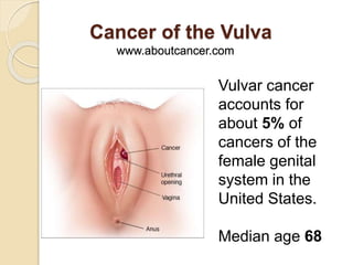
Vulvar cancer accounts for about 5% of cancers of the female genital system in the US. Possible signs include bleeding, itching, lumps or growths on the vulva, skin color changes, or ulcers. Risk factors include HPV infection, smoking, and precancerous skin conditions. Treatment depends on stage but may involve surgery to remove the tumor and nearby lymph nodes, radiation therapy, or chemoradiation. Side effects can include skin irritation in the vulvar area, as well as urinary and bowel issues. Overall survival rates vary from over 80% for local stage to under 20% for distant stage disease.
An UPDATE solid knowledge in Vulval cancer, consisting of 12 years experience form lecture notes of
Professor Basel Obaidat~ FRCOG. Gyne/Onco.
24\3\2016
Vaginal cancer is a rare type of cancer most common in women 60 and older.
Women are more likely to develop vaginal cancer if they have the human papillomavirus (HPV) or if your birth mother took diethylstilbestol (DES) when she was pregnant.
There are several types of vaginal cancer:
Squamous cell carcinoma
About 70 of every 100 cases of vaginal cancer are squamous cell carcinomas. These cancers begin in the squamous cells that make up the epithelial lining of the vagina. These cancers are more common in the upper area of the vagina near the cervix. Squamous cell cancers of the vagina often develop slowly. First, some of the normal cells of the vagina get pre-cancerous changes. Then some of the pre-cancer cells turn into cancer cells. This process can take many years.
The medical term most often used for this pre-cancerous condition is vaginal intraepithelial neoplasia (VAIN). "Intraepithelial" means that the abnormal cells are only found in the surface layer of the vaginal skin (epithelium). There are 3 types of VAIN: VAIN1, VAIN2, and VAIN3, with 3 indicating furthest progression toward a true cancer. VAIN is more common in women who have had their uterus removed (hysterectomy) and in those who were previously treated for cervical cancer or pre-cancer.
In the past, the term dysplasia was used instead of VAIN. This term is used much less now. When talking about dysplasia, there is also a range of increasing progress toward cancer - first, mild dysplasia; next, moderate dysplasia; and then severe dysplasia.
Adenocarcinoma
Cancer that begins in gland cells is called adenocarcinoma. About 15 of every 100 cases of vaginal cancer are adenocarcinomas. The usual type of vaginal adenocarcinoma typically develops in women older than 50. One certain type, called clear cell adenocarcinoma, occurs more often in young women who were exposed to diethylstilbestrol (DES) in utero (when they were in their mother’s womb). (See the section called "What are the risk factors for vaginal cancer?" for more information on DES and clear cell carcinoma.)
Melanoma
Melanomas develop from pigment-producing cells that give skin its color. These cancers usually are found on sun-exposed areas of the skin but can form on the vagina or other internal organs. About 9 of every 100 cases of vaginal cancer are melanomas. Melanoma tends to affect the lower or outer portion of the vagina. The tumors vary greatly in size, color, and growth pattern. More information about melanoma can be found in our document called Melanoma Skin Cancer.
Sarcoma
A sarcoma is a cancer that begins in the cells of bones, muscles, or connective tissue. Up to 4 of every 100 cases of vaginal cancer are sarcomas. These cancers form deep in the wall of the vagina, not on its surface. There are several types of vaginal sarcomas. Rhabdomyosarcoma is the most common type of vaginal sarcoma. It is most often found in children and is rare in adults. A sarcoma called leiomyosarcoma is seen more often in adults.
India is the highest TB burden country accounting for more than one-fourth of the global incidence .Genital TB is found in 5-10% of women with infertility problems, with low rates in Australia (1%) and high rates of up to 19% in India (ICMR,2011)
How useful are advance directives in directing end of life care and do people really understand or want to know the true status of their health as the end nears?
Understanding how intermittent fasting may not only help weight loss but have multiple other health benefits including life prolongation, preventing cancer and dementia
The Importance of Community Nursing Care.pdfAD Healthcare
NDIS and Community 24/7 Nursing Care is a specific type of support that may be provided under the NDIS for individuals with complex medical needs who require ongoing nursing care in a community setting, such as their home or a supported accommodation facility.
Telehealth Psychology Building Trust with Clients.pptxThe Harvest Clinic
Telehealth psychology is a digital approach that offers psychological services and mental health care to clients remotely, using technologies like video conferencing, phone calls, text messaging, and mobile apps for communication.
ICH Guidelines for Pharmacovigilance.pdfNEHA GUPTA
The "ICH Guidelines for Pharmacovigilance" PDF provides a comprehensive overview of the International Council for Harmonisation of Technical Requirements for Pharmaceuticals for Human Use (ICH) guidelines related to pharmacovigilance. These guidelines aim to ensure that drugs are safe and effective for patients by monitoring and assessing adverse effects, ensuring proper reporting systems, and improving risk management practices. The document is essential for professionals in the pharmaceutical industry, regulatory authorities, and healthcare providers, offering detailed procedures and standards for pharmacovigilance activities to enhance drug safety and protect public health.
CHAPTER 1 SEMESTER V - ROLE OF PEADIATRIC NURSE.pdfSachin Sharma
Pediatric nurses play a vital role in the health and well-being of children. Their responsibilities are wide-ranging, and their objectives can be categorized into several key areas:
1. Direct Patient Care:
Objective: Provide comprehensive and compassionate care to infants, children, and adolescents in various healthcare settings (hospitals, clinics, etc.).
This includes tasks like:
Monitoring vital signs and physical condition.
Administering medications and treatments.
Performing procedures as directed by doctors.
Assisting with daily living activities (bathing, feeding).
Providing emotional support and pain management.
2. Health Promotion and Education:
Objective: Promote healthy behaviors and educate children, families, and communities about preventive healthcare.
This includes tasks like:
Administering vaccinations.
Providing education on nutrition, hygiene, and development.
Offering breastfeeding and childbirth support.
Counseling families on safety and injury prevention.
3. Collaboration and Advocacy:
Objective: Collaborate effectively with doctors, social workers, therapists, and other healthcare professionals to ensure coordinated care for children.
Objective: Advocate for the rights and best interests of their patients, especially when children cannot speak for themselves.
This includes tasks like:
Communicating effectively with healthcare teams.
Identifying and addressing potential risks to child welfare.
Educating families about their child's condition and treatment options.
4. Professional Development and Research:
Objective: Stay up-to-date on the latest advancements in pediatric healthcare through continuing education and research.
Objective: Contribute to improving the quality of care for children by participating in research initiatives.
This includes tasks like:
Attending workshops and conferences on pediatric nursing.
Participating in clinical trials related to child health.
Implementing evidence-based practices into their daily routines.
By fulfilling these objectives, pediatric nurses play a crucial role in ensuring the optimal health and well-being of children throughout all stages of their development.
R3 Stem Cells and Kidney Repair A New Horizon in Nephrology.pptxR3 Stem Cell
R3 Stem Cells and Kidney Repair: A New Horizon in Nephrology" explores groundbreaking advancements in the use of R3 stem cells for kidney disease treatment. This insightful piece delves into the potential of these cells to regenerate damaged kidney tissue, offering new hope for patients and reshaping the future of nephrology.
Defecation
Normal defecation begins with movement in the left colon, moving stool toward the anus. When stool reaches the rectum, the distention causes relaxation of the internal sphincter and an awareness of the need to defecate. At the time of defecation, the external sphincter relaxes, and abdominal muscles contract, increasing intrarectal pressure and forcing the stool out
The Valsalva maneuver exerts pressure to expel faeces through a voluntary contraction of the abdominal muscles while maintaining forced expiration against a closed airway. Patients with cardiovascular disease, glaucoma, increased intracranial pressure, or a new surgical wound are at greater risk for cardiac dysrhythmias and elevated blood pressure with the Valsalva maneuver and need to avoid straining to pass the stool.
Normal defecation is painless, resulting in passage of soft, formed stool
CONSTIPATION
Constipation is a symptom, not a disease. Improper diet, reduced fluid intake, lack of exercise, and certain medications can cause constipation. For example, patients receiving opiates for pain after surgery often require a stool softener or laxative to prevent constipation. The signs of constipation include infrequent bowel movements (less than every 3 days), difficulty passing stools, excessive straining, inability to defecate at will, and hard feaces
IMPACTION
Fecal impaction results from unrelieved constipation. It is a collection of hardened feces wedged in the rectum that a person cannot expel. In cases of severe impaction the mass extends up into the sigmoid colon.
DIARRHEA
Diarrhea is an increase in the number of stools and the passage of liquid, unformed feces. It is associated with disorders affecting digestion, absorption, and secretion in the GI tract. Intestinal contents pass through the small and large intestine too quickly to allow for the usual absorption of fluid and nutrients. Irritation within the colon results in increased mucus secretion. As a result, feces become watery, and the patient is unable to control the urge to defecate. Normally an anal bag is safe and effective in long-term treatment of patients with fecal incontinence at home, in hospice, or in the hospital. Fecal incontinence is expensive and a potentially dangerous condition in terms of contamination and risk of skin ulceration
HEMORRHOIDS
Hemorrhoids are dilated, engorged veins in the lining of the rectum. They are either external or internal.
FLATULENCE
As gas accumulates in the lumen of the intestines, the bowel wall stretches and distends (flatulence). It is a common cause of abdominal fullness, pain, and cramping. Normally intestinal gas escapes through the mouth (belching) or the anus (passing of flatus)
FECAL INCONTINENCE
Fecal incontinence is the inability to control passage of feces and gas from the anus. Incontinence harms a patient’s body image
PREPARATION AND GIVING OF LAXATIVESACCORDING TO POTTER AND PERRY,
An enema is the instillation of a solution into the rectum and sig
Deep Leg Vein Thrombosis (DVT): Meaning, Causes, Symptoms, Treatment, and Mor...The Lifesciences Magazine
Deep Leg Vein Thrombosis occurs when a blood clot forms in one or more of the deep veins in the legs. These clots can impede blood flow, leading to severe complications.
1. Cancer of the Vulva
www.aboutcancer.com
Vulvar cancer
accounts for
about 5% of
cancers of the
female genital
system in the
United States.
Median age 68
2. Possible signs of vulvar cancer include bleeding
or itching should lead to an examination by a
physician
3. Possible signs of vulvar cancer include bleeding
or itching.
-A lump or growth on the vulva.
-Changes in the vulvar skin, such as color
changes or growths that look like a wart
or ulcer.
-Itching in the vulvar area, that does not go
away.
-Bleeding
-Tenderness in the vulvar area.
4. Female Cancers in the US in 2014
Cancer Incidence Deaths
Breast 235,030 40,420
Uterus 52,630 8,590
Ovary 21,980 14,270
Cervix 12,360 4,020
Vulva 4,850 1,030
Vagina 3,170 880
5. What causes Vulva Cancer?
Two independent pathways of vulvar
carcinogenesis are felt to currently
exist, the first related to mucosal HPV
(Human Papilloma Virus) infection
second related to chronic
inflammatory (vulvar dystrophy) or
autoimmune processes
6. The risk of developing vulvar cancer
is increased by the following:
Older age
Precancerous changes (dysplasia) in vulvar
tissues
Lichen sclerosus, which causes persistent
itching and scarring of the vulva
Human papillomavirus (HPV) infection
Cancer of the vagina or cervix
Heavy cigarette smoking
Chronic granulomatous disease (a hereditary
disease that impairs the immune system)
8. SEER rates for new vulvar cancer cases have been rising on average
0.5% each year over 2002-2011. Death rates have been rising on
average 0.5% each year over 2001-2010.
9. Histology and Prognosis
About 90% of vulvar carcinomas
are squamous cell cancers.
Survival is dependent on the
pathologic status of the inguinal
nodes and whether spread to
adjacent structures has
occurred.
11. Other Histologies
Non-neoplastic epithelial disorders of skin and
mucosa
Lichen sclerosus (lichen sclerosus et atrophicus).
Squamous cell hyperplasia (formerly hyperplastic dystrophy).
Other dermatoses.
VIN vulvar intraepithelial neoplasias
Usual type (high-grade 2 and 3).
Differentiated type (high-grade 3).
Paget disease of the vulva
Characteristic large pale cells in the epithelium and skin adnexa.
Other histologies
Basal cell carcinoma.
Histiocytosis X.
Malignant melanoma.
Sarcoma.
Verrucous carcinoma.
12. Vulva Anatomy
The vulva is the
area
immediately
external to the
vagina, including
the mons pubis,
labia, clitoris,
Bartholin glands,
and perineum.
13.
14. Vulva Anatomy The labia majora are
the most common site
of vulvar carcinoma
involvement and
account for about
50% of cases.
The labia minora
account for 15% to
20% of vulvar
carcinoma cases.
The clitoris and
Bartholin glands are
less frequently
involved.
Lesions are multifocal
in about 5% of cases.
20. Odds of Lymph Node Spread
If the groin nodes are enlarged the
odds of finding cancer is 59% -
76%
If the groin nodes are not enlarged
the odds 25-35% (16-24%)
If there is cancer in the groin nodes
the odds of cancer in the pelvic
nodes is 28 - 30%
22. Stage I Tumor confined to the vulva.
IA Lesions ≤2 cm in size, confined to
the vulva or perineum and with
stromal invasion ≤1.0 mm, no nodal
metastasis.
IB Lesions >2 cm in size or with stromal
invasion >1.0 mm, confined to the
vulva or perineum, with negative
nodes.
Stage II Tumor of any size with extension to
adjacent perineal structures (1/3
lower urethra, 1/3 lower vagina,
Vulva Stage System
24. Stage III Tumor of any size with or without
extension to adjacent perineal
structures (1/3 lower urethra, 1/3 lower
vagina, anus) with positive inguino-
femoral lymph nodes.
IIIA (i) With 1 lymph node metastasis (≥5
mm), or
(ii) 1–2 lymph node metastasis(es) (<5
mm).
IIIB (i) With 2 or more lymph node
metastases (≥5 mm), or
(ii) 3 or more lymph node metastases
(<5 mm).
IIIC With positive nodes with extracapsular
spread.
27. Stage IV Tumor invades other regional (2/3
upper urethra, 2/3 upper vagina), or
distant structures.
IVA Tumor invades any of the following:
(i) upper urethral and/or vaginal
mucosa, bladder mucosa, rectal
mucosa, or fixed to pelvic bone, or
(ii) fixed or ulcerated inguino-femoral
lymph nodes.
IVB Any distant metastasis including pelvic
lymph nodes.
28. Stage and Survival in the
US (2004-2010)
SEER Stage Incidence 5 Year Relative
Survival
Local 59% 86%
Regional 32% 54%
Distant 5% 16%
32. Five-Year Survival by Stage and Node Status
Clinical FIGO Stage
I 98%
II 85%
III 74%
IV 31%
Node Status
Groin Negative 91%
Groin Positive 52%
Pelvic Positive 11%
35. Surgery
Until the 1980’s, the standard
therapeutic approach was radical
surgery, including complete en bloc
resection of the vulva and regional
lymph nodes.
36. In tumors clinically confined to the vulva or perineum,
radical local excision with a margin of at least 1 cm has
generally replaced radical vulvectomy;
separate incision has replaced en bloc inguinal node
dissection;
ipsilateral inguinal node dissection has replaced
bilateral dissection for laterally localized tumors;
and femoral lymph node dissection has been omitted
in many cases.
37. Modern Treatment
Early Stage: Radical Local Excision
More Advanced: Modified Radical
Excision with Sentinel Node Biopsy
Advanced Stage: Radiation plus
Chemotherapy (chemoradiation)
possibly followed by limited surgery
39. Sentinel Node Biopsy
Node metastases were identified in 26% of sentinel
node procedures, and these patients went on to full
inguinofemoral lymphadenectomy. The patients with
negative sentinel nodes were followed with no further
therapy.
Side Effects Node Dissection Sentinel Nodes
wound breakdown 34% 11.7%
cellulitis 21% 4.5%
lymphedema 25% 2%
40. Frequency of Bilateral Nodes
Midline: 70%
Laterally ambiguous: 58%
Lateral position: 22%
42. Radiation Instead of Surgery for Lymph Nodes
About 20-35% of patients will be found to
have spread to the groin lymph nodes
Small study compared surgery with radiation
to the groin and there were more relapse in
the radiation group (18% versus 0%) so the
study was discontinued
The radiation dose was very low in the study
so the results may not be valid
43. Radiation Instead of Surgery for Lymph Nodes
Women with positive groin nodes were
randomized between pelvic node surgery or
radiation.
Radiation was superior with better survival
(51%/6y versus 41%)
Lower vulva cancer mortality (29% versus
51%)
and less chronic lymphedema (16% s 22%)
45. Indications for
Chemoradiation
-Anorectal, urethral, or bladder
involvement (in an effort to avoid
colostomy and urostomy)
-Disease that is fixed to the bone
-Gross inguinal or femoral node
involvement (regardless of whether
a debulking lymphadenectomy was
performed)
46. Chemoradiation for Squamous
Cancer of the Vulva
Chemotherapy: 5FU plus cisplatin or
mitomycin
Radiation: 40 – 65Gy range
Cure Rate: 25-75% range
47. Locally-Advanced Squamous Cell
Carcinoma of the Vulva Treated With
Definitive Radiation Therapy
Records of all patients treated for
squamous cell carcinoma of the vulva
between 1980 and 2011 were reviewed
International Journal of Radiation
Oncology • Biology • Physics
Volume 87, Issue 2, Supplement,
Pages S129–S130, October 1, 2013
48. Eighty-eight patients were identified whose only
vulvar treatment was radiation therapy (RT) +/-
chemotherapy (CT) due primarily to
unresectable disease or co-morbidities
Median prescribed dose of RT to the vulva was
64 Gy
The median age 67 years
Clinical FIGO stages were T1 (10%), T2 (65%),
or T3 (25%);
70% had clinically positive inguinal nodes. The
Median tumor size was 5 cm.
49. Overall Survival rate for all patients
was 50% at 5 yrs.
Local Recurrence rate in the vulva
for all patients was 25% at 5 yrs.
Incidence of late grade 3 and 4
toxicities was 4% for gastrointestinal
and 10% for genitourinary.
50. CT scan is obtained at the time of
simulation
CT images are then imported
into the treatment planning
computer