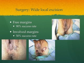
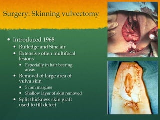
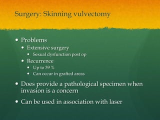
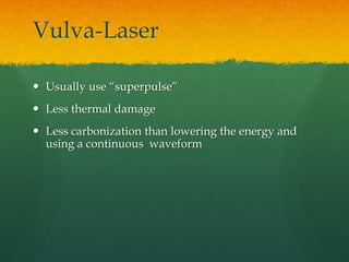
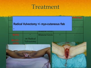
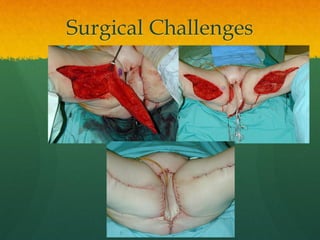
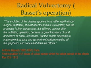
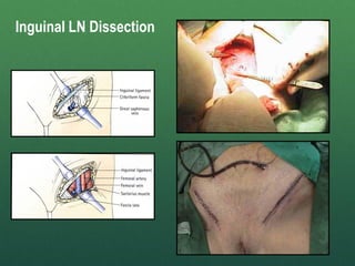
Vulvar Cancer
with special guest
VIN
Professor Khalid Sait (FRCSC)
Chairman of Scientific chair of Prof. Basalamah for
gynecological cancer
Director of Gynecology oncology unit
Faculty of medicine
King Abdulaziz university
Jeddah, Saudi Arabia
Epidemiology
 4% of gential tract malignancies
 Mean age 65 is decreasing
 Two types:
VIN associated Vulvar dystrophy
Prevalence 20-30% 50-60%
Age Younger 30-35 Older 55-85
Histology Poorly diff./ non-keratinizing Well diff./ keratinizing
Lesions Multifocal Unifocal
Epidemiology
 4% of gential tract malignancies
 Mean age 65 is decreasing
 Two types:
VIN associated Vulvar dystrophy
Prevalence 20-30% 50-60%
Age Younger 30-35 Older 55-85
Histology Poorly diff./ non-keratinizing Well diff./ keratinizing
Lesions Multifocal Unifocal
Epidemiology
 4% of gential tract malignancies
 Mean age 65 is decreasing
 Two types:
VIN associated Vulvar dystrophy
Prevalence 20-30% 50-60%
Age Younger 30-35 Older 55-85
Histology Poorly diff./ non-keratinizing Well diff./ keratinizing
Lesions Multifocal Unifocal
Risk Factors
 Smoking
 Vulvar dystrophy
 VIN
 HPV 16 and 18
 Immunodeficiency
 Previous cervical cancer
 Northern European
 Radiation
Presentation
 Symptomatic
 Pruritis
 Soreness
 Dysparunia
 Lumps
 Asymptomatic
20%
Presentation
 Associations
 CIN
 PAIN in up to 30% of cases
Diagnosis
 Vulvoscopy
 Enhances examination
 Useful in selecting sites for biopsy
 Acetic acid
 Can enhance lesions
 Makes clinically unimportant HPV
obvious
 Toludine blue
 Not used clinically
Diagnosis: Biopsy
 Use local anaesthetic
 Xylocaine
 EMLA cream
 Punch biopsy
 Keyes punch
 Cervical biopsy forcep
 Haemostasis
 Apply AGNO3
 Apply monsels soln
 suture
10
Marcaine
0.5 % w/o
Classification: 1989
 Intraepithelial neoplasia
 Squamous
 VIN 1
 VIN 2
 VIN 3
 Nonsquamous intraepithelial neoplasia
 Paget’s disease
 Tumours of the melanocytes, non invasive (melanoma in situ)
ISSVD Int J Gynecol Pathol 1989;8:83
VIN 3 replaces, Bowens disease, Erythroplasia of Queyrat,Carcinoma in situ
,Bowenoid papulosis
VIN
White plaques Pigmented: brown/ red Condyloma
ISVVD 2004
 VIN 1
 VIN 2, 3 = VIN
 VIN
 Usual type VIN ( warty, basaloid) HPV associated (type
16 in >80%)
 Differentiated type VIN
<2-5% of VIN
Older patient
Associated with squamous hyperplasia and/or lichen sclerosus
p53 mutation and/ or over-expression relatively common
Sideri M et al. J Reprod Med. 2005 Nov;50(11):807-10
Basiloid
 Thickened epithelium
 Flat, smooth surface
 Atypical small immature
parabasal type cells
 Numerous mitotic figures
 Enlarged hyperchromatic
nuclei.
Condylomatous
• Least common
• Papillary form: Undulating or
spiking surface
• Hyperkeratosis, parakeratosis
• Mitotic figures and abnormal
maturation
• Surface keratinocytes with
koilocytosis, binucleation or
multinucleation
Parakeratosis: nuclei in surface keratin, sign of high turnover
Koilocytosis: HPV effect, hyperchromatic/ “raisin” nuclei, vacuoles in cytoplasm
Differentiated (simplex) type
• Easy to miss
• Almost looks normal
• Full thickness but large abnormal cells
confined to the parabasal and basal
portion of the rete pegs with
eosinophilic cytoplasm
• Mild nuclear atypia
• Keratin pearl formation may be present
VIN 2
VIN 1
VIN 3
HPV only- not neoplastic
Untreated
Progresses to
Cancer
Variable rates
Women > 30 yrs
Treated
VIN Natural History
Spontaneous regression
Can occur
Women < 30 yrs
Risk of subsequent
malignancy 2.5-7%
Risk of invasion
at treatment 18-22%
Preinvasive neoplasia - VIN
“Early and effective treatment, elimination of smoking and
long-term follow-up of all patients with VIN is imperative.”
Local excision: local 0.5cm margin, epidermis and dermis
Skinning vulvectomy: multifocal, epidermis only
Laser ablation: multifocal 1-3mm depth
5FU: multifocal
Imiquimod:
Only AFTER appropriate biopsies to rule out
invasive disease
•Surface epithelium
• only possible with laser
• Epidermis & papillary dermis
• Condylomata
• Epidermis & part of reticular
dermis
• VIN
• To fascia
• Malignancy
Principles of excision
4th plane
Surgery: Wide local excision
 Commonest surgical
modality
 Excise skin with 5mm
margins
Surgery: Wide local excision
 Free margins
 90% success rate
 Involved margins
 50% success rate
Surgery: Skinning vulvectomy
 Introduced 1968
 Rutledge and Sinclair
 Extensive often multifocal
lesions
 Especially in hair bearing
areas
 Removal of large area of
vulva skin
 5 mm margins
 Shallow layer of skin removed
 Split thickness skin graft
used to fill defect
Surgery: Skinning vulvectomy
 Problems
 Extensive surgery
 Sexual dysfunction post op
 Recurrence
 Up to 39 %
 Can occur in grafted areas
 Does provide a pathological specimen when
invasion is a concern
 Can be used in association with laser
Vulva-Laser
 Usually use “superpulse”
 Less thermal damage
 Less carbonization than lowering the energy and
using a continuous waveform
26
LASER
Therapy 8 Weeks
Post
Laser
Other Treatments
 Photodynamic therapy
 using topically applied 5-aminolevulinic acid.
 Limited experience and availability
 Labour intensive
 5FU (Efudex cream)
 Painful –not recommended
 Imiquimod cream 5% (Aldara)
 promising
 5% cream applied 3X a week for 8 weeks or more,
continue 2 weeks after disappearance
27
Preinvasive neoplasia -
VIN
30% of women will have recurrence
Especially:
- high grade
- multifocal
- positive margins
Therefore long-term follow up is necessary
Paget’s Vulva
 Often multifocal erethematous-whithe plaques
 <10 % have an associated cancer, Colon, Bladder,
endomerium
 10-20% have associated invasive Pagets or vulvar Ca
Pagets
• acanthosis and elongated rete pegs
•Paget’s cells with large nucleus and prominent neucleolus, occur in
clusters
•Hyperplastic epithelium
Approach to Paget’s
 Investigations:
 Colonoscopy
 CT abdomen/ pelvis
 Urinalysis
Paget’s:Therapy
 Excision:
 Wide local excision, 2 cm into subcutaneous tissues
 Suspicion of invasion treated with radical excision
 Medical therapy:
 ? imiquimod
Vulvar Cancer Continued…
 Surgical staging is preferable because the inguinal
femoral lymph node status is the most important
predictor of over all prognosis
 Stage I: tumor confined to the vulva
 IA: lesions ≤ 2 cm in size, confined to the vulva or
perineum and with stromal invasion ≤ 1.0 mm with
negative node.
 IB: lesions > 2 cm in size or with stromal invasion >
1.0 mm, confined to the vulva or perineum with
negative node
FIGO 2010 staging of vulvar
cancers
 Stage II: Tumor of any size with extension to
adjacent perineal structures (1/3 lower urethra, 1/3
lower vagina and/or extension to the anus) with
negative nodes.
Stage III: tumor of any size with or without extension to the
adjacent perineal structures (1/3 lower urethra, 1/3 lower vagina, anus) with positive
inguino-femoral lymph nodes.
 IIIA: (i) 1 lymph node metastasis ≥ 5 mm
(ii) 1–2 lymph node metastasis(es) < 5 mm
 IIIB: (i) 2 or more lymph node metastases ≥ 5 mm
(ii) 3 or more lymph node metastases < 5 mm
 IIIC: Positive node(s) with extracapsular spread
Stage IV: tumor invades other regional (2/3 upper urethra or
vagina), or distant structures.
 IVA: cancer invades any of the following:
(i) upper urethral and/or vaginal mucosa, bladder mucosa,
rectal mucosa, or fixed to pelvic bone.
(ii) fixed or ulcerated inguino-femoral lymph nodes
 IVB: any distant metastasis including pelvic lymph nodes
Principles of treatment
 Treatment should be individualized for each
patient.
 A full pre-operative work-up, including colposcopy
of cervix, vagina and vulva, should be performed to
assess the extent of subclinical disease.
 Preinvasive disease should be removed with the
primary lesion, if feasible.
Principles of treatment
 Surgery attempts to
 1) Stage (i.e., assess the extent of
disease and hence the prognosis).
 2) Debulk a) Primary Tumor
b) Lymph Nodes
 Conservative surgery is preferred
over radical surgery as long as
prognosis is not adversely affected.
Principles of treatment
 Vulvar cancers spread by embolozation, not
permeation so tissue between tumor and LNs need
not be ressected (i.e., en bloc).
 When recurrence occurs locally, it reflects the
behaviour of the disease, not inadequate ressection.
Principles of treatment
 Positive margin need re-excision
 Positive groin node two or more microscopic , one or more
macroscopic, any extra-capsular spread need adjuvant
radiation.
 Elimination of routine pelvic lymphadenectomy in case of positive
groin node i.e. pelvic radiation( GOG).
 Adjuvant radiation required to the vulva in locally advanced
disease after excision.
 Chemotherapy may be useful in the treatment of locally
advanced disease( to avoid urostomy or if there is bone
involvement) /distant metastases and recurrent disease in
groin.
Requirment for unilateral
LND
 UNIFOCAL
 Lateral more than 1 cm from the mid line
 Not located in the ant. Portion of the labia minora
 No palapable groin node in both side
 No l. node mets found at the time of the unilat LND
Treatment
Treatment
Surgery LN Radiation Chemotherapy
IA(T1)
<= 1 mm
invasion
Radical local
Excison
- - -
IB
(T1)
> 1 mm
invasion
Ipsilateral/
Bilateral Groin
- -
II(T2)
III (T3 –
early)
¯
OR Radical
Vulvectomy
Bilateral Groin + -
III (T3 –
late)
IV (T4)
Rad.
Vulvectomy/
Exenteration
N2/ N3 Groin
± Bilateral
Groin & Pelvic
Preop ?
Recurrence Rexcison + ?
Treatment
Radical local excision:
• A wide (1-2 cm margins) and deep (to the inferior fascia of the urogenital diaphragm)
excision of the primary tumor
• Closure: primary two layers
Surgery LN Radiation Chemotherapy
IA(T1)
<= 1 mm
invasion
Radical local
Excison
- - -
IB Ipsilateral/
Bilateral Groin
- -
T2
T3 – early
¯
OR Radical
Vulvectomy
Bilateral Groin + -
T3 – late
T4
Rad.
Vulvectomy/
Exenteration
N2/ N3 Groin
± Bilateral
Groin & Pelvic
Preop ?
Recurrence Rexcison + ?
Treatment
Surgery LN Radiation Chemotherapy
IA Radical local
Excison
- - -
IB
(T1)
> 1 mm
invasion
Ipsilateral/
Bilateral Groin
- -
T2
T3 – early
¯
OR Radical
Vulvectomy
Bilateral Groin + -
T3 – late
T4
Rad.
Vulvectomy/
Exenteration
N2/ N3 Groin
± Bilateral
Groin & Pelvic
Preop +
Recurrence Rexcison + +
Radical local excision:
Radical Vulvectomy
•
•
Treatment
Surgery LN Radiation Chemotherapy
IA Radical local
Excison
- - -
IB Ipsilateral/
Bilateral Groin
- -
II(T2)
III (T3 –
early)
¯
OR Radical
Vulvectomy
Bilateral Groin + -
T3 – late
T4
Rad.
Vulvectomy/
Exenteration
N2/ N3 Groin
± Bilateral
Groin & Pelvic
Preop ?
Recurrence Rexcison + ?
Radical Vulvectomy +/- myo-cutaneous flab
•
Surgical Challenges
Treatment
Surgery LN Radiation Chemotherapy
IA Radical local
Excison
- - -
IB Ipsilateral/
Bilateral Groin
- -
II(T2)
III (T3 –
early)
¯
OR Radical
Vulvectomy
Bilateral Groin + -
III (T3 –
late)
IV (T4)
Rad.
Vulvectomy/
Exenteration
N2/ N3 Groin
± Bilateral
Groin & Pelvic
Preop Cis or 5FU
Recurrence Rexcison ? ?
Exenteration:
• En bloc dissection from:
• the cardinal ligaments laterally
• the broad ligaments of the rectum
posteriorly
• the supralevator attachments of the
bladder, urethra & vagina anteriorly
• the perineum around vagina & anus
caudally
• Reconstruct vagina with gracilis myocutaneous
flap
• 10% operative mortality
•Surface epithelium
• only possible with laser
• Epidermis & papillary dermis
• Condylomata
• Epidermis & part of reticular
dermis
• VIN
• To fascia
• Malignancy
Principles of excision
4th plane
1. Acetic Acid
2. Colposcopy
3. Outline incision
Principles of excision
4. Select appropriate depth
Principles of excision
5. Mobilize adjacent tissues
• Vagina
• Perineum
Principles of excision
6. Primary Closure/ Graft
7. Post operative
• Analgesia
• Sitz baths
• Stool softner
• Bedrest
• DVT prophylaxis
• +/- Drains
Principles of excision
Radical Vulvectomy (
Basset’s operation)
Antoine Basset (1882-1951) Paris
First to publish 147 cases of vulval cancer which he called cancer of the clitoris
Rev Chir 1912
‘’ The evolution of the disease appears to be rather rapid without
surgical treatment, at least after the tumour is ulcerated, and the
prognosis is then always fatal. It is still very somber after
the mutilating operation, because of great frequency of local,
and above all nodal, recurrence. But this seems amenable to
improvement by early and systemic extirpation including all
the lymphatics and nodes that drain the clitoris ‘’
Treatment
Surgery LN Radiation Chemotherapy
IA Radical local
Excison
- - -
IB Ipsilateral/
Bilateral Groin
- -
T2
T3 – early
¯
OR Radical
Vulvectomy
Bilateral Groin + -
T3 – late
T4
Rad.
Vulvectomy/
Exenteration
N2/ N3 Groin
± Bilateral
Groin & Pelvic
Preop ?
Recurrence Rexcison + ?
Groin (Inguinal and Femoral)
LN Dissection:
• Linear incision medial 4/5ths
between ASIS and pubic tubercle
• Dissect between fascia lata and
superficial fascia, preserving tissue
above
• Tie off saphenous vein
• Inguinal: above fascia lata, 2cm
above inguinal ligament, medially by
adductor longus, laterally by sartorius
• Femoral: Below fascial lata, medial
to femoral vein at opening of the
femoral ring
Inguinal LN Dissection
 High perioperative morbidity:
 Requires 3-4 days of bed rest, wound breakdown 50%,
lymphocele, lymphedema, infection, bleeding, DVT
• Can some low-risk
women avoid full LN
dissection?
Sentinel Node Biopsy
• Used in breast CA and melanoma
• Two methods:
– Radiolabelled human albumin and gamma-detecting probe
Possible 91-100% NPV: 100%
– Isosulfan blue dye Possible 56-75% NPV: 96-100%
Key Points
 VIN is a condition of increasing importance with multiple
methods of treatment
 At least two possible etiologies are considered for vulvar
cancer. One is related to infection with oncogenic HPV ,
other is related to maturation disorders
 Management must address local control and regional
( nodal ) disease
 The evolution of management has been driven by desire
to reduce morbidity whilst maintaining disease control
 Vulvar cancer is a challenging disease to treat. Its rarity
has mindered attempts to improve management through
clinical trials
Thank You!
Vulva ca sep course quick revision 2012

Vulva ca sep course quick revision 2012

  • 1.
    Vulvar Cancer with specialguest VIN Professor Khalid Sait (FRCSC) Chairman of Scientific chair of Prof. Basalamah for gynecological cancer Director of Gynecology oncology unit Faculty of medicine King Abdulaziz university Jeddah, Saudi Arabia
  • 2.
    Epidemiology  4% ofgential tract malignancies  Mean age 65 is decreasing  Two types: VIN associated Vulvar dystrophy Prevalence 20-30% 50-60% Age Younger 30-35 Older 55-85 Histology Poorly diff./ non-keratinizing Well diff./ keratinizing Lesions Multifocal Unifocal
  • 3.
    Epidemiology  4% ofgential tract malignancies  Mean age 65 is decreasing  Two types: VIN associated Vulvar dystrophy Prevalence 20-30% 50-60% Age Younger 30-35 Older 55-85 Histology Poorly diff./ non-keratinizing Well diff./ keratinizing Lesions Multifocal Unifocal
  • 4.
    Epidemiology  4% ofgential tract malignancies  Mean age 65 is decreasing  Two types: VIN associated Vulvar dystrophy Prevalence 20-30% 50-60% Age Younger 30-35 Older 55-85 Histology Poorly diff./ non-keratinizing Well diff./ keratinizing Lesions Multifocal Unifocal
  • 5.
    Risk Factors  Smoking Vulvar dystrophy  VIN  HPV 16 and 18  Immunodeficiency  Previous cervical cancer  Northern European  Radiation
  • 6.
    Presentation  Symptomatic  Pruritis Soreness  Dysparunia  Lumps  Asymptomatic 20%
  • 7.
  • 8.
    Diagnosis  Vulvoscopy  Enhancesexamination  Useful in selecting sites for biopsy  Acetic acid  Can enhance lesions  Makes clinically unimportant HPV obvious  Toludine blue  Not used clinically
  • 9.
    Diagnosis: Biopsy  Uselocal anaesthetic  Xylocaine  EMLA cream  Punch biopsy  Keyes punch  Cervical biopsy forcep  Haemostasis  Apply AGNO3  Apply monsels soln  suture
  • 10.
  • 11.
    Classification: 1989  Intraepithelialneoplasia  Squamous  VIN 1  VIN 2  VIN 3  Nonsquamous intraepithelial neoplasia  Paget’s disease  Tumours of the melanocytes, non invasive (melanoma in situ) ISSVD Int J Gynecol Pathol 1989;8:83 VIN 3 replaces, Bowens disease, Erythroplasia of Queyrat,Carcinoma in situ ,Bowenoid papulosis
  • 12.
    VIN White plaques Pigmented:brown/ red Condyloma
  • 13.
    ISVVD 2004  VIN1  VIN 2, 3 = VIN  VIN  Usual type VIN ( warty, basaloid) HPV associated (type 16 in >80%)  Differentiated type VIN <2-5% of VIN Older patient Associated with squamous hyperplasia and/or lichen sclerosus p53 mutation and/ or over-expression relatively common Sideri M et al. J Reprod Med. 2005 Nov;50(11):807-10
  • 14.
    Basiloid  Thickened epithelium Flat, smooth surface  Atypical small immature parabasal type cells  Numerous mitotic figures  Enlarged hyperchromatic nuclei.
  • 15.
    Condylomatous • Least common •Papillary form: Undulating or spiking surface • Hyperkeratosis, parakeratosis • Mitotic figures and abnormal maturation • Surface keratinocytes with koilocytosis, binucleation or multinucleation Parakeratosis: nuclei in surface keratin, sign of high turnover Koilocytosis: HPV effect, hyperchromatic/ “raisin” nuclei, vacuoles in cytoplasm
  • 16.
    Differentiated (simplex) type •Easy to miss • Almost looks normal • Full thickness but large abnormal cells confined to the parabasal and basal portion of the rete pegs with eosinophilic cytoplasm • Mild nuclear atypia • Keratin pearl formation may be present
  • 17.
    VIN 2 VIN 1 VIN3 HPV only- not neoplastic Untreated Progresses to Cancer Variable rates Women > 30 yrs Treated VIN Natural History Spontaneous regression Can occur Women < 30 yrs Risk of subsequent malignancy 2.5-7% Risk of invasion at treatment 18-22%
  • 18.
    Preinvasive neoplasia -VIN “Early and effective treatment, elimination of smoking and long-term follow-up of all patients with VIN is imperative.” Local excision: local 0.5cm margin, epidermis and dermis Skinning vulvectomy: multifocal, epidermis only Laser ablation: multifocal 1-3mm depth 5FU: multifocal Imiquimod: Only AFTER appropriate biopsies to rule out invasive disease
  • 19.
    •Surface epithelium • onlypossible with laser • Epidermis & papillary dermis • Condylomata • Epidermis & part of reticular dermis • VIN • To fascia • Malignancy Principles of excision 4th plane
  • 20.
    Surgery: Wide localexcision  Commonest surgical modality  Excise skin with 5mm margins
  • 21.
    Surgery: Wide localexcision  Free margins  90% success rate  Involved margins  50% success rate
  • 22.
    Surgery: Skinning vulvectomy Introduced 1968  Rutledge and Sinclair  Extensive often multifocal lesions  Especially in hair bearing areas  Removal of large area of vulva skin  5 mm margins  Shallow layer of skin removed  Split thickness skin graft used to fill defect
  • 23.
    Surgery: Skinning vulvectomy Problems  Extensive surgery  Sexual dysfunction post op  Recurrence  Up to 39 %  Can occur in grafted areas  Does provide a pathological specimen when invasion is a concern  Can be used in association with laser
  • 24.
    Vulva-Laser  Usually use“superpulse”  Less thermal damage  Less carbonization than lowering the energy and using a continuous waveform
  • 26.
  • 27.
    Other Treatments  Photodynamictherapy  using topically applied 5-aminolevulinic acid.  Limited experience and availability  Labour intensive  5FU (Efudex cream)  Painful –not recommended  Imiquimod cream 5% (Aldara)  promising  5% cream applied 3X a week for 8 weeks or more, continue 2 weeks after disappearance 27
  • 28.
    Preinvasive neoplasia - VIN 30%of women will have recurrence Especially: - high grade - multifocal - positive margins Therefore long-term follow up is necessary
  • 29.
    Paget’s Vulva  Oftenmultifocal erethematous-whithe plaques  <10 % have an associated cancer, Colon, Bladder, endomerium  10-20% have associated invasive Pagets or vulvar Ca
  • 30.
  • 33.
    • acanthosis andelongated rete pegs •Paget’s cells with large nucleus and prominent neucleolus, occur in clusters •Hyperplastic epithelium
  • 34.
    Approach to Paget’s Investigations:  Colonoscopy  CT abdomen/ pelvis  Urinalysis
  • 35.
    Paget’s:Therapy  Excision:  Widelocal excision, 2 cm into subcutaneous tissues  Suspicion of invasion treated with radical excision  Medical therapy:  ? imiquimod
  • 36.
  • 37.
     Surgical stagingis preferable because the inguinal femoral lymph node status is the most important predictor of over all prognosis
  • 38.
     Stage I:tumor confined to the vulva  IA: lesions ≤ 2 cm in size, confined to the vulva or perineum and with stromal invasion ≤ 1.0 mm with negative node.  IB: lesions > 2 cm in size or with stromal invasion > 1.0 mm, confined to the vulva or perineum with negative node FIGO 2010 staging of vulvar cancers
  • 39.
     Stage II:Tumor of any size with extension to adjacent perineal structures (1/3 lower urethra, 1/3 lower vagina and/or extension to the anus) with negative nodes.
  • 40.
    Stage III: tumorof any size with or without extension to the adjacent perineal structures (1/3 lower urethra, 1/3 lower vagina, anus) with positive inguino-femoral lymph nodes.  IIIA: (i) 1 lymph node metastasis ≥ 5 mm (ii) 1–2 lymph node metastasis(es) < 5 mm  IIIB: (i) 2 or more lymph node metastases ≥ 5 mm (ii) 3 or more lymph node metastases < 5 mm  IIIC: Positive node(s) with extracapsular spread
  • 41.
    Stage IV: tumorinvades other regional (2/3 upper urethra or vagina), or distant structures.  IVA: cancer invades any of the following: (i) upper urethral and/or vaginal mucosa, bladder mucosa, rectal mucosa, or fixed to pelvic bone. (ii) fixed or ulcerated inguino-femoral lymph nodes  IVB: any distant metastasis including pelvic lymph nodes
  • 42.
    Principles of treatment Treatment should be individualized for each patient.  A full pre-operative work-up, including colposcopy of cervix, vagina and vulva, should be performed to assess the extent of subclinical disease.  Preinvasive disease should be removed with the primary lesion, if feasible.
  • 43.
    Principles of treatment Surgery attempts to  1) Stage (i.e., assess the extent of disease and hence the prognosis).  2) Debulk a) Primary Tumor b) Lymph Nodes  Conservative surgery is preferred over radical surgery as long as prognosis is not adversely affected.
  • 44.
    Principles of treatment Vulvar cancers spread by embolozation, not permeation so tissue between tumor and LNs need not be ressected (i.e., en bloc).  When recurrence occurs locally, it reflects the behaviour of the disease, not inadequate ressection.
  • 45.
    Principles of treatment Positive margin need re-excision  Positive groin node two or more microscopic , one or more macroscopic, any extra-capsular spread need adjuvant radiation.  Elimination of routine pelvic lymphadenectomy in case of positive groin node i.e. pelvic radiation( GOG).  Adjuvant radiation required to the vulva in locally advanced disease after excision.  Chemotherapy may be useful in the treatment of locally advanced disease( to avoid urostomy or if there is bone involvement) /distant metastases and recurrent disease in groin.
  • 46.
    Requirment for unilateral LND UNIFOCAL  Lateral more than 1 cm from the mid line  Not located in the ant. Portion of the labia minora  No palapable groin node in both side  No l. node mets found at the time of the unilat LND
  • 47.
  • 48.
    Treatment Surgery LN RadiationChemotherapy IA(T1) <= 1 mm invasion Radical local Excison - - - IB (T1) > 1 mm invasion Ipsilateral/ Bilateral Groin - - II(T2) III (T3 – early) ¯ OR Radical Vulvectomy Bilateral Groin + - III (T3 – late) IV (T4) Rad. Vulvectomy/ Exenteration N2/ N3 Groin ± Bilateral Groin & Pelvic Preop ? Recurrence Rexcison + ?
  • 49.
    Treatment Radical local excision: •A wide (1-2 cm margins) and deep (to the inferior fascia of the urogenital diaphragm) excision of the primary tumor • Closure: primary two layers Surgery LN Radiation Chemotherapy IA(T1) <= 1 mm invasion Radical local Excison - - - IB Ipsilateral/ Bilateral Groin - - T2 T3 – early ¯ OR Radical Vulvectomy Bilateral Groin + - T3 – late T4 Rad. Vulvectomy/ Exenteration N2/ N3 Groin ± Bilateral Groin & Pelvic Preop ? Recurrence Rexcison + ?
  • 50.
    Treatment Surgery LN RadiationChemotherapy IA Radical local Excison - - - IB (T1) > 1 mm invasion Ipsilateral/ Bilateral Groin - - T2 T3 – early ¯ OR Radical Vulvectomy Bilateral Groin + - T3 – late T4 Rad. Vulvectomy/ Exenteration N2/ N3 Groin ± Bilateral Groin & Pelvic Preop + Recurrence Rexcison + + Radical local excision: Radical Vulvectomy • •
  • 51.
    Treatment Surgery LN RadiationChemotherapy IA Radical local Excison - - - IB Ipsilateral/ Bilateral Groin - - II(T2) III (T3 – early) ¯ OR Radical Vulvectomy Bilateral Groin + - T3 – late T4 Rad. Vulvectomy/ Exenteration N2/ N3 Groin ± Bilateral Groin & Pelvic Preop ? Recurrence Rexcison + ? Radical Vulvectomy +/- myo-cutaneous flab •
  • 52.
  • 53.
    Treatment Surgery LN RadiationChemotherapy IA Radical local Excison - - - IB Ipsilateral/ Bilateral Groin - - II(T2) III (T3 – early) ¯ OR Radical Vulvectomy Bilateral Groin + - III (T3 – late) IV (T4) Rad. Vulvectomy/ Exenteration N2/ N3 Groin ± Bilateral Groin & Pelvic Preop Cis or 5FU Recurrence Rexcison ? ? Exenteration: • En bloc dissection from: • the cardinal ligaments laterally • the broad ligaments of the rectum posteriorly • the supralevator attachments of the bladder, urethra & vagina anteriorly • the perineum around vagina & anus caudally • Reconstruct vagina with gracilis myocutaneous flap • 10% operative mortality
  • 54.
    •Surface epithelium • onlypossible with laser • Epidermis & papillary dermis • Condylomata • Epidermis & part of reticular dermis • VIN • To fascia • Malignancy Principles of excision 4th plane
  • 55.
    1. Acetic Acid 2.Colposcopy 3. Outline incision Principles of excision
  • 56.
    4. Select appropriatedepth Principles of excision
  • 57.
    5. Mobilize adjacenttissues • Vagina • Perineum Principles of excision
  • 58.
    6. Primary Closure/Graft 7. Post operative • Analgesia • Sitz baths • Stool softner • Bedrest • DVT prophylaxis • +/- Drains Principles of excision
  • 59.
    Radical Vulvectomy ( Basset’soperation) Antoine Basset (1882-1951) Paris First to publish 147 cases of vulval cancer which he called cancer of the clitoris Rev Chir 1912 ‘’ The evolution of the disease appears to be rather rapid without surgical treatment, at least after the tumour is ulcerated, and the prognosis is then always fatal. It is still very somber after the mutilating operation, because of great frequency of local, and above all nodal, recurrence. But this seems amenable to improvement by early and systemic extirpation including all the lymphatics and nodes that drain the clitoris ‘’
  • 60.
    Treatment Surgery LN RadiationChemotherapy IA Radical local Excison - - - IB Ipsilateral/ Bilateral Groin - - T2 T3 – early ¯ OR Radical Vulvectomy Bilateral Groin + - T3 – late T4 Rad. Vulvectomy/ Exenteration N2/ N3 Groin ± Bilateral Groin & Pelvic Preop ? Recurrence Rexcison + ? Groin (Inguinal and Femoral) LN Dissection: • Linear incision medial 4/5ths between ASIS and pubic tubercle • Dissect between fascia lata and superficial fascia, preserving tissue above • Tie off saphenous vein • Inguinal: above fascia lata, 2cm above inguinal ligament, medially by adductor longus, laterally by sartorius • Femoral: Below fascial lata, medial to femoral vein at opening of the femoral ring
  • 61.
  • 62.
    Inguinal LN Dissection High perioperative morbidity:  Requires 3-4 days of bed rest, wound breakdown 50%, lymphocele, lymphedema, infection, bleeding, DVT • Can some low-risk women avoid full LN dissection? Sentinel Node Biopsy
  • 63.
    Sentinel Node Biopsy •Used in breast CA and melanoma • Two methods: – Radiolabelled human albumin and gamma-detecting probe Possible 91-100% NPV: 100% – Isosulfan blue dye Possible 56-75% NPV: 96-100%
  • 64.
    Key Points  VINis a condition of increasing importance with multiple methods of treatment  At least two possible etiologies are considered for vulvar cancer. One is related to infection with oncogenic HPV , other is related to maturation disorders  Management must address local control and regional ( nodal ) disease  The evolution of management has been driven by desire to reduce morbidity whilst maintaining disease control  Vulvar cancer is a challenging disease to treat. Its rarity has mindered attempts to improve management through clinical trials
  • 65.