RADIOTHERAPY PRINCIPLES AND
TECHNIQUES IN MANAGEMENT OF
VULVAR CANCERS
Dr. ROSHAN N
MODERATOR: PROF. BHAVANA RAI
INTRODUCTION
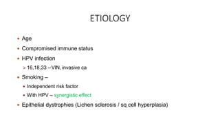
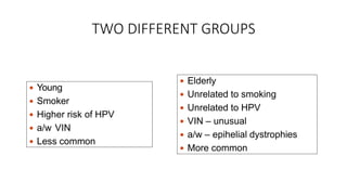
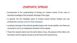
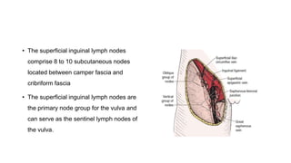
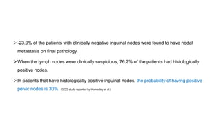
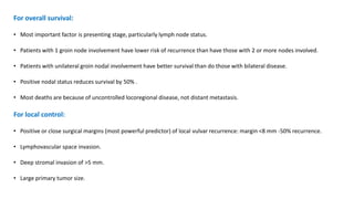
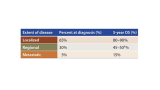
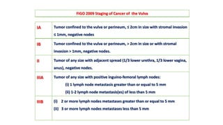
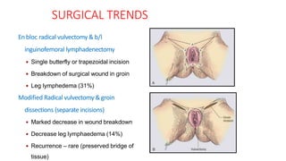
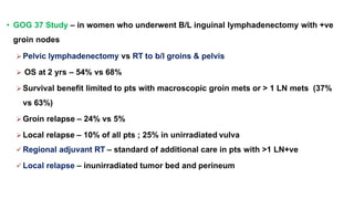
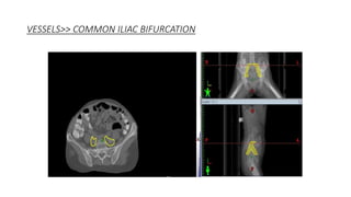
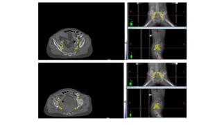
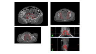
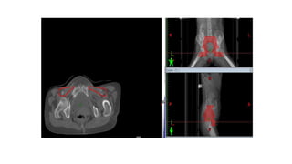
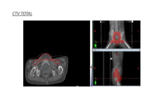
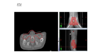
• Vulvar cancer is a rare malignancy that represents <1% of all the
cancers diagnosed in women
• <5% of all gynecologic neoplasms
• Incidence is 2.5 cases per 100,000 women
• Incidence increases to 15.5 per 100,000 in women of age >75 years
ETIOLOGY
 Age
 Compromised immune status
 HPV infection
 16,18,33 –VIN, invasive ca
 Smoking –
 Independent risk factor
 With HPV – synergistic effect
 Epithelial dystrophies (Lichen sclerosis / sq cell hyperplasia)
TWO DIFFERENT GROUPS
 Young
 Smoker
 Higher risk of HPV
 a/w VIN
 Less common
 Elderly
 Unrelated to smoking
 Unrelated to HPV
 VIN – unusual
 a/w – epihelial dystrophies
 More common
ANATOMY
• Composed of the mons pubis,
clitoris, labia majora and minora,
vaginal vestibule, and their
supporting subcutaneous
tissues
• Blends with the urinary meatus
anteriorly and with the perineum
and anus posteriorly
PATHOLOGY
 SCC – MC (80-90%)
 Malignant Melanoma – 10%
 Verrucous ca
 Basal cell ca
 Merkel cell ca
 Transitional cell ca
 Adenocystic
 Bartholin gland tumors
 Sarcomas
PATTERNS OF DISEASE AND SPREAD
• Primary disease can appear anywhere on the vulva.
• Approximately 70% arises primarily on the labia.
• Disease more commonly occurs in labia majora;
however, it may appear on the labia minora, clitoris, and
perineum.
• 15% in the clitoris, 5% in the perineum and fourchette,
5% in the prepuce, Bartholin's glands and urethra
• The disease is usually localized and well demarcated;
although it can occasionally be so extensive that the
primary location cannot be determined.
Primary Site
• Local extension occurs in a stepwise progressive fashion from a distal-to
proximal direction.
• Direct involvement of lower third of urethra, lower third of vagina and/or
extension to the anus is first.
• More extensive cases: further progression proximally involves upper urethral
and/or vaginal mucosa, bladder mucosa, rectal mucosa, or fixation to bony
pelvic structures.
LYMPHATIC SPREAD
• Fundamental to the understanding of therapy for invasive cancer of the vulva is
thorough knowledge of the lymphatic drainage of this organ.
• In general, the four histologic types of invasive cancer behave similarly and use
primarily the lymphatic route for initial metastases.
• Lymphatic drainage of the external genitalia begins with minute papillae, and these are
connected in turn to a multilayered meshwork of fine vessels.
• These fine vessels extend over the entire labium minus, the prepuce of the clitoris, the
fourchette, and the vaginal mucosa up to the level of the hymenal ring.
• Drainage of these lymphatics extends toward the anterior
portion of the labium minus, where they emerge into three
or four collecting trunks whose course is toward the mons
veneris, bypassing the clitoris.
• Vessels from the prepuce anastomose with these
lymphatics.
• Similarly, vessels from the labium majus proceed anteriorly
to the upper part of the vulva and mons veneris, there
joining the vessels of the prepuce and labium minus.
• These lymphatic vessels abruptly change direction, turning
laterally, and terminate in ipsilateral or contralateral
femoral nodes.
The superficial inguinofemoral lymph nodes
are the first echelon.
• The nodes are medial to the great saphenous vein above the cribriform
fascia and in turn may drain secondarily to the deep femoral group.
• The next echelon of nodes is the pelvic/iliac nodes.
• The superficial inguinal lymph nodes
comprise 8 to 10 subcutaneous nodes
located between camper fascia and
cribriform fascia
• The superficial inguinal lymph nodes are
the primary node group for the vulva and
can serve as the sentinel lymph nodes of
the vulva.
• The deep femoral nodes, located beneath
the cribriform fascia, are the secondary
node recipients and are involved before
drainage into the deep pelvic nodes
occurs.
• The Cloquet node, the last node of the
deep femoral group, is located just
beneath the Poupart ligament.
• Lymphatic drainage is specific to the location of a vulvar lesion.
• Labial lesions drain into the superficial inguinal and femoral lymph nodes,
and then penetrate the cribriform fascia and reach the deep femoral nodes
• Lesions of the fourchette and perineum follow the lymphatics of the labia.
• For well-lateralized lesions, metastasis to the contralateral inguinal or
pelvic lymph nodes is unusual in the absence of ipsilateral inguinofemoral
node involvement.
• Lymphatic drainage from the glans clitoris or perineal body enters either
unilateral or bilateral superficial femoral nodes or the deep femoral and
pelvic lymph nodes.
• Some lymphatics originating in the clitoris enter the pelvis directly,
bypassing the femoral nodes to connect with the obturator and external iliac
lymph nodes,
• Pelvic lymph nodes are rarely involved without synchronous involvement of
the inguinal nodes
• The frequency of inguinal lymph node metastasis in surgically staged
patients ranges from 6% to 50%, depending on tumor invasion and
extent of disease.
Primary Tumor Size and
Depth of Invasion Number of Patients
Number of Patients with
Positive Lymph Nodes (%)
Depth
<1 mm 120 0 (0)
1.1–2 mm 121 8 (6.6)
2.1–3 mm 97 8 (8.2)
3.1–4 mm 50 11 (22)
4.1–5 mm 40 10 (25)
Size
>5 mm 32 12 (37.5)
>2 cm 168 77 (45.8)
Any size of primary tumor
extending beyond the vulva
70 38 (54.2)
• Physical examination alone is inaccurate to assess lymph node involvement
• 62% incidence of lymph node metastases in patients with clinically palpable
adenopathy
• 35% involvement in patients without clinically palpable adenopathy. (Plentl and Friedman)
• Approximately 75% of patients with clinically suspicious lymph nodes had nodal
metastasis
• 11% to 43% of patients with clinically negative nodes had metastasis to the
nodes.(Review of clinical staging, Franklin)
-23.9% of the patients with clinically negative inguinal nodes were found to have nodal
metastasis on final pathology.
When the lymph nodes were clinically suspicious, 76.2% of the patients had histologically
positive nodes.
In patients that have histologically positive inguinal nodes, the probability of having positive
pelvic nodes is 30%. (GOG study reported by Homesley et al.)
• Clinical and histologic tumor characteristics identified as predictors of nodal
involvement are :
- clinical node status (palpable vs. Non palpable),
- grade,
- capillary-lymphatic space involvement,
- tumor thickness
- patient's age
- size of the primary tumor
• The probability of having positive inguinal nodes rose from 18.9% for patients with lesions <3 cm to
72.4% for patients with primary tumors ≥3 cm.
• Extension of the primary tumor to the urethra, vagina, and anal area is associated with an
increased incidence of nodal involvement and worsening of prognosis
• Inguinal nodal involvement is a strong predictor for pelvic nodal disease
• Of patients with positive inguinal nodes, the incidence of pelvic node involvement was 28.3%
• Because of rarity of isolated pelvic nodal metastasis, the status of the inguinal nodes determines
the management of the pelvic nodes
Distant Metastases:
• Hematogenous dissemination generally occurs late in the natural history of the disease,
with the most common sites being the lungs, liver, and bones.
• The overall survival is limited in these cases to a median of approximately 6 months
PROGNOSTIC FACTORS:
• Lymph node metastasis is the most important prognostic factor in patients with vulvar cancer.
The presence of inguinal node metastases is accompanied by a 50% reduction in long-term
survival.
• Size, number and ECE of LN affects prognosis
• Pelvic nodal metastasis has an even more profound negative effect on survival
• Tumor size >4 cm
• Capillary lymphatic space involvement
• Depth of invasion> 5 mm
• Surgical margins <8 mm.
• Three publications have reported a clear association between risk of
vulvar recurrence and width of surgical resection margins.
• When microscopic margins are 8 mm or less in formalin-fixed tissue,
local recurrence has been observed in 43 of 145 patients (30%)
•Positive or close margins <5mm have a significant risk of vulvar recurrence after vulvar cancer resection.
• A radiation dose ≥56Gy may reduce the risk of vulvar recurrence
For overall survival:
• Most important factor is presenting stage, particularly lymph node status.
• Patients with 1 groin node involvement have lower risk of recurrence than have those with 2 or more nodes involved.
• Patients with unilateral groin nodal involvement have better survival than do those with bilateral disease.
• Positive nodal status reduces survival by 50% .
• Most deaths are because of uncontrolled locoregional disease, not distant metastasis.
For local control:
• Positive or close surgical margins (most powerful predictor) of local vulvar recurrence: margin <8 mm -50% recurrence.
• Lymphovascular space invasion.
• Deep stromal invasion of >5 mm.
• Large primary tumor size.
IA Tumor confined to the vulva or perineum, ≤ 2cm in size with stromal invasion
≤ 1mm, negative nodes
IB Tumor confined to the vulva or perineum, > 2cm in size or with stromal
invasion > 1mm, negative nodes.
II Tumor of any size with adjacent spread (1/3 lower urethra, 1/3 lower vagina,
anus), negative nodes.
IIIA Tumor of any size with positive inguino-femoral lymph nodes:
(i) 1 lymph node metastasis greater than or equal to 5 mm
(ii) 1-2 lymph node metastasis(es) of less than 5 mm
IIIB (i) 2 or more lymph nodes metastases greater than or equal to 5 mm
(ii) 3 or more lymph nodes metastases less than 5 mm
FIGO 2009 Staging of Cancer of the Vulva
IIIC Positive node(s) with extracapsular spread
IVA (i) Tumor invades other regional structures (2/3 upper urethra, 2/3 upper
vagina), bladder mucosa, rectal mucosa, or fixed to pelvic bone.
(ii) Fixed or ulcerated inguino-femoral lymph nodes
IVB Any distant metastasis including pelvic lymph nodes
• STAGE I: 90%
• STAGE II: 81%
• STAGE III: 68%
• STAGE IV: 20%
5-year survival rates:
GENERAL MANAGEMENT:
The treatment of carcinoma of the vulva is challenging for multiple reasons:
• In general patients with this disease are older and have comorbidities.
• The tumor, by virtue of its location, can easily involve adjacent organs such as the bladder
and the rectum, and the frequency of nodal involvement is high.
• Because of its relatively low incidence, most published reports include rather small and
heterogeneous groups of patients.
• Management of carcinoma of the vulva is further complicated by the major psychosexual
impact that the treatment can have on patients.
• The management of vulvar carcinoma has undergone very significant changes in the last few
decades.
• En bloc resection of the primary tumor and inguinal node dissection, which used to be the
standard of care, have been replaced by multimodality therapy, with the surgery being more
tailored to the extent of the disease.
• This change follows the recognition of the:
- morbidity associated with radical surgery,
- the improved results achieved with multimodality therapy, and
- the recognition of the negative impact that radical surgery can have on sexual
function and body image.
TREATMENT MODALITIES
 Surgery
 Radiation
 Chemotherapy
SURGERY
 Surgical strategy depends on
Primary tumor parameters
Assessment of groin nodes
Status of adjacent tissues
Age of patient
SURGICAL TRENDS
En bloc radical vulvectomy & b/l
inguinofemoral lymphadenectomy
 Single butterfly or trapezoidal incision
 Breakdown of surgical wound in groin
 Leg lymphedema (31%)
Modified Radical vulvectomy &groin
dissections (separate incisions)
 Marked decrease in wound breakdown
 Decrease leg lymphaedema (14%)
 Recurrence – rare (preserved bridge of
tissue)
EXENTERATIVE SURGERY
 Advanced disease
 High morbidity
 Rarely chosen as initial therapy
 Rarely appropriate when regional nodes are present
GROIN DISSECTION
Indications
 Tumor size - >2 cm
 Depth of invasion - >1 mm
 LN +ve
Types
 Radical b/l inguino femoral LN
dissection
 Superficial & deep nodes
 Wound breakdown – 50%
 Lymphedema – 10-15%
 Superficial groin dissection
 Rationale
 Orderly sequence of metastatic
progression in groin nodal chain
 Mets in deep femoral nodes without
involvement of superficial nodes is
uncommon
 Risk of groin recurrence post-superficial
dissection – 16%
 Adq LN dissection : 10 LN
 U/L groin dissection
Lateralized lesions
Groin nodes –ve
If +ve - contralateral groin dissection
 Midline tumors or within 2 cm of midline or involve ant labia minora
b/l superficial groin dissection
 Fixed or ulcerating nodes
Combined modality treatment
SENTINEL LYMPHNODE DISSECTION
• Assessment of the groin nodes may be accomplished via full superficial inguinal dissection or
alternatively via a sentinel lymph node procedure alone
• Optimal lesions for sentinel node procedures are those with small (<4 cm), unifocal lesions
with surgeons who have performed at least 10 prior sentinel procedures under supervision
• The sentinel nodes should be evaluated prior to vulvar resection to avoid disruption of
lymphatic channels emanating from the tumor.
• If no sentinel node is found, a full inguinofemoral lymphadenectomy is recommended
• If sentinel node is found to be positive a full ipsilateral groin dissection and/or radiotherapy to
the groin should be considered
PATTERN OF RECURRENCE – POST SX
 Vulva/perineum – 60%
 Groin – 23%
 Pelvis – 16%
 Distant – 19%
RADIATION THERAPY
ROLE AND ITS TECHNIQUES
RADIATION
 Types
 EBRT
 photons
 Electrons
 BRACHYTHERAPY
 Modalities
Adjuvant post-op RT
Pre-op RT/ CRT
Definitive RT/ CRT
Palliative RT
• Indications of Adjuvant RT to tumor bed
Margin < 8mm
Positive margins
Depth of invasion >5mm
LVI
• Indications of adjuvant RT to groin
>1 positive LN
ECE
Gross residual nodal disease
ADJUVANT POST OPERATIVE RADIOTHERAPY
• GOG 37 Study – in women who underwent B/L inguinal lymphadenectomy with +ve
groin nodes
 Pelvic lymphadenectomy vs RT to b/l groins & pelvis
 OS at 2 yrs – 54% vs 68%
 Survival benefit limited to pts with macroscopic groin mets or > 1 LN mets (37%
vs 63%)
 Groin relapse – 24% vs 5%
 Local relapse – 10% of all pts ; 25% in unirradiated vulva
 Regional adjuvant RT – standard of additional care in pts with >1 LN+ve
 Local relapse – inunirradiated tumor bed and perineum
Faul et al – observational study IJROBP-1997
 62 pts with +ve or close margins were restropectively studied
 Adjuvant RT to vulva vs observation alone
 Local recurrence – 16% vs 58%
 Adjuvant RT to vulva significantly reduces LR in close or +ve margins
Dusenbery– reported central recurrences in 13 of 26 pts (50%) of ca vulva
with LN +ve post Sx treated with adjuvant RT to pelvis and groins with
midline shielding
 the target volume for adjuvant post-op RT for pts with node mets
should include the tumor bed
DEFINITIVE RADIATION THERAPY
• Indications
Advanced/ unresectable
Where even after pre-op RT - disease would be unresectable
If Sx done - pelvic exenteration
Sexually active female with clitoris involvement
Medically inoperable
• The standard of care for early lesions is surgical resection; however, selected
patients with small central lesions may be considered for definitive
radiation,particularly when the lesions are in close proximity to the urethra,
clitoris, or anus
• Selected patients with a low risk of having nodal involvement may be
treated to the vulvar area alone.
• The treatment may be delivered using electrons, low-energy photons, or IMRT
BRACHYTHERAPY
• Brachytherapy has been used for inoperable vulvar cancer and as a boost
to the primary tumor and/or to the lymph nodes.
• The efficacy of this treatment is difficult to evaluate because of the
variability of the clinical situation in which this type of treatment may be
employed
• A series from the Centre Alexis Vautrin Institute in France describes 34 patients, 21 with
primary and 13 with recurrent disease, treated with brachytherapy only.
• The median brachytherapy dose was 60 Gy, with a range of 53 to 88 Gy.
• In the group of 21 patients who underwent brachytherapy as primary treatment, 3
patients developed locoregional recurrence, for a 5-year local control rate of 80% and a
disease-specific survival of 70%.
• In the group of 13 patients who were treated for recurrence, 8 developed
local recurrence, with or without disease at other sites, for a local control rate of 19%.
• Of the entire group of 34 patients, 5 developed VULVAR necrosis.
• Definitive chemoradiation is used for patients with:
- advanced tumors considered unresectable at presentation
- or for patients who are medically inoperable.
• This may be used when the tumor does not become resectable in the midst of preoperative
intent chemoradiation or as an upfront alternative to surgically based treatment.
• In these patients, chemotherapy should be continued throughout the entire course of radiation
for the purpose of radiosensitization of the tumor in the treatment volume and possible
eradication of subclinical disease outside of the radiation field.
DEFINITIVE CHEMORADIATION THERAPY
 Concurrent chemo – cisplatin/ 5-FU/ MMC/ bleomycin
 Improved LC ; CR – 70%
 Retrospective study of t/t outcomes comparing CRT vs RT alone suggests improved LC
with CRT
 Prospective randomized trials – do not exist
 Increased acute reactions – moist desquamations – t/t interruptions
PREOPERATIVE RADIATION THERAPY WITH
CONCURRENT CHEMOTHERAPY
• Indications
 Locally advanced tumors –T3,T4
 Anticipated surgical margin – < 2 cm
 Involvement of anal sphincter, pubic arch, more than distal urethra
 Tumor approaching clitoris or extending more than minimally past vaginal introitus
• Rationale
 Toreduce the scope of Sx
 Toconserve normal tissue structure and function (organ preservation)
 Toconvert the status from inoperable to operable
• The follow-up phase II GOG 205 study enrolled women with T3 or T4
tumors, treated with a total dose of 5,760 cGy (180 cGy/fraction) and weekly
cisplatin (40 mg/m2), with no planned treatment breaks.
• Sixty-nine percent of women were able to complete the protocol per plan, 64%
achieved a complete response, and 50% had a pCR on surgical biopsy.
• Overall survival at 2 years was 40% for those with no or partial clinical response, 62%
for those with a complete clinical response, and 81% for those with a complete
pathologic response
• After initial concurrent chemoradiation and healing of the reaction, the
response to the therapy at the primary site and the lymph nodes is assessed
• If there is complete clinical regression of the disease at the site of the
primary, one option is to perform biopsies of the primary site, foregoing
resection if there is a pathologic complete response
• It is recommended to carry out an inguinal dissection as well, as residual
disease maybe found in lymph nodes
• Recurrences at the primary site are potentially salvageable, nodal
recurrences, particularly after any prior irradiation, are often fatal
RT – TECHNIQUES
Prerequisites
Initial tumor volume
Margin status
Groin status
Pelvic LN status
Volume of RT
The radiation target volume usually encompasses the vulva,
both groins and the lower pelvic nodes.
2-D AND 3-D CONFORMAL TREATMENT
• SIMULATION:
Frog leg positioning may be used to reduce skin folds in the inguinal
regions
Markers should be placed on the primary, lymph nodes and scars from
previous surgery to document the extent of the disesase
With full bladder (to minimize the volume of small bowel in the radiation
field),
with custom immobilizations.
PELVIS + GROIN + VULVA
 Sup. – absent pelvic N- mid SI jt
(includes caudal Ext I N and Int I N)
– pelvic N +ve/ N cephalad to ing lig -L3-L4
(includes Com. I N)
 Lat – pelvis -2 cm lateral to widest point of pelvic
inlet
– groin - extend lateral upto ant iliac crest
 Inf – upper medial thigh/ 5cm below & parallel
to inguinal lig
– extensive skin involvement - additional 5 cm
of skin flap to be included in target volume
• To reduce the dose to the femoral heads while delivering an
adequate dose to inguinal nodes, various techniques are
available:
Large anterior field and narrow posterior field with electron boost
Partial transmission block
Segmental boost
LARGER ANTERIOR FIELD AND SMALL
POST FIELD WITH ELECTRON BOOST
 Field weightage ant : post 3:2
 diff energy –
ant field 6 Mv. Post field 15 Mv
 Use of supplemental electrons to bring
up the dose to lateral ing N
 Adv – dose to femoral head reduced
 Problem – field matcing of photon &
electron fields
MATCHING – PHOTON & ELECTRON FIELD
 Since % of total dose contribution to cover
inguinal nodes by posterior field is less, need for
boost in ant field by electrons
Electron boost field for inguinal region
Steps –
1.find out divergence of post field at ant skin
surface
2.Subtract this length from ant field’s X dimension
3.Divide this by 2
This give the width of 1 electron field
Electron
boost
PARTIAL TRANSMISSION BLOCK TECHNIQUE
 Large anterior field (pelvis + inguinal)
 Encompass entire target volume
 Delivers full dose to groins
 Central partial transmission block – attenuates dose to midplane of the pelvis to 50% of
daily dose
 6 MV photons
 Small posterior field ( pelvis only)
 Remaining 50% dose
 Excludes femoral neck
 15 MV photons
MODIFIED SEGMENTAL BOOST
TECHNIQUE
 Large anterior pelvic field
 Standard posterior pelvic field
 2 anterior oblique fields ( inguinofemoral
region)
 slightly angled to align with the divergence of the posterior
field edge.
 Advantages
 Better dose distribution
 Better coverage of groin region
 Problems
 Difficult planning
DOSE DISTRIBUTION – CONSIDERATIONS
 Treatment of skin
Use of wax bolus
 Depth of prescription for groin RT
CT delineation
 Matching of photon & electron fields
CT planning
IMRT
• Intensity-modulated radiation therapy
(IMRT) has been demonstrated to offer an
advantage over 3D-conformal radiation for
vulvar cancer by improving conformality
and eliminating matching problems .
• IMRT reduces the dose to normal
structures, such as the bladder, rectum,
small bowel, and femoral heads, and
eliminates problems with matching
between electrons and photons.
1. Patient position for simulation and treatment:
• Frog leg position
• Vac-lok
2. Planning CT:
• 3-5 mm slice thickness
 Oral contrast
 IV contrast
 Rectal contrast
• VOI….
Atleast L2 to mid thigh
• Isocenter…
• Centre of pelvis, or…
• Vulva at the level of pubic symphysis
3. Bladder and rectal filling:
Full bladder
Empty rectum
>> ITV bladder and rectal contours be generated for locally
advanced cases (vaginal, urethral and anal extension)
>> Enema on previous day
>> Resimulated if rectal distension > 3-3.5cm
4. Placement of bolus and wire on scars:
• Customised bolus of 0.5-1cm thickness for the vulvar region at
simulation
• Groins >>
Extra-capsular extension of lymph nodes
Skin involvement
Bolus should be placed over scars plus a margin of at least 3cm)
In pre-operative cases>>
1. Large or superficially located lymph nodes, or
2. Skin involvement
• Separate plans are generated with and without bolus at the
time of IMRT planning…
• Documentation of In vivo dosimetry in the event the bolus
needs to be removed during treatment….
• Alternatively, virtual bolus can also be added and used to guide
actual bolus as needed.
5. Contouring:
TARGET VOLUME….
1. Entire vulvar postoperative bed
2. Bilateral Inguino-femoral lymph node regions
3. Pelvic lymph nodes
1. VULVAR PRIMARY:
CTV tumour/ CTV 1>> Entire vulva
>> Vulvar Primary (close or positive margins) :
• Close or positive margins should be well within the CTV with an
approximate 2cm margin.
• Wire placed on scars
>> PRE-OPERATIVE VULVAR PRIMARY:
• CTV = GTV + 1 cm
• Including the entire vulva (+/− perineum, vagina, or urethra if
involved) with editing to exclude uninvolved bone, muscle, and
adjacent organs
1. VAGINAL INVOLVEMENT:
• Gross d/s plus 3 cm in CTV.
• If uncertain of proximal extent or LVSI include entire vagina in CTV
2. URETHRAL INVOLVEMENT:
• Include gross d/s plus 2 cm of urethra for periurethral lesion.
•Entire urethra and bladder neck included if extensive urethral
involvement
3. ANAL INVOLVEMENT:
•Include a 1–2 cm margin if anal verge is involved
•Entire mesorectum if anal canal involvement
2. CTV NODAL:
1. Inguinal-femoral
2. Pelvic lymph nodes
(bilateral obturator and external and internal iliac)
Entire common iliac chain to the aortic bifurcation if pelvic
node positive
Proximal half of the posterior vaginal wall involved >>
pre- sacral LNs (from S1-S3).
VESSELS>> COMMON ILIAC BIFURCATION
INGUINOFEMORAL LYMPH NODES
First echelon lymphatic drainage>>
1. Superficial medial inguinal
2. Deep femoral lymph nodes.
• Anatomical compartment
• Laterally ----medial border of
the iliopsoas
• Medially ---- lateral border of
the adductor longus
• Posteriorly ----iliopsoas muscle
and pectineus
• Anteriorly ---edge of the
sartorius muscle and
rectus femoris
(Kim et al. 2012)
CTV TOTAL
PTV
Organs at risk
RADIATION DOSE IN VULVA CANCER TREATMENT
DOSE ESCALATION
• The GOG/NRG study (GOG 279) to examine vulvar cancer utilizes a dose of
45 to 50 Gy to the elective nodal volume and 64 Gy to the vulva and gross
nodal disease, with a combination of weekly cisplatin and gemcitabine
• Dissection is performed upfront for patients with initially resectable nodal
disease, and further surgery is reserved for any residual disease after 6 to 8
weeks post treatment.
Early stage disease – t/t algorithm
Advanced stage – t/t algorithm
ADVERSE EFFECT OF RT:
 ACUTE:
• The most significant is the skin reaction in the vulva–perineal region and inguinal folds.
• Moist desquamation by the 3rd–5th weeks of treatment is common; a treatment break is usually
necessary
• Diarrhea and cystitis are other common acute side effects
• Acute hematologic toxicity is common and depends on the type and intensity of the chemotherapy
used
LATE:
• Common late toxicities include telangiectasia, atrophy of skin, fibrosis,
dryness, and shortening/narrowing of the vagina
• Avascular necrosis of the femoral head has been reported
• Femoral neck fracture is associated with osteoporosis and smoking
• Groin radiation can cause lymphocyst formation, lymphedema, and
infection
CONCLUSION
• Treatment of vulvar cancer is complex given the rarity of the disease,
the sensitivity of the tissues, the irregularities in shape, and the
requirements for differential dosing of many different target regions
• Radiation therapy has a major role in curative treatment of vulvar
cancer patients in both postoperative and preoperative settings
• Combined modality treatment gives improved local control and DFS

CA VULVA.pptx

  • 1.
    RADIOTHERAPY PRINCIPLES AND TECHNIQUESIN MANAGEMENT OF VULVAR CANCERS Dr. ROSHAN N MODERATOR: PROF. BHAVANA RAI
  • 2.
    INTRODUCTION • Vulvar canceris a rare malignancy that represents <1% of all the cancers diagnosed in women • <5% of all gynecologic neoplasms • Incidence is 2.5 cases per 100,000 women • Incidence increases to 15.5 per 100,000 in women of age >75 years
  • 3.
    ETIOLOGY  Age  Compromisedimmune status  HPV infection  16,18,33 –VIN, invasive ca  Smoking –  Independent risk factor  With HPV – synergistic effect  Epithelial dystrophies (Lichen sclerosis / sq cell hyperplasia)
  • 4.
    TWO DIFFERENT GROUPS Young  Smoker  Higher risk of HPV  a/w VIN  Less common  Elderly  Unrelated to smoking  Unrelated to HPV  VIN – unusual  a/w – epihelial dystrophies  More common
  • 5.
    ANATOMY • Composed ofthe mons pubis, clitoris, labia majora and minora, vaginal vestibule, and their supporting subcutaneous tissues • Blends with the urinary meatus anteriorly and with the perineum and anus posteriorly
  • 6.
    PATHOLOGY  SCC –MC (80-90%)  Malignant Melanoma – 10%  Verrucous ca  Basal cell ca  Merkel cell ca  Transitional cell ca  Adenocystic  Bartholin gland tumors  Sarcomas
  • 7.
    PATTERNS OF DISEASEAND SPREAD • Primary disease can appear anywhere on the vulva. • Approximately 70% arises primarily on the labia. • Disease more commonly occurs in labia majora; however, it may appear on the labia minora, clitoris, and perineum. • 15% in the clitoris, 5% in the perineum and fourchette, 5% in the prepuce, Bartholin's glands and urethra • The disease is usually localized and well demarcated; although it can occasionally be so extensive that the primary location cannot be determined. Primary Site
  • 8.
    • Local extensionoccurs in a stepwise progressive fashion from a distal-to proximal direction. • Direct involvement of lower third of urethra, lower third of vagina and/or extension to the anus is first. • More extensive cases: further progression proximally involves upper urethral and/or vaginal mucosa, bladder mucosa, rectal mucosa, or fixation to bony pelvic structures.
  • 9.
    LYMPHATIC SPREAD • Fundamentalto the understanding of therapy for invasive cancer of the vulva is thorough knowledge of the lymphatic drainage of this organ. • In general, the four histologic types of invasive cancer behave similarly and use primarily the lymphatic route for initial metastases. • Lymphatic drainage of the external genitalia begins with minute papillae, and these are connected in turn to a multilayered meshwork of fine vessels. • These fine vessels extend over the entire labium minus, the prepuce of the clitoris, the fourchette, and the vaginal mucosa up to the level of the hymenal ring.
  • 10.
    • Drainage ofthese lymphatics extends toward the anterior portion of the labium minus, where they emerge into three or four collecting trunks whose course is toward the mons veneris, bypassing the clitoris. • Vessels from the prepuce anastomose with these lymphatics. • Similarly, vessels from the labium majus proceed anteriorly to the upper part of the vulva and mons veneris, there joining the vessels of the prepuce and labium minus. • These lymphatic vessels abruptly change direction, turning laterally, and terminate in ipsilateral or contralateral femoral nodes. The superficial inguinofemoral lymph nodes are the first echelon.
  • 11.
    • The nodesare medial to the great saphenous vein above the cribriform fascia and in turn may drain secondarily to the deep femoral group. • The next echelon of nodes is the pelvic/iliac nodes.
  • 12.
    • The superficialinguinal lymph nodes comprise 8 to 10 subcutaneous nodes located between camper fascia and cribriform fascia • The superficial inguinal lymph nodes are the primary node group for the vulva and can serve as the sentinel lymph nodes of the vulva.
  • 13.
    • The deepfemoral nodes, located beneath the cribriform fascia, are the secondary node recipients and are involved before drainage into the deep pelvic nodes occurs. • The Cloquet node, the last node of the deep femoral group, is located just beneath the Poupart ligament.
  • 14.
    • Lymphatic drainageis specific to the location of a vulvar lesion. • Labial lesions drain into the superficial inguinal and femoral lymph nodes, and then penetrate the cribriform fascia and reach the deep femoral nodes • Lesions of the fourchette and perineum follow the lymphatics of the labia. • For well-lateralized lesions, metastasis to the contralateral inguinal or pelvic lymph nodes is unusual in the absence of ipsilateral inguinofemoral node involvement.
  • 15.
    • Lymphatic drainagefrom the glans clitoris or perineal body enters either unilateral or bilateral superficial femoral nodes or the deep femoral and pelvic lymph nodes. • Some lymphatics originating in the clitoris enter the pelvis directly, bypassing the femoral nodes to connect with the obturator and external iliac lymph nodes, • Pelvic lymph nodes are rarely involved without synchronous involvement of the inguinal nodes
  • 16.
    • The frequencyof inguinal lymph node metastasis in surgically staged patients ranges from 6% to 50%, depending on tumor invasion and extent of disease. Primary Tumor Size and Depth of Invasion Number of Patients Number of Patients with Positive Lymph Nodes (%) Depth <1 mm 120 0 (0) 1.1–2 mm 121 8 (6.6) 2.1–3 mm 97 8 (8.2) 3.1–4 mm 50 11 (22) 4.1–5 mm 40 10 (25) Size >5 mm 32 12 (37.5) >2 cm 168 77 (45.8) Any size of primary tumor extending beyond the vulva 70 38 (54.2)
  • 17.
    • Physical examinationalone is inaccurate to assess lymph node involvement • 62% incidence of lymph node metastases in patients with clinically palpable adenopathy • 35% involvement in patients without clinically palpable adenopathy. (Plentl and Friedman) • Approximately 75% of patients with clinically suspicious lymph nodes had nodal metastasis • 11% to 43% of patients with clinically negative nodes had metastasis to the nodes.(Review of clinical staging, Franklin)
  • 18.
    -23.9% of thepatients with clinically negative inguinal nodes were found to have nodal metastasis on final pathology. When the lymph nodes were clinically suspicious, 76.2% of the patients had histologically positive nodes. In patients that have histologically positive inguinal nodes, the probability of having positive pelvic nodes is 30%. (GOG study reported by Homesley et al.)
  • 19.
    • Clinical andhistologic tumor characteristics identified as predictors of nodal involvement are : - clinical node status (palpable vs. Non palpable), - grade, - capillary-lymphatic space involvement, - tumor thickness - patient's age - size of the primary tumor
  • 20.
    • The probabilityof having positive inguinal nodes rose from 18.9% for patients with lesions <3 cm to 72.4% for patients with primary tumors ≥3 cm. • Extension of the primary tumor to the urethra, vagina, and anal area is associated with an increased incidence of nodal involvement and worsening of prognosis • Inguinal nodal involvement is a strong predictor for pelvic nodal disease • Of patients with positive inguinal nodes, the incidence of pelvic node involvement was 28.3% • Because of rarity of isolated pelvic nodal metastasis, the status of the inguinal nodes determines the management of the pelvic nodes
  • 21.
    Distant Metastases: • Hematogenousdissemination generally occurs late in the natural history of the disease, with the most common sites being the lungs, liver, and bones. • The overall survival is limited in these cases to a median of approximately 6 months
  • 22.
    PROGNOSTIC FACTORS: • Lymphnode metastasis is the most important prognostic factor in patients with vulvar cancer. The presence of inguinal node metastases is accompanied by a 50% reduction in long-term survival. • Size, number and ECE of LN affects prognosis • Pelvic nodal metastasis has an even more profound negative effect on survival • Tumor size >4 cm • Capillary lymphatic space involvement • Depth of invasion> 5 mm • Surgical margins <8 mm.
  • 23.
    • Three publicationshave reported a clear association between risk of vulvar recurrence and width of surgical resection margins. • When microscopic margins are 8 mm or less in formalin-fixed tissue, local recurrence has been observed in 43 of 145 patients (30%)
  • 24.
    •Positive or closemargins <5mm have a significant risk of vulvar recurrence after vulvar cancer resection. • A radiation dose ≥56Gy may reduce the risk of vulvar recurrence
  • 25.
    For overall survival: •Most important factor is presenting stage, particularly lymph node status. • Patients with 1 groin node involvement have lower risk of recurrence than have those with 2 or more nodes involved. • Patients with unilateral groin nodal involvement have better survival than do those with bilateral disease. • Positive nodal status reduces survival by 50% . • Most deaths are because of uncontrolled locoregional disease, not distant metastasis. For local control: • Positive or close surgical margins (most powerful predictor) of local vulvar recurrence: margin <8 mm -50% recurrence. • Lymphovascular space invasion. • Deep stromal invasion of >5 mm. • Large primary tumor size.
  • 27.
    IA Tumor confinedto the vulva or perineum, ≤ 2cm in size with stromal invasion ≤ 1mm, negative nodes IB Tumor confined to the vulva or perineum, > 2cm in size or with stromal invasion > 1mm, negative nodes. II Tumor of any size with adjacent spread (1/3 lower urethra, 1/3 lower vagina, anus), negative nodes. IIIA Tumor of any size with positive inguino-femoral lymph nodes: (i) 1 lymph node metastasis greater than or equal to 5 mm (ii) 1-2 lymph node metastasis(es) of less than 5 mm IIIB (i) 2 or more lymph nodes metastases greater than or equal to 5 mm (ii) 3 or more lymph nodes metastases less than 5 mm FIGO 2009 Staging of Cancer of the Vulva
  • 28.
    IIIC Positive node(s)with extracapsular spread IVA (i) Tumor invades other regional structures (2/3 upper urethra, 2/3 upper vagina), bladder mucosa, rectal mucosa, or fixed to pelvic bone. (ii) Fixed or ulcerated inguino-femoral lymph nodes IVB Any distant metastasis including pelvic lymph nodes
  • 29.
    • STAGE I:90% • STAGE II: 81% • STAGE III: 68% • STAGE IV: 20% 5-year survival rates:
  • 30.
  • 31.
    The treatment ofcarcinoma of the vulva is challenging for multiple reasons: • In general patients with this disease are older and have comorbidities. • The tumor, by virtue of its location, can easily involve adjacent organs such as the bladder and the rectum, and the frequency of nodal involvement is high. • Because of its relatively low incidence, most published reports include rather small and heterogeneous groups of patients. • Management of carcinoma of the vulva is further complicated by the major psychosexual impact that the treatment can have on patients.
  • 32.
    • The managementof vulvar carcinoma has undergone very significant changes in the last few decades. • En bloc resection of the primary tumor and inguinal node dissection, which used to be the standard of care, have been replaced by multimodality therapy, with the surgery being more tailored to the extent of the disease. • This change follows the recognition of the: - morbidity associated with radical surgery, - the improved results achieved with multimodality therapy, and - the recognition of the negative impact that radical surgery can have on sexual function and body image.
  • 33.
    TREATMENT MODALITIES  Surgery Radiation  Chemotherapy
  • 34.
    SURGERY  Surgical strategydepends on Primary tumor parameters Assessment of groin nodes Status of adjacent tissues Age of patient
  • 35.
    SURGICAL TRENDS En blocradical vulvectomy & b/l inguinofemoral lymphadenectomy  Single butterfly or trapezoidal incision  Breakdown of surgical wound in groin  Leg lymphedema (31%) Modified Radical vulvectomy &groin dissections (separate incisions)  Marked decrease in wound breakdown  Decrease leg lymphaedema (14%)  Recurrence – rare (preserved bridge of tissue)
  • 36.
    EXENTERATIVE SURGERY  Advanceddisease  High morbidity  Rarely chosen as initial therapy  Rarely appropriate when regional nodes are present
  • 37.
    GROIN DISSECTION Indications  Tumorsize - >2 cm  Depth of invasion - >1 mm  LN +ve Types  Radical b/l inguino femoral LN dissection  Superficial & deep nodes  Wound breakdown – 50%  Lymphedema – 10-15%  Superficial groin dissection  Rationale  Orderly sequence of metastatic progression in groin nodal chain  Mets in deep femoral nodes without involvement of superficial nodes is uncommon  Risk of groin recurrence post-superficial dissection – 16%  Adq LN dissection : 10 LN
  • 38.
     U/L groindissection Lateralized lesions Groin nodes –ve If +ve - contralateral groin dissection  Midline tumors or within 2 cm of midline or involve ant labia minora b/l superficial groin dissection  Fixed or ulcerating nodes Combined modality treatment
  • 39.
    SENTINEL LYMPHNODE DISSECTION •Assessment of the groin nodes may be accomplished via full superficial inguinal dissection or alternatively via a sentinel lymph node procedure alone • Optimal lesions for sentinel node procedures are those with small (<4 cm), unifocal lesions with surgeons who have performed at least 10 prior sentinel procedures under supervision • The sentinel nodes should be evaluated prior to vulvar resection to avoid disruption of lymphatic channels emanating from the tumor. • If no sentinel node is found, a full inguinofemoral lymphadenectomy is recommended • If sentinel node is found to be positive a full ipsilateral groin dissection and/or radiotherapy to the groin should be considered
  • 40.
    PATTERN OF RECURRENCE– POST SX  Vulva/perineum – 60%  Groin – 23%  Pelvis – 16%  Distant – 19%
  • 41.
  • 42.
    RADIATION  Types  EBRT photons  Electrons  BRACHYTHERAPY  Modalities Adjuvant post-op RT Pre-op RT/ CRT Definitive RT/ CRT Palliative RT
  • 43.
    • Indications ofAdjuvant RT to tumor bed Margin < 8mm Positive margins Depth of invasion >5mm LVI • Indications of adjuvant RT to groin >1 positive LN ECE Gross residual nodal disease ADJUVANT POST OPERATIVE RADIOTHERAPY
  • 44.
    • GOG 37Study – in women who underwent B/L inguinal lymphadenectomy with +ve groin nodes  Pelvic lymphadenectomy vs RT to b/l groins & pelvis  OS at 2 yrs – 54% vs 68%  Survival benefit limited to pts with macroscopic groin mets or > 1 LN mets (37% vs 63%)  Groin relapse – 24% vs 5%  Local relapse – 10% of all pts ; 25% in unirradiated vulva  Regional adjuvant RT – standard of additional care in pts with >1 LN+ve  Local relapse – inunirradiated tumor bed and perineum
  • 45.
    Faul et al– observational study IJROBP-1997  62 pts with +ve or close margins were restropectively studied  Adjuvant RT to vulva vs observation alone  Local recurrence – 16% vs 58%  Adjuvant RT to vulva significantly reduces LR in close or +ve margins Dusenbery– reported central recurrences in 13 of 26 pts (50%) of ca vulva with LN +ve post Sx treated with adjuvant RT to pelvis and groins with midline shielding  the target volume for adjuvant post-op RT for pts with node mets should include the tumor bed
  • 46.
    DEFINITIVE RADIATION THERAPY •Indications Advanced/ unresectable Where even after pre-op RT - disease would be unresectable If Sx done - pelvic exenteration Sexually active female with clitoris involvement Medically inoperable
  • 47.
    • The standardof care for early lesions is surgical resection; however, selected patients with small central lesions may be considered for definitive radiation,particularly when the lesions are in close proximity to the urethra, clitoris, or anus • Selected patients with a low risk of having nodal involvement may be treated to the vulvar area alone. • The treatment may be delivered using electrons, low-energy photons, or IMRT
  • 48.
    BRACHYTHERAPY • Brachytherapy hasbeen used for inoperable vulvar cancer and as a boost to the primary tumor and/or to the lymph nodes. • The efficacy of this treatment is difficult to evaluate because of the variability of the clinical situation in which this type of treatment may be employed
  • 49.
    • A seriesfrom the Centre Alexis Vautrin Institute in France describes 34 patients, 21 with primary and 13 with recurrent disease, treated with brachytherapy only. • The median brachytherapy dose was 60 Gy, with a range of 53 to 88 Gy. • In the group of 21 patients who underwent brachytherapy as primary treatment, 3 patients developed locoregional recurrence, for a 5-year local control rate of 80% and a disease-specific survival of 70%. • In the group of 13 patients who were treated for recurrence, 8 developed local recurrence, with or without disease at other sites, for a local control rate of 19%. • Of the entire group of 34 patients, 5 developed VULVAR necrosis.
  • 50.
    • Definitive chemoradiationis used for patients with: - advanced tumors considered unresectable at presentation - or for patients who are medically inoperable. • This may be used when the tumor does not become resectable in the midst of preoperative intent chemoradiation or as an upfront alternative to surgically based treatment. • In these patients, chemotherapy should be continued throughout the entire course of radiation for the purpose of radiosensitization of the tumor in the treatment volume and possible eradication of subclinical disease outside of the radiation field. DEFINITIVE CHEMORADIATION THERAPY
  • 51.
     Concurrent chemo– cisplatin/ 5-FU/ MMC/ bleomycin  Improved LC ; CR – 70%  Retrospective study of t/t outcomes comparing CRT vs RT alone suggests improved LC with CRT  Prospective randomized trials – do not exist  Increased acute reactions – moist desquamations – t/t interruptions
  • 52.
    PREOPERATIVE RADIATION THERAPYWITH CONCURRENT CHEMOTHERAPY • Indications  Locally advanced tumors –T3,T4  Anticipated surgical margin – < 2 cm  Involvement of anal sphincter, pubic arch, more than distal urethra  Tumor approaching clitoris or extending more than minimally past vaginal introitus • Rationale  Toreduce the scope of Sx  Toconserve normal tissue structure and function (organ preservation)  Toconvert the status from inoperable to operable
  • 53.
    • The follow-upphase II GOG 205 study enrolled women with T3 or T4 tumors, treated with a total dose of 5,760 cGy (180 cGy/fraction) and weekly cisplatin (40 mg/m2), with no planned treatment breaks. • Sixty-nine percent of women were able to complete the protocol per plan, 64% achieved a complete response, and 50% had a pCR on surgical biopsy. • Overall survival at 2 years was 40% for those with no or partial clinical response, 62% for those with a complete clinical response, and 81% for those with a complete pathologic response
  • 54.
    • After initialconcurrent chemoradiation and healing of the reaction, the response to the therapy at the primary site and the lymph nodes is assessed • If there is complete clinical regression of the disease at the site of the primary, one option is to perform biopsies of the primary site, foregoing resection if there is a pathologic complete response • It is recommended to carry out an inguinal dissection as well, as residual disease maybe found in lymph nodes • Recurrences at the primary site are potentially salvageable, nodal recurrences, particularly after any prior irradiation, are often fatal
  • 55.
    RT – TECHNIQUES Prerequisites Initialtumor volume Margin status Groin status Pelvic LN status Volume of RT The radiation target volume usually encompasses the vulva, both groins and the lower pelvic nodes.
  • 56.
    2-D AND 3-DCONFORMAL TREATMENT • SIMULATION: Frog leg positioning may be used to reduce skin folds in the inguinal regions Markers should be placed on the primary, lymph nodes and scars from previous surgery to document the extent of the disesase With full bladder (to minimize the volume of small bowel in the radiation field), with custom immobilizations.
  • 57.
    PELVIS + GROIN+ VULVA  Sup. – absent pelvic N- mid SI jt (includes caudal Ext I N and Int I N) – pelvic N +ve/ N cephalad to ing lig -L3-L4 (includes Com. I N)  Lat – pelvis -2 cm lateral to widest point of pelvic inlet – groin - extend lateral upto ant iliac crest  Inf – upper medial thigh/ 5cm below & parallel to inguinal lig – extensive skin involvement - additional 5 cm of skin flap to be included in target volume
  • 58.
    • To reducethe dose to the femoral heads while delivering an adequate dose to inguinal nodes, various techniques are available: Large anterior field and narrow posterior field with electron boost Partial transmission block Segmental boost
  • 59.
    LARGER ANTERIOR FIELDAND SMALL POST FIELD WITH ELECTRON BOOST  Field weightage ant : post 3:2  diff energy – ant field 6 Mv. Post field 15 Mv  Use of supplemental electrons to bring up the dose to lateral ing N  Adv – dose to femoral head reduced  Problem – field matcing of photon & electron fields
  • 60.
    MATCHING – PHOTON& ELECTRON FIELD  Since % of total dose contribution to cover inguinal nodes by posterior field is less, need for boost in ant field by electrons Electron boost field for inguinal region Steps – 1.find out divergence of post field at ant skin surface 2.Subtract this length from ant field’s X dimension 3.Divide this by 2 This give the width of 1 electron field Electron boost
  • 61.
    PARTIAL TRANSMISSION BLOCKTECHNIQUE  Large anterior field (pelvis + inguinal)  Encompass entire target volume  Delivers full dose to groins  Central partial transmission block – attenuates dose to midplane of the pelvis to 50% of daily dose  6 MV photons  Small posterior field ( pelvis only)  Remaining 50% dose  Excludes femoral neck  15 MV photons
  • 62.
    MODIFIED SEGMENTAL BOOST TECHNIQUE Large anterior pelvic field  Standard posterior pelvic field  2 anterior oblique fields ( inguinofemoral region)  slightly angled to align with the divergence of the posterior field edge.  Advantages  Better dose distribution  Better coverage of groin region  Problems  Difficult planning
  • 63.
    DOSE DISTRIBUTION –CONSIDERATIONS  Treatment of skin Use of wax bolus  Depth of prescription for groin RT CT delineation  Matching of photon & electron fields CT planning
  • 64.
    IMRT • Intensity-modulated radiationtherapy (IMRT) has been demonstrated to offer an advantage over 3D-conformal radiation for vulvar cancer by improving conformality and eliminating matching problems . • IMRT reduces the dose to normal structures, such as the bladder, rectum, small bowel, and femoral heads, and eliminates problems with matching between electrons and photons.
  • 65.
    1. Patient positionfor simulation and treatment: • Frog leg position • Vac-lok 2. Planning CT: • 3-5 mm slice thickness  Oral contrast  IV contrast  Rectal contrast • VOI…. Atleast L2 to mid thigh • Isocenter… • Centre of pelvis, or… • Vulva at the level of pubic symphysis
  • 66.
    3. Bladder andrectal filling: Full bladder Empty rectum >> ITV bladder and rectal contours be generated for locally advanced cases (vaginal, urethral and anal extension) >> Enema on previous day >> Resimulated if rectal distension > 3-3.5cm
  • 67.
    4. Placement ofbolus and wire on scars: • Customised bolus of 0.5-1cm thickness for the vulvar region at simulation • Groins >> Extra-capsular extension of lymph nodes Skin involvement Bolus should be placed over scars plus a margin of at least 3cm) In pre-operative cases>> 1. Large or superficially located lymph nodes, or 2. Skin involvement
  • 68.
    • Separate plansare generated with and without bolus at the time of IMRT planning… • Documentation of In vivo dosimetry in the event the bolus needs to be removed during treatment…. • Alternatively, virtual bolus can also be added and used to guide actual bolus as needed.
  • 69.
    5. Contouring: TARGET VOLUME…. 1.Entire vulvar postoperative bed 2. Bilateral Inguino-femoral lymph node regions 3. Pelvic lymph nodes
  • 70.
    1. VULVAR PRIMARY: CTVtumour/ CTV 1>> Entire vulva
  • 72.
    >> Vulvar Primary(close or positive margins) : • Close or positive margins should be well within the CTV with an approximate 2cm margin. • Wire placed on scars >> PRE-OPERATIVE VULVAR PRIMARY: • CTV = GTV + 1 cm • Including the entire vulva (+/− perineum, vagina, or urethra if involved) with editing to exclude uninvolved bone, muscle, and adjacent organs
  • 73.
    1. VAGINAL INVOLVEMENT: •Gross d/s plus 3 cm in CTV. • If uncertain of proximal extent or LVSI include entire vagina in CTV 2. URETHRAL INVOLVEMENT: • Include gross d/s plus 2 cm of urethra for periurethral lesion. •Entire urethra and bladder neck included if extensive urethral involvement 3. ANAL INVOLVEMENT: •Include a 1–2 cm margin if anal verge is involved •Entire mesorectum if anal canal involvement
  • 74.
    2. CTV NODAL: 1.Inguinal-femoral 2. Pelvic lymph nodes (bilateral obturator and external and internal iliac) Entire common iliac chain to the aortic bifurcation if pelvic node positive Proximal half of the posterior vaginal wall involved >> pre- sacral LNs (from S1-S3).
  • 75.
  • 78.
    INGUINOFEMORAL LYMPH NODES Firstechelon lymphatic drainage>> 1. Superficial medial inguinal 2. Deep femoral lymph nodes.
  • 79.
    • Anatomical compartment •Laterally ----medial border of the iliopsoas • Medially ---- lateral border of the adductor longus • Posteriorly ----iliopsoas muscle and pectineus • Anteriorly ---edge of the sartorius muscle and rectus femoris (Kim et al. 2012)
  • 82.
  • 83.
  • 84.
  • 86.
    RADIATION DOSE INVULVA CANCER TREATMENT
  • 87.
    DOSE ESCALATION • TheGOG/NRG study (GOG 279) to examine vulvar cancer utilizes a dose of 45 to 50 Gy to the elective nodal volume and 64 Gy to the vulva and gross nodal disease, with a combination of weekly cisplatin and gemcitabine • Dissection is performed upfront for patients with initially resectable nodal disease, and further surgery is reserved for any residual disease after 6 to 8 weeks post treatment.
  • 88.
    Early stage disease– t/t algorithm
  • 89.
    Advanced stage –t/t algorithm
  • 90.
    ADVERSE EFFECT OFRT:  ACUTE: • The most significant is the skin reaction in the vulva–perineal region and inguinal folds. • Moist desquamation by the 3rd–5th weeks of treatment is common; a treatment break is usually necessary • Diarrhea and cystitis are other common acute side effects • Acute hematologic toxicity is common and depends on the type and intensity of the chemotherapy used
  • 91.
    LATE: • Common latetoxicities include telangiectasia, atrophy of skin, fibrosis, dryness, and shortening/narrowing of the vagina • Avascular necrosis of the femoral head has been reported • Femoral neck fracture is associated with osteoporosis and smoking • Groin radiation can cause lymphocyst formation, lymphedema, and infection
  • 92.
    CONCLUSION • Treatment ofvulvar cancer is complex given the rarity of the disease, the sensitivity of the tissues, the irregularities in shape, and the requirements for differential dosing of many different target regions • Radiation therapy has a major role in curative treatment of vulvar cancer patients in both postoperative and preoperative settings • Combined modality treatment gives improved local control and DFS

Editor's Notes

  • #66 If treating lymph nodes If only vulva is being treated
  • #67 Full bladder to possibly limit the radiation dose to the bowel Empty rectum represents the most conservative posterior location for rectal filling effects on interfraction motion
  • #68  Groin>> bolus NOT ROUTINELY RECOMMENDED Bolus should be carefully tailored to to cover only the specific area of concern and not the entire vulvar surface in the PTV
  • #69 If the patient develops a brisk skin reaction, treatment can be continued without bolus without any treatment interruption FOR DOCUMENTING SKIN DOSES
  • #71 EXCLUDING BONE AND MUSCLE
  • #72 Coronal images can be useful for identifying the lateral extent of the vulva (white arrow), and on the sagittal images extension into the vagina is specifically included within the CTV (red arrows).
  • #73 For extension of superior or inferior margins
  • #77 TILL THE APPEARANCE OF FEMORAL HEADS
  • #78 7MM MARGIN AROUND THE VESSELS SHAVING OFF FROM THE BONE AND MUSCLES 1.5CM BRUSH JOINING EI AND II OBTURATOR LNS TILL THE VESSELS EXIT THE PELVIS OR THE UPPER BORDER OF THE OBTURATOR FORAMEN
  • #80 is to the superficial medial inguinal and deep femoral lymph nodes.
  • #81 space deep to the femoral vessels was not included in the CTV
  • #82 The inguinal-femoral compartment should extend caudally approximately 2 cm from the saphenous-femoral junction or to the level of the lesser trochanter
  • #84 PTV EXPANSION INTO THE CUSTOM BOLUS FOR IMRT OPTIMIZATION