ARRO CASE
Operable VulvarCancer
April 17, 2015
Jessica Schuster and Emma Fields
Virginia Commonwealth Unversity
Richmond, VA
2.
Case Presentation: History
April17, 2015
• 62 yo female with HIV on HAART
• “Several week history of a growth in my groin”
• Denies change in size, pruritis, or bleeding from site
• PMH: HIV+ - on HAART, Hep C cirrhosis - no meds, HTN, Depression/Anxiety
• PSH: TAH
• Past Gyn History:
– G1P1, SVD x 1, post-menopausal
– Prior abnormal paps: many LGSIL paps with HPV+
– Not currently sexually active
• SH: + smokes 1ppd x 49yrs, drinks beer on weekends, no current illegal drug use,
past use of IV heroin 20-30 yrs ago
3.
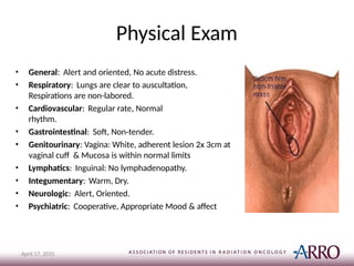
Physical Exam
• General:Alert and oriented, No acute distress.
• Respiratory: Lungs are clear to auscultation,
Respirations are non-labored.
• Cardiovascular: Regular rate, Normal
rhythm.
• Gastrointestinal: Soft, Non-tender.
• Genitourinary: Vagina: White, adherent lesion 2x 3cm at
vaginal cuff & Mucosa is within normal limits
• Lymphatics: Inguinal: No lymphadenopathy.
• Integumentary: Warm, Dry.
• Neurologic: Alert, Oriented.
• Psychiatric: Cooperative, Appropriate Mood & affect
April 17, 2015
4.
Case Presentation
April 17,2015
Pathology
• Colposcopy with biopsies
• Vaginal cuff, condylomatous lesion 10:00: High-grade squamous intraepithelial
lesion (VAIN II)
• Vaginal cuff, epithelial scrapings 11:00: Pronounced HPV cytopathic effect,
low-
grade squamous intraepithelial lesion (VAIN I)
• Left vulva, necrotic lesion (specimen #3); punch biopsy: Squamous cell carcinoma,
moderately to well-differentiated
Laboratory Studies
• HIV viral load undetectable
• CD4 count 1200
• CBC and CMP within normal limits
– Hgb 13.3
5.
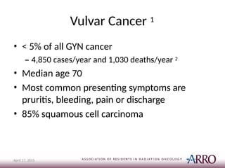
Vulvar Cancer 1
April17, 2015
• < 5% of all GYN cancer
– 4,850 cases/year and 1,030 deaths/year 2
• Median age 70
• Most common presenting symptoms are
pruritis, bleeding, pain or discharge
• 85% squamous cell carcinoma
6.
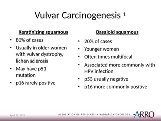
Vulvar Carcinogenesis 1
April17, 2015
Keratinizing squamous
• 80% of cases
• Usually in older women
with vulvar dystrophy,
lichen sclerosis
• May have p53
mutation
• p16 rarely positive
Basaloid squamous
• 20% of cases
• Younger women
• Often times multifocal
• Associated more commonly with
HPV infection
• p53 usually negative
• p16 more commonly positive
7.
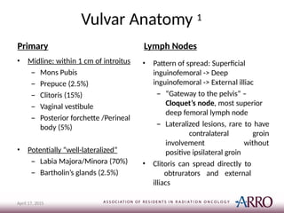
Vulvar Anatomy 1
April17, 2015
Primary
• Midline: within 1 cm of introitus
– Mons Pubis
– Prepuce (2.5%)
– Clitoris (15%)
– Vaginal vestibule
– Posterior forchette /Perineal
body (5%)
• Potentially “well-lateralized”
– Labia Majora/Minora (70%)
– Bartholin’s glands (2.5%)
Lymph Nodes
• Pattern of spread: Superficial
inguinofemoral -> Deep
inguinofemoral -> External illiac
– “Gateway to the pelvis” –
Cloquet’s node, most superior
deep femoral lymph node
– Lateralized lesions, rare to have
contralateral groin
involvement without
positive ipsilateral groin
• Clitoris can spread directly to
obtrurators and external
illiacs
8.
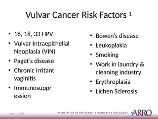
Vulvar Cancer RiskFactors 1
April 17, 2015
• 16, 18, 33 HPV
• Vulvar Intraepithelial
Neoplasia (VIN)
• Paget’s disease
• Chronic irritant
vaginitis
• Immunosuppr
ession
• Bowen’s disease
• Leukoplakia
• Smoking
• Work in laundry &
cleaning industry
• Erythroplasia
• Lichen Sclerosis
9.
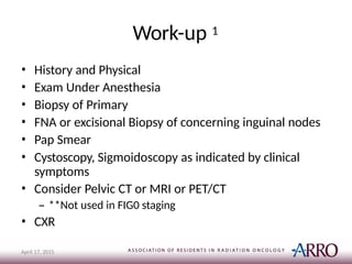
Work-up 1
April 17,2015
• History and Physical
• Exam Under Anesthesia
• Biopsy of Primary
• FNA or excisional Biopsy of concerning inguinal nodes
• Pap Smear
• Cystoscopy, Sigmoidoscopy as indicated by clinical
symptoms
• Consider Pelvic CT or MRI or PET/CT
– **Not used in FIG0 staging
• CXR
10.
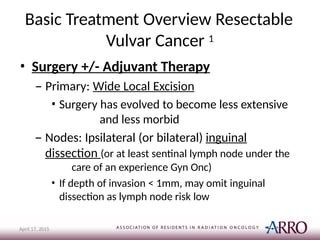
Basic Treatment OverviewResectable
Vulvar Cancer 1
April 17, 2015
• Surgery +/- Adjuvant Therapy
– Primary: Wide Local Excision
• Surgery has evolved to become less extensive
and less morbid
– Nodes: Ipsilateral (or bilateral) inguinal
dissection (or at least sentinal lymph node under the
care of an experience Gyn Onc)
• If depth of invasion < 1mm, may omit inguinal
dissection as lymph node risk low
11.
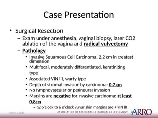
Case Presentation
April 17,2015
• Surgical Resection
– Exam under anesthesia, vaginal biopsy, laser CO2
ablation of the vagina and radical vulvectomy
– Pathology
• Invasive Squamous Cell Carcinoma, 2.2 cm in greatest
dimension
• Multifocal, moderately differentiated, keratinizing
type
• Associated VIN III, warty type
• Depth of stromal invasion by carcinoma: 0.7 cm
• No lymphovascular or perineural invasion
• Margins are negative for invasive carcinoma: at least
0.8cm
– 12 o'clock to 6 o'clock vulvar skin margins are + VIN III
12.
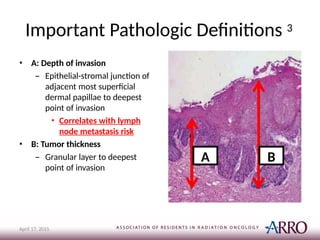
Important Pathologic Definitions3
• A: Depth of invasion
– Epithelial-stromal junction of
adjacent most superficial
dermal papillae to deepest
point of invasion
• Correlates with lymph
node metastasis risk
• B: Tumor thickness
– Granular layer to deepest
point of invasion
A
April 17, 2015
B
13.
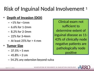
Risk of InguinalNodal Involvement 1
April 17, 2015
• Depth of Invasion (DOI)
– <1% for <1mm
– 6.6% for 1-2mm
– 8.2% for 2-3mm
– 22% for 3-4mm
– At least 25% for > 4 mm
• Tumor Size
– 37.5% > 5 mm
– 45.8% > 2 cm
– 54.2% any extension beyond vulva
Clinical exam not
sufficient to
determine extent of
inguinal disease as 11-
43% of clinically node
negative patients are
pathologically node
positive
14.
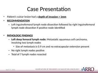
Case Presentation
April 17,2015
• Patient’s vulvar lesion had a depth of invasion > 1mm
• RECOMMENDATION
– Left inguinofemoral lymph node dissection followed by right inguinofemoral
lymph node dissection if positive node identified
• PATHOLOGIC FINDINGS
– Left deep femoral lymph node: Metastatic squamous cell carcinoma,
involving two lymph nodes
• Size of metastasis is 0.9 cm and no extracapsular extension present
– No right lymph nodes positive
– Total of 7 lymph nodes resected
15.
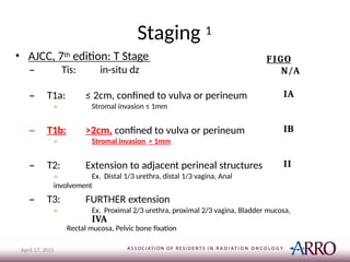
Staging 1
• AJCC,7th edition: T Stage
– Tis: in-situ dz
April 17, 2015
– T1a: ≤ 2cm, confined to vulva or perineum
» Stromal invasion ≤ 1mm
– T1b: >2cm, confined to vulva or perineum
» Stromal invasion > 1mm
– T2: Extension to adjacent perineal structures
» Ex. Distal 1/3 urethra, distal 1/3 vagina, Anal
involvement
FIGO
N/A
IA
IB
II
– T3: FURTHER extension
» Ex. Proximal 2/3 urethra, proximal 2/3 vagina, Bladder mucosa,
IVA
Rectal mucosa, Pelvic bone fixation
16.
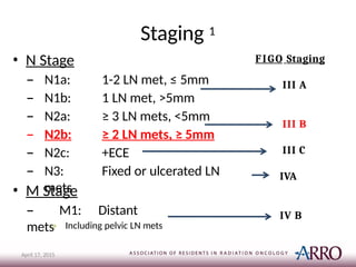
Staging 1
• NStage
– N1a: 1-2 LN met, ≤ 5mm
– N1b: 1 LN met, >5mm
– N2a: ≥ 3 LN mets, <5mm
– N2b: ≥ 2 LN mets, ≥ 5mm
– N2c: +ECE
– N3: Fixed or ulcerated LN
mets
• M Stage
– M1: Distant
mets» Including pelvic LN mets
FIGO Staging
III A
III B
III C
IVA
IV B
April 17, 2015
17.
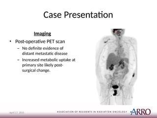
Case Presentation
Imaging
• Post-operativePET scan
– No definite evidence of
distant metastatic disease
– Increased metabolic uptake at
primary site likely post-
surgical change.
April 17, 2015
18.
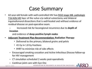
Case Summary
April 17,2015
• 62-year-old female with well-controlled HIV has FIGO stage IIIB, pathologic
T1b N2b M0 Sqcc of the vulva s/p radical vulvectomy and bilateral
inguinofemoral dissections that is well-healed and without evidence of
residual disease on post-operative exam.
– Increased risk for locoregional recurrence due to depth of
invasion
and evidence of deep positive lymph nodes
• Adjuvant Treatment Plan Recommendation: Radiation Therapy
• Delivered to the primary, bilateral groins and pelvis
• 45 Gy in 1.8 Gy fractions
• IMRT to minimize risk of side effects
– Encouraged smoking cessation and Active Infectious Disease follow-up
for HIV management
– CT simulation scheduled 2 weeks post-operatively
– Continue joint care with Gyn Onc
19.
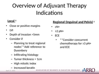
Overview of AdjuvantTherapy
Indications
April 17, 2015
Local 3
• Close or positive margins
• LVI
• Depth of invasion >5mm
• Consider if
– Planning to treat regional
nodes**Add reference to
Dusenbery
– Infiltrating histology
– Tumor thickness > 1cm
– High mitotic index
– Increased keratin
Regional (Inguinal and Pelvis) 5
• cN+
• ≥2 pN+
• ECE
• **Consider concurrent
chemotherapy for ≥3 pN+
and ECE
20.
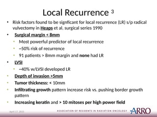
Local Recurrence 3
April17, 2015
• Risk factors found to be signficant for local recurrence (LR) s/p radical
vulvectomy in Heaps et al. surgical series 1990
• Surgical margin < 8mm
• Most powerful predictor of local recurrence
• ~50% risk of recurrence
• 91 patients > 8mm margin and none had LR
• LVSI
• ~40% w/LVSI developed LR
• Depth of invasion >5mm
• Tumor thickness: < 10mm
• Infiltrating growth pattern increase risk vs. pushing border growth
pattern
• Increasing keratin and > 10 mitoses per high power field
21.
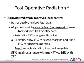
Post-Operative Radiation 4
April17, 2015
• Adjuvant radiation improves local control
– Retrospective review, Faul et al.
– 62 patients with close (<8mm) or +margins were
treated with XRT or observed
• Referral for XRT at surgeon discretion
– XRT: AP/PA, 4867 cGy for close margins and 5854
cGy for positive margins
• Target: vulva, bilateral inguinals, and low pelvis
– 58% local recurrence without XRT vs. 16% with
XRT
22.
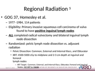
Regional Radiation 5
April17, 2015
• GOG 37, Homesley et al.
– 1977 -1984, 114 patients
– Eligibility: Primary invasive squamous cell carcinoma of vulva
found to have positive inguinal lymph nodes
– ALL completed radical vulvectomy and bilateral inguinal lymph
node dissection
– Randomized: pelvic lymph node dissection vs. adjuvant
radiation
• Pelvic Dissection: Common, External and Internal Iliacs, and Obturator
• XRT: 4500-5000 cGy to midplane and 2-3 cm depth at inguinal and
femoral
lymph nodes
– XRT Target: Common, External, and Internal Iliacs, Obturator, Femoral and Inguinal
Nodes, NO XRT to VULVA
23.
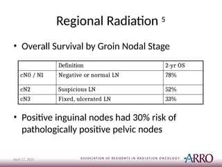
Regional Radiation 5
April17, 2015
• Overall Survival by Groin Nodal Stage
• Positive inguinal nodes had 30% risk of
pathologically positive pelvic nodes
Definition 2-yr OS
cN0 / N1 Negative or normal LN 78%
cN2 Suspicious LN 52%
cN3 Fixed, ulcerated LN 33%
24.
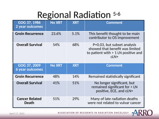
Regional Radiation 5-6
April17, 2015
GOG 37, 1986
2 year outcomes
No XRT XRT Comment
Groin Recurrence 23.6% 5.1% This benefit thought to be main
contributor to OS improvement
Overall Survival 54% 68% P=0.03, but subset analysis
showed that benefit was limited
to patient with > 1 LN positive and
cLN+
GOG 37, 2009
6 year outcomes
No XRT XRT Comment
Groin Recurrence 48% 14% Remained statistically significant
Overall Survival 41% 51% No longer significant, but
remained significant for > LN
positive, ECE, and cLN+
Cancer Related
Death
51% 29% Many of late radiation deaths
were not related to vulvar cancer
25.
Local Recurrence afterRegional XRT
April 17, 2015
• GOG 37 5-6
– Coverage of vulva not required
– 23% of recurrences were local in vulva
• Dusenbery et al. 7
– Reported vulvar recurrence rate of 48% in patients
treated with midline block while receiving
adjuvant nodal radiation
• Recommend vulva local radiation, if treating
regional lymph nodes
26.
Case Presentation
CT SIMULATION
•Supine in frog leg position
• Arms up on a wing board and in an
immobilization device
• Wire on all scars
• Anal bb placed
• No bolus placed as IMRT utilized, but in
vivo dosimetry with
thermoluminescent dosimeters
on day 1 (optional)
• Consider IV contrast
• Full bladder for CT simulation and daily
treatment
• Consider ITV full and empty bladder
– in order to compensate for variable
bladder fill
April 17, 2015
27.
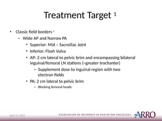
Treatment Target 1
April17, 2015
• Classic field borders 1
– Wide AP and Narrow PA
• Superior: Mid – Sacroiliac Joint
• Inferior: Flash Vulva
• AP: 2 cm lateral to pelvic brim and encompassing bilateral
inguinal/femoral LN stations (~greater trochanter)
– Supplement dose to inguinal region with two
electron fields
• PA: 2 cm lateral to pelvic brim
– Blocking femoral heads
28.
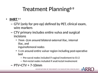
Treatment Planning8-9
• IMRT8-9
– GTV (only for pre-op) defined by PET, clinical exam,
wire markers
– CTV primary includes entire vulva and surgical
incisions
• 7mm -2cm around bilateral external iliac, internal
iliac, and
inguinofemoral nodes
• 1 cm around entire vulvar region including post-operative
bed
– Pre-sacral nodes included if vaginal involvment to S1-2
– Peri-rectal nodes included if anal/rectal involvement
– PTV=CTV + 7-10mm
29.
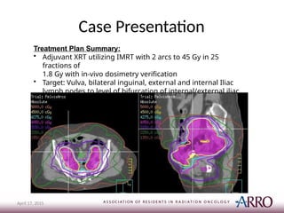
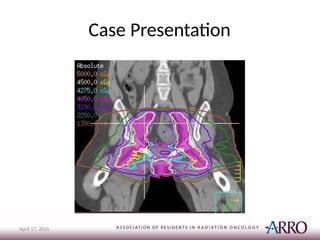
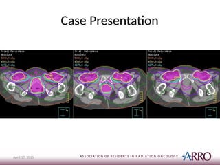
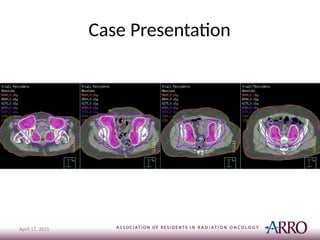
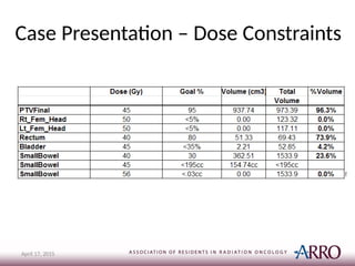
Case Presentation
Treatment PlanSummary:
• Adjuvant XRT utilizing IMRT with 2 arcs to 45 Gy in 25
fractions of
1.8 Gy with in-vivo dosimetry verification
• Target: Vulva, bilateral inguinal, external and internal Iliac
lymph nodes to level of bifurcation of internal/external iliac
April 17, 2015
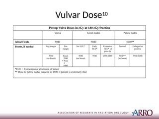
Postop Vulva Dosesin cGy at 180 cGy/fraction
Vulva Groin nodes Pelvic nodes
Initial Fields 5040 5040 5040**
Boosts, if needed Neg margin Pos
margin
No ECE* Early
ECE*
Extensive
ECE* or
gross dz
Normal Enlarged or
positive
5040
(no boost)
Focal:
5940
> Foca
l:
6480
5040
(no boost)
5940 6300-6480 5040**
(no boost)
5940-6480
*ECE = Extracapsular extension of tumor
** Dose to pelvic nodes reduced to 4500 if patient is extremely frail
Vulvar Dose10
37.
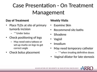
Case Presentation -On Treatment
Management
Day of Treatment Weekly Visits
• Place TLDs at site of primary
tumor& incision
– **Under bolus
• Check positioning of legs
– May need extra tattoos or
set-up marks on legs to get
correct angle
• Check bolus placement
• Examine Skin
• Recommend sitz baths
• Silvadene
• Vagisil
• Imodium
• May need temporary catheter
– **when treating definitive doses
• Vaginal dilator for late stenosis
38.
Case Counseling 1
April17, 2015
• Potential side effects of treatment
– Radiation dermatitis
• Increased with increasing BMI 6
– Fatigue
– Cystitis
– Proctopathy and Diarrhea
– Vaginal Stenosis
– Lymphedema
• ~16% per GOG 37 5-6
39.
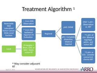
Treatment Algorithm 1
Resectable
Well
Lateralized
VulvarCancer:
Wide Local
Excision
< 1mm DOI:
Observation
>1mm DOI:
Ispilateral
Inguinal LN
Evaluation
Regional
pN0: DONE
pN+: Complete
dissection,
bilateral
inguinal
ONLY 1 pN+:
Observation
vs. XRT
>1 pN+ of
cN+: Bilateral
inguingal,
pelvic, and
vulvar XRT
>2 pN+ or
ECE: consider
concurrent
chemo-XRT
Local
If margins <
8mm, LVSI, or
DOI > 5mm:
vulvar RT
April 17, 2015
* May consider adjuvant
RT
40.
Treatment Algorithm 1
Resectable
Midline
Vulvar
Cancer:
WideLocal
Excision
< 1mm DOI:
Observation
>1mm DOI:
Bilateral
Inguinal LN
Dissection
Regional
pN0:
Observation
ONLY 1 pN+:
Observation vs.
XRT
> 1 pN+:
Bilateral
inguingal, pelvic,
and vulvar XRT
>2 pN+ or ECE:
consider
concurrent
chemo-XRT
Local
If margins <
8mm, LVSI,
or DOI >
5mm: vulvar
RT
April 17, 2015
41.
References
1.
2.
3.
4.
5.
6.
7.
8.
9.
10.
Halperin EC, PerezCA, Brady LW: Perez and Brady's principles and practice of radiation oncology (ed 5th). Philadelphia,
Wolters Kluwer Health/Lippincott Williams & Wilkins, 2008
American Cancer Society's (ACS) publication, Cancer Facts & Figures 2014: http://www.cancer.net/cancer-types/vulvar-
cancer/statistics
Heaps JM, Fu YS, Montz FJ, et al: Surgical-pathologic variables predictive of local recurrence in squamous cell carcinoma
of the vulva. Gynecologic oncology 38:309-14, 1990
Faul CM, Mirmow D, Huang Q, et al: Adjuvant radiation for vulvar carcinoma: improved local control. International
journal
of radiation oncology, biology, physics 38:381-9, 1997
Homesley HD, Bundy BN, Sedlis A, et al: Radiation therapy versus pelvic node resection for carcinoma of the vulva with
positive groin nodes. Obstetrics and gynecology 68:733-40, 1986
Kunos C, Simpkins F, Gibbons H, et al: Radiation therapy compared with pelvic node resection for node-positive vulvar
cancer: a randomized controlled trial. Obstetrics and gynecology 114:537-46, 2009
Dusenbery KE, Carlson JW, LaPorte RM, et al: Radical vulvectomy with postoperative irradiation for vulvar cancer:
therapeutic implications of a central block. International journal of radiation oncology, biology, physics 29:989-98, 1994
Beriwal S, Heron DE, Kim H, et al: Intensity-modulated radiotherapy for the treatment of vulvar carcinoma: a comparative
dosimetric study with early clinical outcome. International journal of radiation oncology, biology, physics 64:1395-400,
2006
Beriwal S, Coon D, Heron DE, et al: Preoperative intensity-modulated radiotherapy and chemotherapy for locally
advanced vulvar carcinoma. Gynecologic oncology 109:291-5, 2008
Hoppe, R, Phillips, Mach III, R: Leibel and Phillips Textbook of Radiation Oncology (ed 3rd). Philadelphia, Elsevier Saunder,
2004
Please provide feedback regarding this case or other ARROcases to arrocase@gmail.com
April 17, 2015