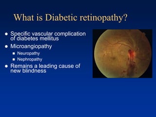
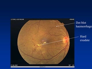
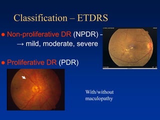
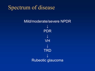
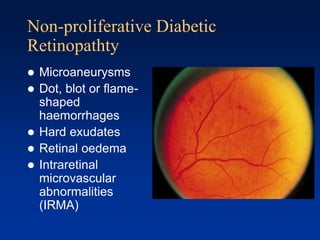
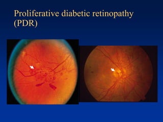
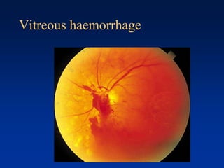
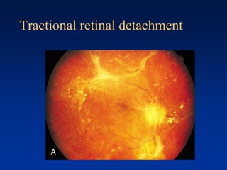
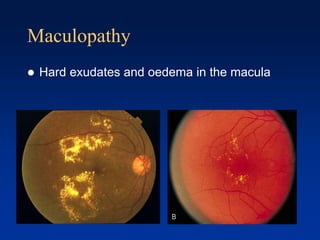
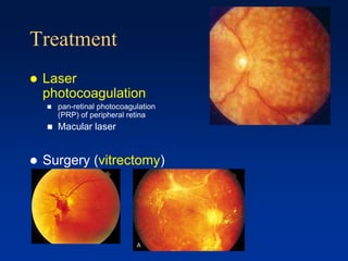
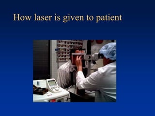
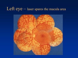
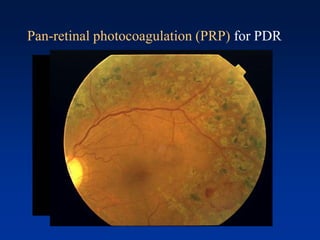
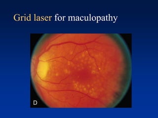
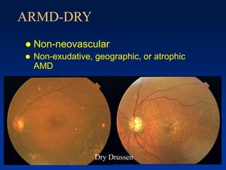
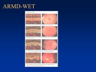
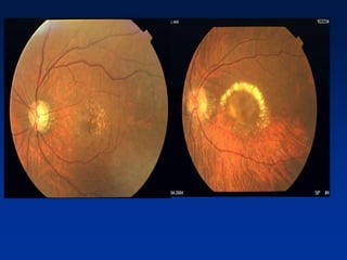
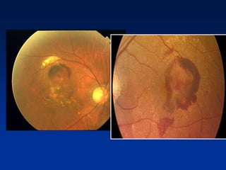
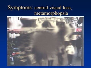
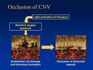
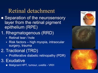
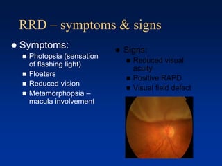
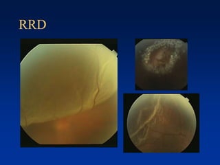
Common retinal diseases
Dr. Mushawiahti Mustapha
Department. Of Ophthalmology
Universiti Kebangsaan Malaysia
Retinal diseases - outline
 Diabetes mellitus
 Hypertension
 Other retinal vascular problem: CRVO/CRAO
 Age related macular degeneration (AMD)
 Retinal Detachment
10 layers of
retina
Normal fundus
What is Diabetic retinopathy?
 Specific vascular complication
of diabetes mellitus
 Microangiopathy
 Neuropathy
 Nephropathy
 Remains a leading cause of
new blindness
Diabetic Retinopathy
 Duration of diabetes,
 after 20 years:have some degree of retinopathy
 nearly all patients with type 1 diabetes mellitus
 more than 60% of patients with type 2 diabetes mellitus
 Other risk factors: pregnancy, hypertension,
renal disease, obesity, hyperlipidaemia,
smoking, anaemia
Pathogenesis
 Weak capillary wall →
saccular outpouching of wall
(microaneurysm)-may leak
or thrombosed
 Breakdown of inner BRB
Leakage: capillary changesOcclusion
1. Capillary changes
2. Haematological
changes
↓
→Less O2
→ retinal ischaemia/ hypoxia
↓
Release of VEGF
(Vascular Endothelial Growth Factor)
Occlusion….
Symptoms
 Asymptomatic/painless
 Gradual blurring of vision
 diabetic maculopathy
 Sudden blurring of vision
 vitreous haemorrhage
 Painful gradual blurring of vision
 Rubeotic glaucoma
Retinal hypoxia Manifested as:
- AV shunt
* intraretinal microvascular
abnormalities (IRMA) -
opening of pre-existing
vascular channel
- Neovascularization
* Attempt to revascularize
hypoxic retina
- Rubeosis iridis, NVD, NVE
- CWS
- microinfarction of NFL
causing axoplasmic
swelling
Cotton wool spot
Cotton wool spot
Dot & blot
haemorrhage
Pre-retinal
haemorrhage
Hard exudates
Pathogenesis
 Weak capillary wall →
saccular outpouching of wall
(microaneurysm)-may leak
or thrombosed
 Breakdown of inner BRB
Leakage: capillary changesOcclusion
1. Capillary changes
2. Haematological
changes
Microvascular leakage
Consequence…
 Intraretinal hemorrhage
(dot and blot)
 Oedema-diffuse or focal
(hard exudate-
lipoprotein and lipid
macrophages
surrounding leaking
microvascular lesion in
circinate pattern)
Dot blot
haemorrhage
Hard
exudate
Classification – ETDRS
 Non-proliferative DR (NPDR) –
→ mild, moderate, severe
 Proliferative DR (PDR)
With/without
maculopathy
Spectrum of disease
Mild/moderate/severe NPDR
↓
PDR
↓
VH
↓
TRD
↓
Rubeotic glaucoma
Non-proliferative Diabetic
Retinopathty
 Microaneurysms
 Dot, blot or flame-
shaped
haemorrhages
 Hard exudates
 Retinal oedema
 Intraretinal
microvascular
abnormalities
(IRMA)
Proliferative diabetic retinopathy
(PDR)
Vitreous haemorrhage
Tractional retinal detachment
Maculopathy
 Hard exudates and oedema in the macula
Treatment
 Laser
photocoagulation
 pan-retinal photocoagulation
(PRP) of peripheral retina
 Macular laser
 Surgery (vitrectomy)
How laser is given to patient
Left eye – laser spares the macula area
Pan-retinal photocoagulation (PRP) for PDR
Grid laser for maculopathy
Why DR has to be detected
 Diabetes mellitus is the single most
important cause of
Causes for the blindness in DR
 Non-resolving vitreous hemorrhage
 Tractional retinal detachment
 Diabetic maculopathy
 Cataract
 Rubeotic Glaucoma
How to detect DR?
Using direct ophthalmoscope
When to screen for DR
 All NIDDM at the time of diagnosis
(once a year)
 IDDM after at least 5 years of diagnosis
 Diabetic women
 planning for pregnancy
 need to have complete eye examination
before conception
 Once becomes pregnant
 should be examined for retinopathy early in the
first trimester
Prevention
 Screening
 Control of blood sugar
 Control of hypertension
 Control of hyperlipidaemia
Retinal diseases - outline
 Diabetes mellitus
 Hypertension
 Other retinal vascular problem: CRVO/CRAO
 Age related macular degeneration (AMD)
 Retinal Detachment
HYPERTENSION
AND THE EYE
HYPERTENSION AND EYE
 The eye is the only place in the body
where the vessels can be directly
observed
 Patients with hypertensive retinopathy
are usually asymptomatic
Hypertensive changes in the eye
 Acute hypertension (suddden rise of BP)
 Grade III and IV hypertensive retinopathy
 Choroidopathy
 Optic neuropathy
 Chronic hypertension
 Arterial changes
Pathophysiology
 Arteriolar narrowing due to
vasoconstriction and arteriolosclerosis
(increase in elastic tissue and
musculature)
 Focal closure of retinal vasculature lead
to microinfarcts (cotton wool spots) and
haemorrhages
 Leakage lead to oedema and exudates
 Disc oedema develops in advanced
stage)
Classification
(modified Keith-Wagener-Barker / Scheie)
Grade Haemorr-
hage
Exudate Disc
swelling
AV ratio Light
reflex
AV
crossing
changes
Normal none none none 3:4 Fine
yellow
None
Grade 1 none none none 1:2 Broad
yellow
Mild vein
depression
Grade 2 none none none 1:3 Copper
wiring
A-V
nipping
Grade 3
+ + none 1:4 Silver
wiring
Right angle
Deviation /
Distal
dilatation
Grade 4
+ + + fine
cords
as stage 3 as stage 3
Grade 2 hypertensive retinopathy
Asymptomatic: poorly controlled
HPT
Poor vision:
Grade IV Hypertensive retinopathy
Keith, Wagener, and Barker 1939
 The changes seen in groups I and II are
typically chronic
 groups III and IV are seen with more
acute rises in blood pressure.
 Retinal haemorrhages and hard exudates,
cotton wool spots, optic disc swelling
Is there a need to refer all
hypertensive patients to
ophthalmologist? No, if there is no visual impairment
 We do not monitor the fundus of HPT
patients
 If BP is high: refer physician
Management HT retinopathy
 Referral to physicians for control of the blood
pressure: urgency depends on the stage
 Stage 1 n 2: BP monitoring if on treatment
 Stage 3 n 4: urgent referral
Retinal diseases - outline
 Diabetes mellitus
 Hypertension
 Other retinal vascular problem: CRVO/CRAO
 Age related macular degeneration (AMD)
 Retinal Detachment
Other retinal vascular disorders
 Retinal vein occlusion
 CRVO
 BRVO
 Retinal artery occlusion
 CRAO
 BRAO
CRVO - central retinal vein occlusion
BRVO – branch retinal vein occlusion
CRAO – central retinal artery occlusion
Central retinal artery occlusion
(CRAO)
 Anatomy:
 Internal carotid artery
 Ophthalmic artery
 Central retinal artery
 Mainly supply of blood to
the inner half of
neurosensory retina
CRAO – risk factors
 Risk factors:
 Age older than 70 years old
 Atherosclerosis – DM / HPT /
Hypercholesterolaemia / smoking
 Emboli – cholesterol, calcific, fibrin-platelet
 Others – arteritis, thrombophilic disorders,
migraine
 Young: Connective tissue disease, cardiac
valvular disease
Retinal diseases - outline
 Diabetes mellitus
 Hypertension
 Other retinal vascular problem: CRVO/CRAO
 Age related macular degeneration (AMD)
 Retinal Detachment
AGE RELATED MACULAR
DEGENERATION
(AMD)
DRY (90%) versus WET (10%)
ARMD-DRY
 Non-neovascular
 Non-exudative, geographic, or atrophic
AMD
 Neovascular AMD (Wet)
 Exudative, disciform, or serous AMD
Dry Drussen
ARMD-WET
Symptoms: central visual loss,
metamorphopsia
Diagnostic assessment
 Examination
 Visual acuity
 Fundus biomicroscopy via dilated pupil
 Amsler chart
 Color photography
 Fluorescein angiography
Detection: The Amsler grid
 Amsler grids can
facilitate early detection
 Signs suggestive of CNV
include:
 Distortion
 Blurring
 Darkening or discoloration
of the grid lines
 Inability to fix on the
central dot
Treatment option for CNV
 Argon laser photocoagulation
 Intravitreal anti-VEGF injection
 Photodynamic therapy
Visudyne therapy: a two step process
Step 1
Step 2
Occlusion of CNV
Reactive oxygen
products
Occlusion of abnormal
vessels
Light-activation of Visudyne
Endothelial cell damage
and thrombus formation
Retinal diseases - outline
 Diabetes mellitus
 Hypertension
 Other retinal vascular problem: CRVO/CRAO
 Age related macular degeneration (AMD)
 Retinal Detachment
Retinal detachment
 Separation of the neurosensory
layer from the retinal pigment
epithelium (RPE)
1. Rhegmatogenous (RRD)
 Retinal tear / hole
 Risk factors – high myopia, intraocular
surgery, trauma
2. Tractional (TRD)
 Proliferative diabetic retinopathy (PDR)
3. Exudative
 Malignant HPT, tumour, uveitis - VKH
RRD – symptoms & signs
 Signs:
 Reduced visual
acuity
 Positive RAPD
 Visual field defect
 Symptoms:
 Photopsia (sensation
of flashing light)
 Floaters
 Reduced vision
 Metamorphopsia –
macula involvement
RRD
RRD - management
 Urgent / early intervention to flatten the
retina……
 Pars plana vitrectomy (PPV)
 Scleral buckle surgery
Acknowledgement
 Dr. Aida Zairani
 Senior lecturer / Ophthalmologist, PPUKM
Thank you

Retinal disease lecture. Optometry. Optometri.

  • 1.
    Common retinal diseases Dr.Mushawiahti Mustapha Department. Of Ophthalmology Universiti Kebangsaan Malaysia
  • 2.
    Retinal diseases -outline  Diabetes mellitus  Hypertension  Other retinal vascular problem: CRVO/CRAO  Age related macular degeneration (AMD)  Retinal Detachment
  • 4.
  • 5.
  • 6.
    What is Diabeticretinopathy?  Specific vascular complication of diabetes mellitus  Microangiopathy  Neuropathy  Nephropathy  Remains a leading cause of new blindness
  • 7.
    Diabetic Retinopathy  Durationof diabetes,  after 20 years:have some degree of retinopathy  nearly all patients with type 1 diabetes mellitus  more than 60% of patients with type 2 diabetes mellitus  Other risk factors: pregnancy, hypertension, renal disease, obesity, hyperlipidaemia, smoking, anaemia
  • 8.
    Pathogenesis  Weak capillarywall → saccular outpouching of wall (microaneurysm)-may leak or thrombosed  Breakdown of inner BRB Leakage: capillary changesOcclusion 1. Capillary changes 2. Haematological changes
  • 9.
    ↓ →Less O2 → retinalischaemia/ hypoxia ↓ Release of VEGF (Vascular Endothelial Growth Factor) Occlusion….
  • 10.
    Symptoms  Asymptomatic/painless  Gradualblurring of vision  diabetic maculopathy  Sudden blurring of vision  vitreous haemorrhage  Painful gradual blurring of vision  Rubeotic glaucoma
  • 11.
    Retinal hypoxia Manifestedas: - AV shunt * intraretinal microvascular abnormalities (IRMA) - opening of pre-existing vascular channel - Neovascularization * Attempt to revascularize hypoxic retina - Rubeosis iridis, NVD, NVE - CWS - microinfarction of NFL causing axoplasmic swelling Cotton wool spot
  • 12.
    Cotton wool spot Dot& blot haemorrhage Pre-retinal haemorrhage Hard exudates
  • 13.
    Pathogenesis  Weak capillarywall → saccular outpouching of wall (microaneurysm)-may leak or thrombosed  Breakdown of inner BRB Leakage: capillary changesOcclusion 1. Capillary changes 2. Haematological changes
  • 14.
    Microvascular leakage Consequence…  Intraretinalhemorrhage (dot and blot)  Oedema-diffuse or focal (hard exudate- lipoprotein and lipid macrophages surrounding leaking microvascular lesion in circinate pattern)
  • 15.
  • 16.
    Classification – ETDRS Non-proliferative DR (NPDR) – → mild, moderate, severe  Proliferative DR (PDR) With/without maculopathy
  • 17.
    Spectrum of disease Mild/moderate/severeNPDR ↓ PDR ↓ VH ↓ TRD ↓ Rubeotic glaucoma
  • 18.
    Non-proliferative Diabetic Retinopathty  Microaneurysms Dot, blot or flame- shaped haemorrhages  Hard exudates  Retinal oedema  Intraretinal microvascular abnormalities (IRMA)
  • 19.
  • 20.
  • 21.
  • 22.
    Maculopathy  Hard exudatesand oedema in the macula
  • 23.
    Treatment  Laser photocoagulation  pan-retinalphotocoagulation (PRP) of peripheral retina  Macular laser  Surgery (vitrectomy)
  • 24.
    How laser isgiven to patient
  • 25.
    Left eye –laser spares the macula area
  • 26.
  • 27.
    Grid laser formaculopathy
  • 28.
    Why DR hasto be detected  Diabetes mellitus is the single most important cause of
  • 29.
    Causes for theblindness in DR  Non-resolving vitreous hemorrhage  Tractional retinal detachment  Diabetic maculopathy  Cataract  Rubeotic Glaucoma
  • 30.
    How to detectDR? Using direct ophthalmoscope
  • 31.
    When to screenfor DR  All NIDDM at the time of diagnosis (once a year)  IDDM after at least 5 years of diagnosis  Diabetic women  planning for pregnancy  need to have complete eye examination before conception  Once becomes pregnant  should be examined for retinopathy early in the first trimester
  • 32.
    Prevention  Screening  Controlof blood sugar  Control of hypertension  Control of hyperlipidaemia
  • 33.
    Retinal diseases -outline  Diabetes mellitus  Hypertension  Other retinal vascular problem: CRVO/CRAO  Age related macular degeneration (AMD)  Retinal Detachment
  • 34.
  • 35.
    HYPERTENSION AND EYE The eye is the only place in the body where the vessels can be directly observed  Patients with hypertensive retinopathy are usually asymptomatic
  • 36.
    Hypertensive changes inthe eye  Acute hypertension (suddden rise of BP)  Grade III and IV hypertensive retinopathy  Choroidopathy  Optic neuropathy  Chronic hypertension  Arterial changes
  • 37.
    Pathophysiology  Arteriolar narrowingdue to vasoconstriction and arteriolosclerosis (increase in elastic tissue and musculature)  Focal closure of retinal vasculature lead to microinfarcts (cotton wool spots) and haemorrhages  Leakage lead to oedema and exudates  Disc oedema develops in advanced stage)
  • 38.
    Classification (modified Keith-Wagener-Barker /Scheie) Grade Haemorr- hage Exudate Disc swelling AV ratio Light reflex AV crossing changes Normal none none none 3:4 Fine yellow None Grade 1 none none none 1:2 Broad yellow Mild vein depression Grade 2 none none none 1:3 Copper wiring A-V nipping Grade 3 + + none 1:4 Silver wiring Right angle Deviation / Distal dilatation Grade 4 + + + fine cords as stage 3 as stage 3
  • 39.
  • 40.
  • 41.
    Poor vision: Grade IVHypertensive retinopathy
  • 42.
    Keith, Wagener, andBarker 1939  The changes seen in groups I and II are typically chronic  groups III and IV are seen with more acute rises in blood pressure.  Retinal haemorrhages and hard exudates, cotton wool spots, optic disc swelling
  • 43.
    Is there aneed to refer all hypertensive patients to ophthalmologist? No, if there is no visual impairment  We do not monitor the fundus of HPT patients  If BP is high: refer physician
  • 44.
    Management HT retinopathy Referral to physicians for control of the blood pressure: urgency depends on the stage  Stage 1 n 2: BP monitoring if on treatment  Stage 3 n 4: urgent referral
  • 45.
    Retinal diseases -outline  Diabetes mellitus  Hypertension  Other retinal vascular problem: CRVO/CRAO  Age related macular degeneration (AMD)  Retinal Detachment
  • 46.
    Other retinal vasculardisorders  Retinal vein occlusion  CRVO  BRVO  Retinal artery occlusion  CRAO  BRAO
  • 47.
    CRVO - centralretinal vein occlusion
  • 48.
    BRVO – branchretinal vein occlusion
  • 49.
    CRAO – centralretinal artery occlusion
  • 50.
    Central retinal arteryocclusion (CRAO)  Anatomy:  Internal carotid artery  Ophthalmic artery  Central retinal artery  Mainly supply of blood to the inner half of neurosensory retina
  • 51.
    CRAO – riskfactors  Risk factors:  Age older than 70 years old  Atherosclerosis – DM / HPT / Hypercholesterolaemia / smoking  Emboli – cholesterol, calcific, fibrin-platelet  Others – arteritis, thrombophilic disorders, migraine  Young: Connective tissue disease, cardiac valvular disease
  • 52.
    Retinal diseases -outline  Diabetes mellitus  Hypertension  Other retinal vascular problem: CRVO/CRAO  Age related macular degeneration (AMD)  Retinal Detachment
  • 53.
  • 54.
    ARMD-DRY  Non-neovascular  Non-exudative,geographic, or atrophic AMD  Neovascular AMD (Wet)  Exudative, disciform, or serous AMD Dry Drussen
  • 55.
  • 58.
    Symptoms: central visualloss, metamorphopsia
  • 59.
    Diagnostic assessment  Examination Visual acuity  Fundus biomicroscopy via dilated pupil  Amsler chart  Color photography  Fluorescein angiography
  • 61.
    Detection: The Amslergrid  Amsler grids can facilitate early detection  Signs suggestive of CNV include:  Distortion  Blurring  Darkening or discoloration of the grid lines  Inability to fix on the central dot
  • 62.
    Treatment option forCNV  Argon laser photocoagulation  Intravitreal anti-VEGF injection  Photodynamic therapy
  • 63.
    Visudyne therapy: atwo step process Step 1 Step 2
  • 64.
    Occlusion of CNV Reactiveoxygen products Occlusion of abnormal vessels Light-activation of Visudyne Endothelial cell damage and thrombus formation
  • 65.
    Retinal diseases -outline  Diabetes mellitus  Hypertension  Other retinal vascular problem: CRVO/CRAO  Age related macular degeneration (AMD)  Retinal Detachment
  • 66.
    Retinal detachment  Separationof the neurosensory layer from the retinal pigment epithelium (RPE) 1. Rhegmatogenous (RRD)  Retinal tear / hole  Risk factors – high myopia, intraocular surgery, trauma 2. Tractional (TRD)  Proliferative diabetic retinopathy (PDR) 3. Exudative  Malignant HPT, tumour, uveitis - VKH
  • 67.
    RRD – symptoms& signs  Signs:  Reduced visual acuity  Positive RAPD  Visual field defect  Symptoms:  Photopsia (sensation of flashing light)  Floaters  Reduced vision  Metamorphopsia – macula involvement
  • 68.
  • 69.
    RRD - management Urgent / early intervention to flatten the retina……  Pars plana vitrectomy (PPV)  Scleral buckle surgery
  • 70.
    Acknowledgement  Dr. AidaZairani  Senior lecturer / Ophthalmologist, PPUKM
  • 71.