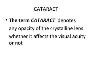
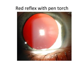
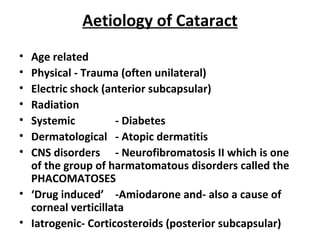
CATARACT AND
REFRACTIVE SURGERY
Ms. A. Bobart Hone
2009
CATARACT
• The term CATARACT denotes
any opacity of the crystalline lens
whether it affects the visual acuity
or not
Anatomy
• Newborn - 3.5mm antero-posteriorly and 5mm equatorially
• Unique to the lens is continued growth throughout life
• Adult - 5mm antero-posteriorly and 9 - 10mm equatorially-i.e.. top to bottom
• With age the lens becomes larger, more compact and less elastic
• The lens is a biconvex optical structure behind iris and in front of vitreous in
posterior chamber of the eye
• It is avascular and not innervated
• Encircled by ciliary processes from which the zonules (suspensory ligament)
radiate to the lens surface
• Zonules hold the lens in place and mediate the accommodative movements of
the ciliary muscle, therefore altering the lens shape.
• Outmost layer is an acellular capsule (basement membrane) that surrounds the
lens
• Anteriorly and just under the anterior capsule is the lens epithelium
• Inner body of lens composed of tightly packed highly organised lens fibres
• Innermost layer called the nucleus
• Outer layers cortex
Red reflex with pen torch
Nuclear cataract
( N.B. Pseudoexfoliation)
Brunescent nuclear sclerosis
Cortical cataract and Posterior
subcapsular cataract
Cortical Cataract
Cortical cataract- multiple white
spokes
Aetiology of Cataract
• Age related
• Physical - Trauma (often unilateral)
• Electric shock (anterior subcapsular)
• Radiation
• Systemic - Diabetes
• Dermatological - Atopic dermatitis
• CNS disorders - Neurofibromatosis II which is one
of the group of harmatomatous disorders called the
PHACOMATOSES
• ‘Drug induced’ -Amiodarone and- also a cause of
corneal verticillata
• Iatrogenic- Corticosteroids (posterior subcapsular)
Corneal verticillata
Secondary
• Associated with Retinitis Pigmentosa
• Uveitis- intraocular inflammation
• Glaucoma, Corneal graft, vitreoretinal and any intraocular
surgery
• Congenital
• Hereditary disorders
• Maternal rubella
• Systemic disease e.g. galactosemia
• Myotonic Dystrophy
• Chromosomal
• Trisomy 13 (Patau’s syndrome)
• Trisomy 18 (Edward’s syndrome)
• Trisomy 21 (Down’s syndrome)
Symptoms of cataract
• Glare
• Gradual loss of vision unless traumatic
• Reduced near vision i.e. difficulty reading - seen in
posterior subcapsular type
• Central lens opacities may decrease pinhole vision
• Second sight - nuclear cataracts cause changes in
refractive index of lens so that they become more
myopic and one may not need reading lasses
• (Normally when one ages one becomes more
presbyopic, i.e. near-sighted requiring more plus lens)
Examination
• Pen torch- red reflex
• Ophthalmoscopy (direct)- red reflex vs. black
opacities in pupil area
• Slit lamp examination distinguishes the
cataract subtype.
Red reflex with pen torch and note the
white cortical lens opacities
Treatment
• Surgical treatment is the only definitive treatment to
remove a cataract
• Indications for and against surgery
• The aim of cataract surgery which is for the most part an
elective procedure is visual rehabilitation
• Reasons ‘AGAINST’ surgery
• Anaesthetic risk e.g. if unsuitable for LA but GA risk high
• Severe amblyopia already investigated and documented
• Extensive age-related macular degeneration or other retinal
pathology
• Total afferent pupillary defect
Other reasons for lens extraction
• Clear lens extraction may be performed- i.e. no significant
cataract present but lens may need to be removed
• Severe myopia
• Patients may elect to have this done at an early age rather than wait
to have cataract develop therefore avoiding need for thick
spectacles or contact lenses
• Dislocated lenses
• Pseudoexfoliation- can sometimes have associated glaucoma and
systemic manifestations with this
• Trauma
• Systemic conditions
» Marfan’s- typically upward dislocation of lens
» Homocysteinuria typically downward dislocation of lens
» Weill-Marchesani
» Sulphite-oxidase deficiency
Dislocated lens in Trauma
Downward dislocation of lens can be
seen in homocysteinuria
Types of Anaesthesia
• Local- most common
• Topical anaesthetic drops
• Retrobulbar, peribulbar, sub-Tenon’s injections
of local anaesthetic
• General (e.g.)
• Children
• Handicapped
• Parkinson’s disease
• Nystagmus
Types of cataract surgery
• Intracapsular
– Rarely done
– Removal of lens and capsule
– Anterior chamber intraocular lens used
• Extracapsular cataract extraction
– Removal of lens but capsule left behind
Phacoemulsification
• The most common type of extracapsular surgery today is :
Phacoemulsification- ultrasonic lens fragmentation with
simultaneous aspiration of lens fragments
– Sophisticated small incision type (approx. 2.7-3.5mm wound
incision)
– Faster visual recover with fewer complications
– Suture not routinely place
• In Extracapsular surgery:
– The intraocular lens is inserted into the capsule either by injection or
unfolding
– If posterior capsule significantly damaged during surgery or deemed
to be unstable lens may be inserted in the sulcus- space between
iris and anterior capsule or alternatively into the anterior chamber.
The latter must occur in conjunction with a peripheral iridotomy
Intraocular lens
Other
• Can combine cataract surgery with:
– Glaucoma surgery
– Corneal graft surgery
– Other surgery
Post-operative care and visual
rehabilitation
• Combined steroid and antibiotic eye drops to be
tapered over 4 weeks
• See at 1 day, 2 weeks and 1 month post-
operatively
• If have artificial lens (pseudophakic) will need
reading glasses post-operatively as no
accommodation
• If no lens in eye (aphakic) correct with
spectacles or contact lens
• Thickening of posterior capsule i.e. an ‘after
cataract’ treated with YAG laser
Posterior capsular thickening with YAG
laser capsulotomy
Laser Eye Surgery
• Excimer laser
• Therapeutic- removal or superficial cornea in
recurrent erosions
• Refractive
– LASIK or Laser assisted in situ keratomileusis
– LASEK or Laser assisted epithelial keratomileusis
– PRK OR ASA or Photorefractive keratectomy or Advanced
Surface Ablation
Refractive Surgery may be used to correct Myopia,
Hyperopia and Astigmatism
VIDEO OF PHACOEMULSIFICATION

Cataract and refractive surgery

  • 1.
  • 2.
    CATARACT • The termCATARACT denotes any opacity of the crystalline lens whether it affects the visual acuity or not
  • 3.
    Anatomy • Newborn -3.5mm antero-posteriorly and 5mm equatorially • Unique to the lens is continued growth throughout life • Adult - 5mm antero-posteriorly and 9 - 10mm equatorially-i.e.. top to bottom • With age the lens becomes larger, more compact and less elastic • The lens is a biconvex optical structure behind iris and in front of vitreous in posterior chamber of the eye • It is avascular and not innervated • Encircled by ciliary processes from which the zonules (suspensory ligament) radiate to the lens surface • Zonules hold the lens in place and mediate the accommodative movements of the ciliary muscle, therefore altering the lens shape. • Outmost layer is an acellular capsule (basement membrane) that surrounds the lens • Anteriorly and just under the anterior capsule is the lens epithelium • Inner body of lens composed of tightly packed highly organised lens fibres • Innermost layer called the nucleus • Outer layers cortex
  • 4.
    Red reflex withpen torch
  • 5.
    Nuclear cataract ( N.B.Pseudoexfoliation)
  • 6.
  • 7.
    Cortical cataract andPosterior subcapsular cataract
  • 8.
  • 9.
  • 10.
    Aetiology of Cataract •Age related • Physical - Trauma (often unilateral) • Electric shock (anterior subcapsular) • Radiation • Systemic - Diabetes • Dermatological - Atopic dermatitis • CNS disorders - Neurofibromatosis II which is one of the group of harmatomatous disorders called the PHACOMATOSES • ‘Drug induced’ -Amiodarone and- also a cause of corneal verticillata • Iatrogenic- Corticosteroids (posterior subcapsular)
  • 11.
  • 12.
    Secondary • Associated withRetinitis Pigmentosa • Uveitis- intraocular inflammation • Glaucoma, Corneal graft, vitreoretinal and any intraocular surgery • Congenital • Hereditary disorders • Maternal rubella • Systemic disease e.g. galactosemia • Myotonic Dystrophy • Chromosomal • Trisomy 13 (Patau’s syndrome) • Trisomy 18 (Edward’s syndrome) • Trisomy 21 (Down’s syndrome)
  • 13.
    Symptoms of cataract •Glare • Gradual loss of vision unless traumatic • Reduced near vision i.e. difficulty reading - seen in posterior subcapsular type • Central lens opacities may decrease pinhole vision • Second sight - nuclear cataracts cause changes in refractive index of lens so that they become more myopic and one may not need reading lasses • (Normally when one ages one becomes more presbyopic, i.e. near-sighted requiring more plus lens)
  • 14.
    Examination • Pen torch-red reflex • Ophthalmoscopy (direct)- red reflex vs. black opacities in pupil area • Slit lamp examination distinguishes the cataract subtype.
  • 15.
    Red reflex withpen torch and note the white cortical lens opacities
  • 16.
    Treatment • Surgical treatmentis the only definitive treatment to remove a cataract • Indications for and against surgery • The aim of cataract surgery which is for the most part an elective procedure is visual rehabilitation • Reasons ‘AGAINST’ surgery • Anaesthetic risk e.g. if unsuitable for LA but GA risk high • Severe amblyopia already investigated and documented • Extensive age-related macular degeneration or other retinal pathology • Total afferent pupillary defect
  • 17.
    Other reasons forlens extraction • Clear lens extraction may be performed- i.e. no significant cataract present but lens may need to be removed • Severe myopia • Patients may elect to have this done at an early age rather than wait to have cataract develop therefore avoiding need for thick spectacles or contact lenses • Dislocated lenses • Pseudoexfoliation- can sometimes have associated glaucoma and systemic manifestations with this • Trauma • Systemic conditions » Marfan’s- typically upward dislocation of lens » Homocysteinuria typically downward dislocation of lens » Weill-Marchesani » Sulphite-oxidase deficiency
  • 18.
  • 19.
    Downward dislocation oflens can be seen in homocysteinuria
  • 20.
    Types of Anaesthesia •Local- most common • Topical anaesthetic drops • Retrobulbar, peribulbar, sub-Tenon’s injections of local anaesthetic • General (e.g.) • Children • Handicapped • Parkinson’s disease • Nystagmus
  • 21.
    Types of cataractsurgery • Intracapsular – Rarely done – Removal of lens and capsule – Anterior chamber intraocular lens used • Extracapsular cataract extraction – Removal of lens but capsule left behind
  • 22.
    Phacoemulsification • The mostcommon type of extracapsular surgery today is : Phacoemulsification- ultrasonic lens fragmentation with simultaneous aspiration of lens fragments – Sophisticated small incision type (approx. 2.7-3.5mm wound incision) – Faster visual recover with fewer complications – Suture not routinely place • In Extracapsular surgery: – The intraocular lens is inserted into the capsule either by injection or unfolding – If posterior capsule significantly damaged during surgery or deemed to be unstable lens may be inserted in the sulcus- space between iris and anterior capsule or alternatively into the anterior chamber. The latter must occur in conjunction with a peripheral iridotomy
  • 23.
  • 24.
    Other • Can combinecataract surgery with: – Glaucoma surgery – Corneal graft surgery – Other surgery
  • 25.
    Post-operative care andvisual rehabilitation • Combined steroid and antibiotic eye drops to be tapered over 4 weeks • See at 1 day, 2 weeks and 1 month post- operatively • If have artificial lens (pseudophakic) will need reading glasses post-operatively as no accommodation • If no lens in eye (aphakic) correct with spectacles or contact lens • Thickening of posterior capsule i.e. an ‘after cataract’ treated with YAG laser
  • 26.
    Posterior capsular thickeningwith YAG laser capsulotomy
  • 27.
    Laser Eye Surgery •Excimer laser • Therapeutic- removal or superficial cornea in recurrent erosions • Refractive – LASIK or Laser assisted in situ keratomileusis – LASEK or Laser assisted epithelial keratomileusis – PRK OR ASA or Photorefractive keratectomy or Advanced Surface Ablation Refractive Surgery may be used to correct Myopia, Hyperopia and Astigmatism
  • 28.