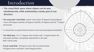
VISUAL FIELD
By Group 6 (Medical Physiology)
Meet the Team
BASSEY Esther
UCHENNA Maryann
ABRAHAM Victory
OKEH Amarachi
ETUKUDO Miracle
AKPAN Emediong
DAVIES Ephraim
AKPAN Ekemini
GODWIN Mberekeobong
UMOH Godswill
ASUQUO Harvest
Navigating this
Presentation
C O N T E N T C O M PA S S :
01
02
03
04
05
Anatomy and Physiology of Visual Field
Central And Peripheral Vision
Visual Field Testing
Clinical Correlation
Introduction
• The monocular visual field: central vision (inner 30 degrees) and peripheral
vision (100 degrees laterally, 60 degrees medially, 60 degrees upward, 75 degrees
downward).
• A vertical line at central fixation divides it into nasal and temporal hemifields.
• The blind spot: 12 to 17 degrees from fixation and 1.5 degrees below the
horizontal meridian, corresponds anatomically to the optic
disk's scleral canal.
• Tyical visual field: 90 degrees to the side of central fixation,
50 degrees above and below, and 60 degrees below.
• The visual field: space where objects can be seen
simultaneously while maintaining a steady gaze in one
direction.
03
Introduction
End-to-End
Retail
Customer satisfaction, from
choice to checkout
Full Customer
Support
Multi-channel
help hotlines
Parts of the Eyes Involved in the Visual Field
• Cornea
2. Lens
3. Retina:
-Photoreceptor Cells
-Rods
-Cones
-Macula
-Peripheral Retina
4. Optic Nerve
5. Visual Cortex (in the Brain
6. Iris
7. Pupil
Anatomy and Physiology of Visual Field
Online
Marketplace
A platform for
borderless selling
Central and Peripheral Vision
• Central Vision: This is the vision within the fovea. It is the most important part of
a person's vision. It is used to read, drive and see pictures or faces. The Macula
provides the central vision.
• Peripheral Vision: This is the vision which occurs outside the fovea. It is outside
the point of fixation.
Importance
Central Vision:
1. Object recognition
2. Acute pattern vision.
Peripheral Vision:
1. Scene gist recognition
2. Recognition of abstract events
Visual Field Testing
06
• Visual field test measures your peripheral vision, it’s also called a
perimetry test. Visual field testing is important for many conditions,
i. including glaucoma, stroke etc.
• Why is visual field test performed?
• As part of a routine eye exam, this test can help diagnose early signs of
diseases.
The two most basic types of visual field test include:
• Standard automated perimetry (SAP)- it uses a computer-controlled
device to present light of varying intensity at different locations in the
field. The patient is asked to press a button whenever they see a light.
• Confrontation visual field (CVF)-
The person giving the test sits 3-4 feels away from the person having the test.
It measures only the outer edge of the visual field.
Clinical
Correlation
DEFECTS
Central Vision Loss: is the loss of central vision sight. The
loss can appear gradually or suddenly and usually appears as a dark
spot, blurred area or distorted area in the centre of the visual fields.
Peripheral Vision Loss: is the loss of the peripheral vision
which results in inability to see to the side without turning your
head.
Diseases associated with central and Peripheral
Vision:
• Coloboma
• Macular edema
• HIV/ AIDS
• Pigment Dispersion Syndrome
• Macular Pucker
* Glaucoma
* Macular Edema
* Macular Pucker: diabetic retinopathy, Cataracts
Conclusion
User
Testing
08
Focus
Groups
Learning
from
discussions
The physiology of the visual field is a highly intricate process that involves multiple
structures and pathways in the eye and brain. Understanding this process is essential for
diagnosing and treating visual disorders and for appreciating the complexity of human
vision.
References
1. Strasburger, H., Rentschler, I., & Juttner, M. (2011). Peripheral vision and pattern recognition: A review. *Journal of Vision, 11*(5), 13.
https://doi.org/10.1167/11.5.13
2. Larson, A. M., & Loschky, L. (2009). The contributions of central versus peripheral vision to scene gist recognition. *Journal of
Vision.* https://jov.arvojournals.org/article.aspx?articleid=2122327
3. American Academy of Ophthalmology. (2020). Preferred practice pattern guidelines: Visual field testing. *American Academy of
Ophthalmology.*
4. Larson, A. M., & Loschky, L. C. (n.d.). The contributions of central versus peripheral vision to scene gist recognition. *Journal of
Vision.* https://jov.arvojournals.org/article.aspx?articleid=2122327
5. Spector, R. H. (1990). Visual Fields. In H. K. Walker, W. D. Hall, & J. W. Hurst (Eds.), *Clinical Methods: The History, Physical, and
Laboratory Examinations* (3rd ed., Chapter 116). Butterworths. https://www.ncbi.nlm.nih.gov/books/NBK220/
6. Snell, R. S. *Clinical Anatomy of the Eye.*
7. Saladin, K. S. . *Anatomy & Physiology: The Unity of Form and Function.

A Visual field Seminar Presentation.pptx

  • 1.
    VISUAL FIELD By Group6 (Medical Physiology)
  • 2.
    Meet the Team BASSEYEsther UCHENNA Maryann ABRAHAM Victory OKEH Amarachi ETUKUDO Miracle AKPAN Emediong DAVIES Ephraim AKPAN Ekemini GODWIN Mberekeobong UMOH Godswill ASUQUO Harvest
  • 3.
    Navigating this Presentation C ON T E N T C O M PA S S : 01 02 03 04 05 Anatomy and Physiology of Visual Field Central And Peripheral Vision Visual Field Testing Clinical Correlation Introduction
  • 4.
    • The monocularvisual field: central vision (inner 30 degrees) and peripheral vision (100 degrees laterally, 60 degrees medially, 60 degrees upward, 75 degrees downward). • A vertical line at central fixation divides it into nasal and temporal hemifields. • The blind spot: 12 to 17 degrees from fixation and 1.5 degrees below the horizontal meridian, corresponds anatomically to the optic disk's scleral canal. • Tyical visual field: 90 degrees to the side of central fixation, 50 degrees above and below, and 60 degrees below. • The visual field: space where objects can be seen simultaneously while maintaining a steady gaze in one direction. 03 Introduction
  • 5.
    End-to-End Retail Customer satisfaction, from choiceto checkout Full Customer Support Multi-channel help hotlines Parts of the Eyes Involved in the Visual Field • Cornea 2. Lens 3. Retina: -Photoreceptor Cells -Rods -Cones -Macula -Peripheral Retina 4. Optic Nerve 5. Visual Cortex (in the Brain 6. Iris 7. Pupil Anatomy and Physiology of Visual Field Online Marketplace A platform for borderless selling
  • 6.
    Central and PeripheralVision • Central Vision: This is the vision within the fovea. It is the most important part of a person's vision. It is used to read, drive and see pictures or faces. The Macula provides the central vision. • Peripheral Vision: This is the vision which occurs outside the fovea. It is outside the point of fixation. Importance Central Vision: 1. Object recognition 2. Acute pattern vision. Peripheral Vision: 1. Scene gist recognition 2. Recognition of abstract events
  • 7.
    Visual Field Testing 06 •Visual field test measures your peripheral vision, it’s also called a perimetry test. Visual field testing is important for many conditions, i. including glaucoma, stroke etc. • Why is visual field test performed? • As part of a routine eye exam, this test can help diagnose early signs of diseases. The two most basic types of visual field test include: • Standard automated perimetry (SAP)- it uses a computer-controlled device to present light of varying intensity at different locations in the field. The patient is asked to press a button whenever they see a light. • Confrontation visual field (CVF)- The person giving the test sits 3-4 feels away from the person having the test. It measures only the outer edge of the visual field.
  • 8.
    Clinical Correlation DEFECTS Central Vision Loss:is the loss of central vision sight. The loss can appear gradually or suddenly and usually appears as a dark spot, blurred area or distorted area in the centre of the visual fields. Peripheral Vision Loss: is the loss of the peripheral vision which results in inability to see to the side without turning your head. Diseases associated with central and Peripheral Vision: • Coloboma • Macular edema • HIV/ AIDS • Pigment Dispersion Syndrome • Macular Pucker * Glaucoma * Macular Edema * Macular Pucker: diabetic retinopathy, Cataracts
  • 9.
    Conclusion User Testing 08 Focus Groups Learning from discussions The physiology ofthe visual field is a highly intricate process that involves multiple structures and pathways in the eye and brain. Understanding this process is essential for diagnosing and treating visual disorders and for appreciating the complexity of human vision.
  • 10.
    References 1. Strasburger, H.,Rentschler, I., & Juttner, M. (2011). Peripheral vision and pattern recognition: A review. *Journal of Vision, 11*(5), 13. https://doi.org/10.1167/11.5.13 2. Larson, A. M., & Loschky, L. (2009). The contributions of central versus peripheral vision to scene gist recognition. *Journal of Vision.* https://jov.arvojournals.org/article.aspx?articleid=2122327 3. American Academy of Ophthalmology. (2020). Preferred practice pattern guidelines: Visual field testing. *American Academy of Ophthalmology.* 4. Larson, A. M., & Loschky, L. C. (n.d.). The contributions of central versus peripheral vision to scene gist recognition. *Journal of Vision.* https://jov.arvojournals.org/article.aspx?articleid=2122327 5. Spector, R. H. (1990). Visual Fields. In H. K. Walker, W. D. Hall, & J. W. Hurst (Eds.), *Clinical Methods: The History, Physical, and Laboratory Examinations* (3rd ed., Chapter 116). Butterworths. https://www.ncbi.nlm.nih.gov/books/NBK220/ 6. Snell, R. S. *Clinical Anatomy of the Eye.* 7. Saladin, K. S. . *Anatomy & Physiology: The Unity of Form and Function.