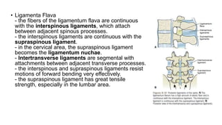
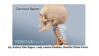
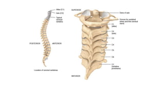
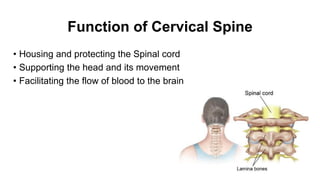
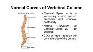
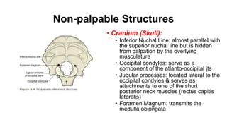
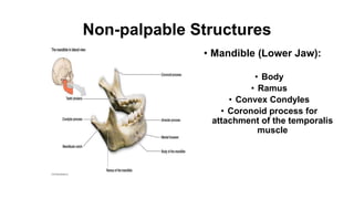
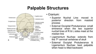
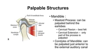
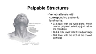
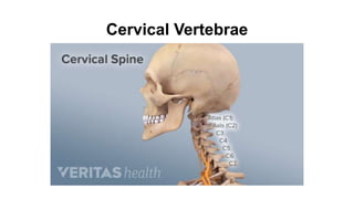
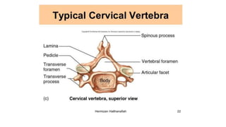
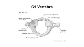
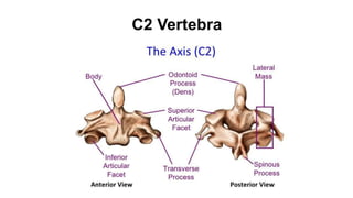
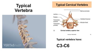
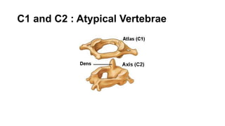
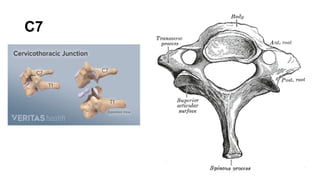
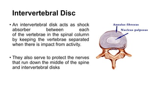
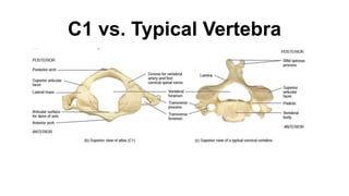
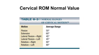
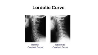
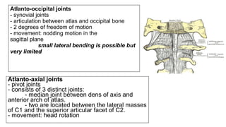
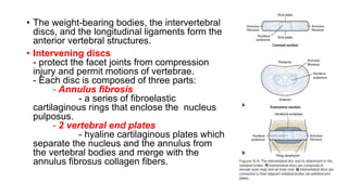
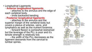
by: Aubrey Vale Sagun, Lady Leanne Esteban, Kamille Chloie Ferrer
Function of Cervical Spine
• Housing and protecting the Spinal cord
• Supporting the head and its movement
• Facilitating the flow of blood to the brain
Normal Curves of Vertebral Column
• Cervical Spine – is a
secondary curve; convex
anteriorly and concave
posteriorly
• Normal Curvature of
Cervical Spine: 20 – 40
degrees
• COG of head – falls on the
concave side of the curves
Non-palpable Structures
• Cranium (Skull):
• Inferior Nuchal Line: almost parallel with
the superior nuchal line but is hidden
from palpation by the overlying
musculature
• Occipital condyles: serve as a
component of the atlanto-occipital jts
• Jugular processes: located lateral to the
occipital condyles & serves as
attachments to one of the short
posterior neck muscles (rectus capitis
lateralis)
• Foramen Magnum: transmits the
medulla oblongata
Non-palpable Structures
• Mandible (Lower Jaw):
• Body
• Ramus
• Convex Condyles
• Coronoid process for
attachment of the temporalis
muscle
Palpable Structures
• Cranium:
• Superior Nuchal Line: moved in
posterior direction from mastoid
process
• External Occipital Protuberance: small
eminence when the two superior
nuchal lines of R & L sides meet at the
median line
• Ligamentum Nuchae: extends from
the 7th cervical vertebrae of skull
• External Occiptal Protuberance &
Ligamentum Nuchae: best palpable
when head is tilted backward
Palpable Structures
• Mandible :
• Mastoid Process: can be
palpated behind the
earlobes
• Cervical Flexion – best felt
• Cervical Extension – only
part of the process is
palpated
• Condyles of Mandible: can
be palpated just anterior to
the external auditory canal
Palpable Structures
• Vertebral levels with
corresponding anatomic
landmarks:
• C-3: level with the hyoid bone, which
can be palpated anteriorly just below
the mandible
• C-4 & C-5: level with thyroid cartilage
• C-6: level with the arch of the cricoid
cartilage
Cervical Vertebrae
C1 Vertebra
C2 Vertebra
C7
Typical
Vertebra
Typical vertebra have:
C3-C6
C1 and C2 : Atypical Vertebrae
C7
Intervertebral Disc
• An intervertebral disk acts as shock
absorber between each
of the vertebrae in the spinal column
by keeping the vertebrae separated
when there is impact from activity.
• They also serve to protect the nerves
that run down the middle of the spine
and intervertebral disks
C1 vs. Typical Vertebra
C2 Superior and Lateral View
Vertebral Motion
• Motion segment – consist of two adjacent vertebrae, three
intervertebral joints, the soft tissues of intervertebral disc,
longitudinal and intersegmental ligaments, facet joint capsules
• Disc and L & R facet joints – form a triangle whereby motion at
one joint always produces motion at the other two joints
• Osteokinematic Motions – flexion & extension (sagittal plane), L
& R lateral flexion (frontal plane), and L & R rotation (transverse
plane)
• Arthrokinematic Motions – anterior/posterior shear/slide, lateral
shear/slide, and distraction & compression
Coupling Motions
• Combined motions of spine
• It occurs because of the orientation of the planes of the left and right facet
joints and limitation of motion provided by the disc, vertebral ligaments,
fascia, and muscles
• The greatest complexity of coupling in the spine is with side bending and
rotation. After one or two degrees of motion, side bending is always
accompanied by rotation, and rotation is always accompanied by side
bending
• It has been traditionally viewed that coupling is influenced by the position
the spine assumes in the sagittal plane.
• When the spine is in neutral position and moves in the sagittal plane,
rotation and side bending occur contralaterally. However, when the spine is
either flexed or extended, rotation and side bending occur in an ipsilateral
fashion
Cervical Region
• Craniovertebral area – form by the occiput (0), atlas (C1), and
axis (C2); the facet joints here are specialized (two to three
degrees of freedom exist), and the planes of motion are nearly
horizontal
• Atlanto-occipital joint (0-C1) – movement is mainly nodding in
the sagittal plane; small lateral bending are also possible but
are quite limited
• Atlanto-axial joint (C1-C2) – axis of motion is vertical through
the dens, the motion occuring is rotation; Approximately 50% of
rotation in cervical occurs in AA jt.
Cervical Region
• Typical cervical vertebral articulations (C2–C3 through C6–
C7):
• facet joints change from horizontal to a 45° angle between the
horizontal and frontal planes;
• The superior articular facets slide superior and anterior in forward
bending (flexion) and inferior and posterior in backward bending
(extension);
• In side bending right, the left superior facet moves superiorly and
anterior while the right superior facet moves inferior and posterior,
producing a rotation of the vertebral body to the right and the spinous
process to the left.
Movements in the Cervical Region
Cervical ROM Normal Value
Lordotic Curve
JOINTS AND
LIGAMENTS
Atlanto-occipital joints
- synovial joints
- articulation between atlas and occipital bone
- 2 degrees of freedom of motion
- movement: nodding motion in the
sagittal plane
small lateral bending is possible but
very limited
Atlanto-axial joints
- pivot joints
- consists of 3 distinct joints:
- median joint between dens of axis and
anterior arch of atlas.
- two are located between the lateral masses
of C1 and the superior articular facet of C2.
- movement: head rotation
• The weight-bearing bodies, the intervertebral
discs, and the longitudinal ligaments form the
anterior vertebral structures.
• Intervening discs
- protect the facet joints from compression
injury and permit motions of vertebrae.
- Each disc is composed of three parts:
- Annulus fibrosis
- a series of fibroelastic
cartilaginous rings that enclose the nucleus
pulposus.
- 2 vertebral end plates
- hyaline cartilaginous plates which
separate the nucleus and the annulus from
the vertebral bodies and merge with the
annulus fibrosus collagen fibers.
• Longitudinal Ligaments
- Anterior longitudinal ligaments
- attaches to the annulus and the edge of
each vertebral body.
- limits backward bending.
- Posterior longitudinal ligaments
- attaches to the annulus and the
superior margin of the vertebral body but
covers a plexus of arteries, veins, and
lymphatics as well as the nutrient foramina.
- forward flexion is somewhat restrained
but the leverage of the PLL is poor and it’s
tensile strength is relatively low.
- the width of the PLL decreases as the
ligament descends from the cervical to
lumbar spines.
Posterior Vertebral Joint Elements
• Posterior vertebral structures comprise the arches, the
transverse and spinous processes, the bilateral facet
joints, joint capsules, and ligaments.
• The facet joints (apophyseal or zygapophyseal joints)
are formed by inferior articulating process of one vertebra
with the superior articulating process of the vertebra
below.
• Major functions of the facet joints are to control vertebra
motions and to protect the disc from excessive shear,
flexion, side bending, and rotation.
• The direction and amount of motion permitted are
determined by the planes of the joint surfaces, which
change in their orientation from the cervical through the
lumbar areas.
Temporomandibular Joint (TMJ)
Motion
• Like a hinge joint in movable base
• Opening the mouth – rotation of the mandibular condyles around the
lower joint space, followed by translation of the articular disc on the
upper joint space down to the articular eminence.
• Closing of mouth – requires reversal of the translatory and rotational
motions.
• Other motions possible in mandible – protrusion (movement of the jaw
forward), retrusion (movement of the jaw posteriorly), and lateral shift to
the left and to the right, which includes mediolateral translatory motion.
• Functional movements of the jaw are combinations of these motions. In
chewing, these motions are called incision for cutting food and
mastication for crushing and grinding.
•Ligaments
- Ligamenta Flava (L., flavus, yellow)
- series of 23 intersegmental ligaments that connect the
lamina of the adjacent vertebrae from C2 to the sacrum.
- its yellow color reflects the high elastic content, which
contributes to the prestress of the disc and resistance to
forward flexion.
- the ligamentum flava fibers cover the anterior surface of
the capsule of the facet joint and provide tension on the capsule
to prevent nipping and injury by the facet joints during
movement.
• Ligamenta Flava
- the fibers of the ligamentum flava are continuous
with the interspinous ligaments, which attach
between adjacent spinous processes.
- the interspinous ligaments are continuous with the
supraspinous ligament.
- in the cervical area, the supraspinous ligament
becomes the ligamentum nuchae.
- Intertransverse ligaments are segmental with
attachments between adjacent transverse processes.
- the interspinous and supraspinous ligaments resist
motions of forward bending very effectively.
- the supraspinous ligament has great tensile
strength, especially in the lumbar area.
MUSCLES OF
THE CERVICAL
SPINE
ANTERIOR CERVICAL MUSCLES
M: Rectus capitis anterior
O: Base of the skull, immediately
ant. To the occipital condyle
I: Anterior surface of C1
N: C1-C2
A: Flexes Head, Head stabilizer
and provider of proprioception
ANTERIOR CERVICAL MUSCLES
M: Rectus capitis lateralis
O: Occipital’s bone jugular process
I: C1 transverse process
N: C1-C2
A: Stabilizes head and may provide
propoprioceptive feedback, medial-
lateral control of the head
ANTERIOR CERVICAL MUSCLES
M: Longus Capitis
O: Basilar portion of the occipital
bone
I: Transverse processes of C3-C6
N: C1-C3
A: Head and neck flexion, lateral
bending and rotation and significant
joint compression
ANTERIOR CERVICAL MUSCLES
M: Longus Colli
O: Ant. Tubercle of C1, bodies of C1-C3 and
transverse processes of C3-C6
I: Bodies of C5 through T3, transverse
processes of C3-C5
N: C2-C6
A: Cervical flexion, stabilizes head
ANTERIOR CERVICAL MUSCLES
M: Anterior Scalene
O: C4-C6 transverse processes
I: First rib
N: C4-C6
A: Forward flex the cervical spine,
laterally flexes the neck and rotates
to same side
ANTERIOR CERVICAL MUSCLES
M: Middle Scalene
O: Transverse processes of upper 6
cervical vertebrae
I: 1st rib
N: C3-C6
A: Forward flex cervical spine, laterally
flexes the neck and rotates to same side,
elevates rib during forces inspiration
ANTERIOR CERVICAL MUSCLES
M: Posterior Scalene
O: Transverse processes of lower
cervical vertebrae
I: 1st rib
N: C7-C8
A: Flexes neck forward, laterally flexes
neck and rotates to same side, elevates
second rib during forces inspiration
ANTERIOR CERVICAL MUSCLES
M: Sternocleidomastoid
O: Manubrium sterni and medial third of
clavicle
I: Mastoid process of temporal bone and
occipital bone
N: Spinal accessory nerve (XI) , C1-C3
A: Forward flexes the neck, rotates neck to
the opposite side, lateral flexion to same side
and extension of head and neck
POSTERIOR CERVICAL SUBOCCIPITAL
MUSCLES
M: Rectus capitis posterior major
O: Base of the skull, immediately
ant. To the occipital condyle
I: Anterior surface of C1
N: C1 suboccipital nerve
A: Head extension and lateral flexion
POSTERIOR CERVICAL SUBOCCIPITAL
MUSCLES
M: Rectus capitis posterior minor
O: Just above the atlas
I: Inferior nuchal line of occiput,
Inferior and superior oblique: occiput
N: C1 suboccipital nerve
A: Head extension and lateral flexion
POSTERIOR TRANSVERSOSPINAL
MUSCLES
M: Rotators
O: Transverse process of 1 vertebrae
I: Base of the spinous process above
N: Dorsal rami of spinal nerves
A: Extend head and neck, laterally flex and
rotate head to same side
POSTERIOR TRANSVERSOSPINAL
MUSCLES
M: Multifidus
O: Transverse processes of 2-5 vertebrae
I: Spinous processes above
N: Dorsal rami of spinal nerves
A: Extend head and neck, laterally flex and rotate head to
same side
POSTERIOR TRANSVERSOSPINAL
MUSCLES
M: Semispinalis capitis
O: Transverse processes of T1-T6
I: Above the inferior nuchal line
N: Dorsal rami of spinal nerves
A: Extend head and neck, laterally flex and rotate head to
same side
POSTERIOR TRANSVERSOSPINAL
MUSCLES
M: Semispinalis cervicis
O: Transverse processes of T1-T6
I: C2-C5 Spinous processes
N: Dorsal rami of spinal nerves
A: Extend head and neck, laterally flex and rotate head to
same side

Cervical spine

  • 1.
    by: Aubrey ValeSagun, Lady Leanne Esteban, Kamille Chloie Ferrer
  • 3.
    Function of CervicalSpine • Housing and protecting the Spinal cord • Supporting the head and its movement • Facilitating the flow of blood to the brain
  • 4.
    Normal Curves ofVertebral Column • Cervical Spine – is a secondary curve; convex anteriorly and concave posteriorly • Normal Curvature of Cervical Spine: 20 – 40 degrees • COG of head – falls on the concave side of the curves
  • 5.
    Non-palpable Structures • Cranium(Skull): • Inferior Nuchal Line: almost parallel with the superior nuchal line but is hidden from palpation by the overlying musculature • Occipital condyles: serve as a component of the atlanto-occipital jts • Jugular processes: located lateral to the occipital condyles & serves as attachments to one of the short posterior neck muscles (rectus capitis lateralis) • Foramen Magnum: transmits the medulla oblongata
  • 6.
    Non-palpable Structures • Mandible(Lower Jaw): • Body • Ramus • Convex Condyles • Coronoid process for attachment of the temporalis muscle
  • 7.
    Palpable Structures • Cranium: •Superior Nuchal Line: moved in posterior direction from mastoid process • External Occipital Protuberance: small eminence when the two superior nuchal lines of R & L sides meet at the median line • Ligamentum Nuchae: extends from the 7th cervical vertebrae of skull • External Occiptal Protuberance & Ligamentum Nuchae: best palpable when head is tilted backward
  • 8.
    Palpable Structures • Mandible: • Mastoid Process: can be palpated behind the earlobes • Cervical Flexion – best felt • Cervical Extension – only part of the process is palpated • Condyles of Mandible: can be palpated just anterior to the external auditory canal
  • 9.
    Palpable Structures • Vertebrallevels with corresponding anatomic landmarks: • C-3: level with the hyoid bone, which can be palpated anteriorly just below the mandible • C-4 & C-5: level with thyroid cartilage • C-6: level with the arch of the cricoid cartilage
  • 10.
  • 12.
  • 13.
  • 14.
  • 15.
  • 16.
    C1 and C2: Atypical Vertebrae
  • 17.
  • 18.
    Intervertebral Disc • Anintervertebral disk acts as shock absorber between each of the vertebrae in the spinal column by keeping the vertebrae separated when there is impact from activity. • They also serve to protect the nerves that run down the middle of the spine and intervertebral disks
  • 19.
  • 20.
    C2 Superior andLateral View
  • 21.
    Vertebral Motion • Motionsegment – consist of two adjacent vertebrae, three intervertebral joints, the soft tissues of intervertebral disc, longitudinal and intersegmental ligaments, facet joint capsules • Disc and L & R facet joints – form a triangle whereby motion at one joint always produces motion at the other two joints • Osteokinematic Motions – flexion & extension (sagittal plane), L & R lateral flexion (frontal plane), and L & R rotation (transverse plane) • Arthrokinematic Motions – anterior/posterior shear/slide, lateral shear/slide, and distraction & compression
  • 22.
    Coupling Motions • Combinedmotions of spine • It occurs because of the orientation of the planes of the left and right facet joints and limitation of motion provided by the disc, vertebral ligaments, fascia, and muscles • The greatest complexity of coupling in the spine is with side bending and rotation. After one or two degrees of motion, side bending is always accompanied by rotation, and rotation is always accompanied by side bending • It has been traditionally viewed that coupling is influenced by the position the spine assumes in the sagittal plane. • When the spine is in neutral position and moves in the sagittal plane, rotation and side bending occur contralaterally. However, when the spine is either flexed or extended, rotation and side bending occur in an ipsilateral fashion
  • 23.
    Cervical Region • Craniovertebralarea – form by the occiput (0), atlas (C1), and axis (C2); the facet joints here are specialized (two to three degrees of freedom exist), and the planes of motion are nearly horizontal • Atlanto-occipital joint (0-C1) – movement is mainly nodding in the sagittal plane; small lateral bending are also possible but are quite limited • Atlanto-axial joint (C1-C2) – axis of motion is vertical through the dens, the motion occuring is rotation; Approximately 50% of rotation in cervical occurs in AA jt.
  • 24.
    Cervical Region • Typicalcervical vertebral articulations (C2–C3 through C6– C7): • facet joints change from horizontal to a 45° angle between the horizontal and frontal planes; • The superior articular facets slide superior and anterior in forward bending (flexion) and inferior and posterior in backward bending (extension); • In side bending right, the left superior facet moves superiorly and anterior while the right superior facet moves inferior and posterior, producing a rotation of the vertebral body to the right and the spinous process to the left.
  • 25.
    Movements in theCervical Region
  • 26.
  • 27.
  • 28.
  • 29.
    Atlanto-occipital joints - synovialjoints - articulation between atlas and occipital bone - 2 degrees of freedom of motion - movement: nodding motion in the sagittal plane small lateral bending is possible but very limited Atlanto-axial joints - pivot joints - consists of 3 distinct joints: - median joint between dens of axis and anterior arch of atlas. - two are located between the lateral masses of C1 and the superior articular facet of C2. - movement: head rotation
  • 30.
    • The weight-bearingbodies, the intervertebral discs, and the longitudinal ligaments form the anterior vertebral structures. • Intervening discs - protect the facet joints from compression injury and permit motions of vertebrae. - Each disc is composed of three parts: - Annulus fibrosis - a series of fibroelastic cartilaginous rings that enclose the nucleus pulposus. - 2 vertebral end plates - hyaline cartilaginous plates which separate the nucleus and the annulus from the vertebral bodies and merge with the annulus fibrosus collagen fibers.
  • 31.
    • Longitudinal Ligaments -Anterior longitudinal ligaments - attaches to the annulus and the edge of each vertebral body. - limits backward bending. - Posterior longitudinal ligaments - attaches to the annulus and the superior margin of the vertebral body but covers a plexus of arteries, veins, and lymphatics as well as the nutrient foramina. - forward flexion is somewhat restrained but the leverage of the PLL is poor and it’s tensile strength is relatively low. - the width of the PLL decreases as the ligament descends from the cervical to lumbar spines.
  • 32.
    Posterior Vertebral JointElements • Posterior vertebral structures comprise the arches, the transverse and spinous processes, the bilateral facet joints, joint capsules, and ligaments. • The facet joints (apophyseal or zygapophyseal joints) are formed by inferior articulating process of one vertebra with the superior articulating process of the vertebra below. • Major functions of the facet joints are to control vertebra motions and to protect the disc from excessive shear, flexion, side bending, and rotation. • The direction and amount of motion permitted are determined by the planes of the joint surfaces, which change in their orientation from the cervical through the lumbar areas.
  • 33.
    Temporomandibular Joint (TMJ) Motion •Like a hinge joint in movable base • Opening the mouth – rotation of the mandibular condyles around the lower joint space, followed by translation of the articular disc on the upper joint space down to the articular eminence. • Closing of mouth – requires reversal of the translatory and rotational motions. • Other motions possible in mandible – protrusion (movement of the jaw forward), retrusion (movement of the jaw posteriorly), and lateral shift to the left and to the right, which includes mediolateral translatory motion. • Functional movements of the jaw are combinations of these motions. In chewing, these motions are called incision for cutting food and mastication for crushing and grinding.
  • 34.
    •Ligaments - Ligamenta Flava(L., flavus, yellow) - series of 23 intersegmental ligaments that connect the lamina of the adjacent vertebrae from C2 to the sacrum. - its yellow color reflects the high elastic content, which contributes to the prestress of the disc and resistance to forward flexion. - the ligamentum flava fibers cover the anterior surface of the capsule of the facet joint and provide tension on the capsule to prevent nipping and injury by the facet joints during movement.
  • 35.
    • Ligamenta Flava -the fibers of the ligamentum flava are continuous with the interspinous ligaments, which attach between adjacent spinous processes. - the interspinous ligaments are continuous with the supraspinous ligament. - in the cervical area, the supraspinous ligament becomes the ligamentum nuchae. - Intertransverse ligaments are segmental with attachments between adjacent transverse processes. - the interspinous and supraspinous ligaments resist motions of forward bending very effectively. - the supraspinous ligament has great tensile strength, especially in the lumbar area.
  • 36.
  • 37.
    ANTERIOR CERVICAL MUSCLES M:Rectus capitis anterior O: Base of the skull, immediately ant. To the occipital condyle I: Anterior surface of C1 N: C1-C2 A: Flexes Head, Head stabilizer and provider of proprioception
  • 38.
    ANTERIOR CERVICAL MUSCLES M:Rectus capitis lateralis O: Occipital’s bone jugular process I: C1 transverse process N: C1-C2 A: Stabilizes head and may provide propoprioceptive feedback, medial- lateral control of the head
  • 39.
    ANTERIOR CERVICAL MUSCLES M:Longus Capitis O: Basilar portion of the occipital bone I: Transverse processes of C3-C6 N: C1-C3 A: Head and neck flexion, lateral bending and rotation and significant joint compression
  • 40.
    ANTERIOR CERVICAL MUSCLES M:Longus Colli O: Ant. Tubercle of C1, bodies of C1-C3 and transverse processes of C3-C6 I: Bodies of C5 through T3, transverse processes of C3-C5 N: C2-C6 A: Cervical flexion, stabilizes head
  • 41.
    ANTERIOR CERVICAL MUSCLES M:Anterior Scalene O: C4-C6 transverse processes I: First rib N: C4-C6 A: Forward flex the cervical spine, laterally flexes the neck and rotates to same side
  • 42.
    ANTERIOR CERVICAL MUSCLES M:Middle Scalene O: Transverse processes of upper 6 cervical vertebrae I: 1st rib N: C3-C6 A: Forward flex cervical spine, laterally flexes the neck and rotates to same side, elevates rib during forces inspiration
  • 43.
    ANTERIOR CERVICAL MUSCLES M:Posterior Scalene O: Transverse processes of lower cervical vertebrae I: 1st rib N: C7-C8 A: Flexes neck forward, laterally flexes neck and rotates to same side, elevates second rib during forces inspiration
  • 44.
    ANTERIOR CERVICAL MUSCLES M:Sternocleidomastoid O: Manubrium sterni and medial third of clavicle I: Mastoid process of temporal bone and occipital bone N: Spinal accessory nerve (XI) , C1-C3 A: Forward flexes the neck, rotates neck to the opposite side, lateral flexion to same side and extension of head and neck
  • 45.
    POSTERIOR CERVICAL SUBOCCIPITAL MUSCLES M:Rectus capitis posterior major O: Base of the skull, immediately ant. To the occipital condyle I: Anterior surface of C1 N: C1 suboccipital nerve A: Head extension and lateral flexion
  • 46.
    POSTERIOR CERVICAL SUBOCCIPITAL MUSCLES M:Rectus capitis posterior minor O: Just above the atlas I: Inferior nuchal line of occiput, Inferior and superior oblique: occiput N: C1 suboccipital nerve A: Head extension and lateral flexion
  • 47.
    POSTERIOR TRANSVERSOSPINAL MUSCLES M: Rotators O:Transverse process of 1 vertebrae I: Base of the spinous process above N: Dorsal rami of spinal nerves A: Extend head and neck, laterally flex and rotate head to same side
  • 48.
    POSTERIOR TRANSVERSOSPINAL MUSCLES M: Multifidus O:Transverse processes of 2-5 vertebrae I: Spinous processes above N: Dorsal rami of spinal nerves A: Extend head and neck, laterally flex and rotate head to same side
  • 49.
    POSTERIOR TRANSVERSOSPINAL MUSCLES M: Semispinaliscapitis O: Transverse processes of T1-T6 I: Above the inferior nuchal line N: Dorsal rami of spinal nerves A: Extend head and neck, laterally flex and rotate head to same side
  • 50.
    POSTERIOR TRANSVERSOSPINAL MUSCLES M: Semispinaliscervicis O: Transverse processes of T1-T6 I: C2-C5 Spinous processes N: Dorsal rami of spinal nerves A: Extend head and neck, laterally flex and rotate head to same side