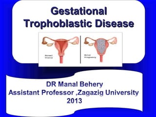
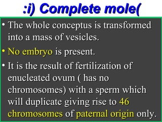
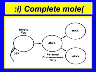
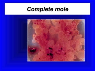
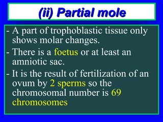
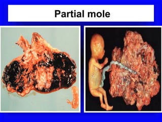
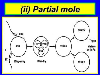
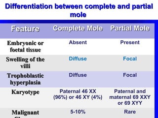
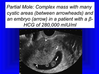
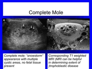
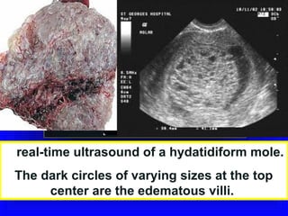
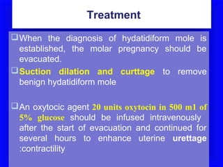
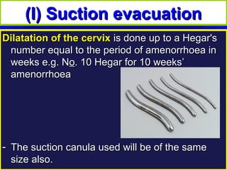
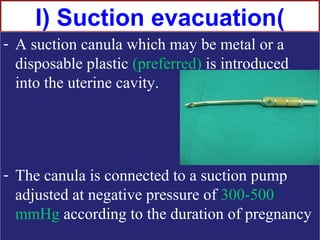
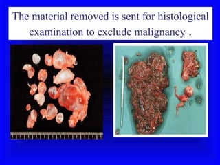
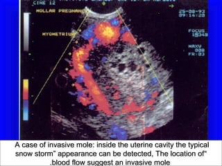
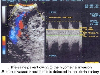
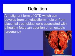
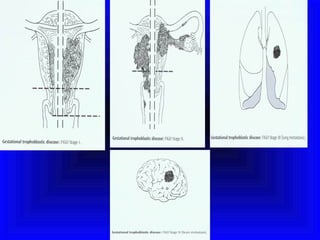
Gestational
Trophoblastic Disease
Definition

A clinical spectrum including all
  neoplasm derived from
  abnormal trophoblastic
  proliferation
Classification

1. Hydatidiform (vesicular )mole
   Complete and Partial
2. Invasive mole
3. Choriocarcinoma
4. Placental-site trophoblastic
   tumor
Vesicular mole
Vesicular mole

Vesicular mole
Vesicular mole
It is a benign neoplasm of the chorionic villi
                characterized by
1. Marked proliferation of the trophoplast,both the
   syncytium & cytotrophoplast are affected.

2. Oedema or hydropic degeneration of the
   connective tissue stroma of the villi which leads
   to their distension and formation of vesicles.

3. Avascularity of the villi: the blood vessels
   disappear from villi explaining early death of
   the embryo
The microscopic appearance of hydatidiform
: mole
Hyperplasia of trophobasitc cells •
Hydropic swelling of all villi •
Vessles are usually absent •
Incidence
• 1:2000 pregnancies in United States and
  Europe, but 10 times more in Asia.

• Predisposing factors include :
 Race, deficiency of protein or carotene
• The incidence is higher toward the
  beginning and more toward the end of the
  childbearing period.
• It is 10 times more in women over 45 years
  old.
Pathology
The uterus is distended by
 thin walled, translucent,
grape-like vesicles of
different sizes.
• These are degenerated chorionic villi
  filled with fluid.
• There is no vasculature in the chorionic
  villi leads to early death of the embryo.
Pathology
• High hCG causes multiple theca lutein
  cysts in the ovaries in about 50% of cases.
• Cysts may reach a large size (10 cm or
  more.


• Cysts disappear
• within few months(2-3),
• after evacuation of the mole.
:i) Complete mole(
• The whole conceptus is transformed
  into a mass of vesicles.
• No embryo is present.
• It is the result of fertilization of
  enucleated ovum ( has no
  chromosomes) with a sperm which
  will duplicate giving rise to 46
  chromosomes of paternal origin only.
:i) Complete mole(
Complete mole
(ii) Partial mole
- A part of trophoblastic tissue only
  shows molar changes.
- There is a foetus or at least an
  amniotic sac.
- It is the result of fertilization of an
  ovum by 2 sperms so the
  chromosomal number is 69
  chromosomes
Partial mole
(ii) Partial mole
Differentiation between complete and partial
                    mole

 Feature          Complete Mole         Partial Mole

Embryonic or            Absent             Present
 foetal tissue
Swelling of the         Diffuse              Focal
     villi
Trophoblastic           Diffuse              Focal
 hyperplasia
  Karyotype          Paternal 46 XX      Paternal and
                  (96%) or 46 XY (4%)   maternal 69 XXY
                                          or 69 XYY
  Malignant             5-10%                Rare
Diagnosis
(A) Symptoms
1.Amenorrhoea: usually of short period
  (2-3 months).

2.Exaggerated symptoms of pregnancy
  especially vomiting.

3.Symptoms of preeclampsia may be
  present as headache, and oedema
(A) Symptoms
4. Vaginal bleeding :
• The main complaint, due to separation of
   vesicles from uterine wall, there may be a
   blood stained watery discharge, the watery
   part is from ruptured vesicles.
• Prune juice disharge may occur.
•   The blood is brown because it has retained
    for sometime in the uterine cavity.
•   passage of vesicles is diagnostic.
•   The blood may be concealed causing
    enlargment & tenderness of the uterus.
(A) Symptoms
5. Abdominal pain : may be
- dull-aching due to rapid distension of the
  uterus by the mole or by cocealed
  haemorrhage.
- Colicky due to starting expulsion,
- Sudden And Severe due to perforating mole
- Ovarian pain due to stretching of the ovarian
  capsule or complication in the cystic ovary as
  torsion
Signs
General examination
1.Pre-eclampsia in 20-30% of cases, usually
  before 20 weeks’ gestation.

2.Pallor indicating anemia may be present.

3.Hyperthyroidism in 3-10% of cases
  manifested by enlarged thyroid gland,
  tachycardia (due to chorionic thyrotropin
  secreted by trophoplast &HCG also has a
  thyroid stimulating effect.
4.Breast signs of pregnancy.
Abdominal examination
1.The uterus is >the period of amenorrhoea in
  50% of cases, corresponds to it in 25% and
  smaller in 25% with inactive or dead mole.

2.The uterus is doughy in consistency due to
  absence of amniotic fluid and its distension
  with vesicles.

3.Fetal parts and heart sound cannot be detected
  except in partial mole.
Local examination
1. Passage of vesicles (sure sign).


2. Bilateral ovarian cysts in 50%
   of cases.
3. No internal ballottement.
(C) Investigations
1.Urine pregnancy test:
  is positive in high dilution.
• 1/200 is highly suggestive,
• 1/500 is surely diagnostic.
• In normal pregnancy it is positive in
  dilutions up to 1/100.
2. Serum b -hCG level
is highly elevated ( > 100.000 mIU/m1).
(C) Investigations
3. Ultrasonography reveals:
• The characteristic intrauterine " snow
  storm" appearance,
• no identifiable foetus,
• bilateral ovarian cysts may be detected.
4. X-ray to the abdomen: shows no foetal
  skeleton.
5. X-ray of the chest: should be performed in
  every case of trophoplastic tumour.
Partial Mole: Complex mass with many
cystic areas (between arrowheads) and
an embryo (arrow) in a patient with a β-
        HCG of 280,000 mIU/ml
Complete Mole


Complete mole: “snowstorm”      Corresponding T1 weighted
appearance with multiple        MRI (MRI can be helpful
cystic areas, no fetal tissue   in determining extent of
present                         )trophoblastic disease
A real-time ultrasound of a hydatidiform mole.
 The dark circles of varying sizes at the top
      center are the edematous villi.
Complications
1. Haemorrhage.
2. Infection due to absence of the amniotic sac and
   due to the large surface area left after expulsion
   or evacuation of the mole.
3. Perforation of the uterus. Spontaneous by a
   perforating mole or during evacuation.
4. Pregnancy induced hypertension
5. Hyperthyroidism.
6. Subsequent development of choriocarcinoma in
   about 5% of cases and invasive mole in about
   10% of cases.
7. Recurrent mole may occur(1-2%).
Treatment
 When the diagnosis of hydatidiform mole is
  established, the molar pregnancy should be
  evacuated.
 Suction dilation and curttage to remove
  benign hydatidiform mole

 An oxytocic agent 20 units oxytocin in 500 m1 of
  5% glucose should be infused intravenously
  after the start of evacuation and continued for
  several hours to enhance uterine urettage
  :contractility
(I) Suction evacuation
Dilatation of the cervix is done up to a Hegar's
 number equal to the period of amenorrhoea in
 weeks e.g. No. 10 Hegar for 10 weeks’
 amenorrhoea


- The suction canula used will be of the same
  size also.
I) Suction evacuation(
- A suction canula which may be metal or a
  disposable plastic (preferred) is introduced
  into the uterine cavity.


- The canula is connected to a suction pump
  adjusted at negative pressure of 300-500
  mmHg according to the duration of pregnancy
The material removed is sent for histological
   examination to exclude malignancy .
Curettage
• After evacuation ,
• the uterus is gently curetted with a
  sharp curette.
• Some advise curettage one week after
  evacuation to ensure complete
  removal, but the is not the routine
  practice.
Theca lutein cysts
• They are hormone dependent.

• Disappear spontaneously after
  evacuation of the mole.

• So, they are not removed surgically
  unless complication occur as torsion or
  rupture.
LargeLarge bilateral theca lutein cysts resembling ovarian germ cell
      bilateral theca lutein cysts resembling ovarian germ cell
tumors. With resolution of the human chorionic gonadotropin(HCG)
    tumors. With resolution of the human chorionic
                   .stimulation, they
gonadotropin(HCG) stimulation,return to normal-appearing ovaries
                                       they return to normal-
.appearing ovaries
(II)Hysterotomy
It may be needed for evacuation of
    a large mole to minimize and
    facilitate control of bleeding.
(III) Hysterectomy
It should be considered in women over
  40 years who have completed their
  family for fear of
developing
choriocarcinoma.
IV) Medical induction(
Oxytocins and / or prostaglandins may
 be used to encourage expulsion of
 the mole but must always be followed
 by surgical evacuation.
Follow up
 As choriocarcinoma may complicate
  the vesicular mole after its
  evacuation, detection of serum ß-
  hCG by radioimmunoassay is
  essential
 Normally B –subunit reach normal
  level 8-12 wks after evacuation
Follow up
•  ß-hCG is measured by
• radioimmunoassay every week till the
  test becomes negative for 3 successive
  weeks, then the test is repeated every
  month for one year.

• Pregnancy is allowed if the test remains
  negative for one year.
Follow up
- Persistent high level indicates remnants of
  molar tissues which necessitate
  chemotherapy ( methotrexate) with or
  without curettage. Hysterectomy is
  indicated if women had enough children.
- Rising hCG level after disappearance
  means developing of choriocarcinoma or
  a new pregnancy.
Follow up
It is expected that urine pregnancy
  test is negative 4 weeks after
  evacuation
* Serum B-hCG is undetectable 4
  months after evacuation.
Contraception during follow up

• The combined pill is started when the
  beta-HCG becomes negative.
• Till this happens, the condom can be
  used.
• If the pill is used early the beta-HCG
  will take a longer time to become
  negative as oestrogen stimulates the
  growth of trophoplast.
IUD during follow up
The intrauterine device is not used
 because it may lead to irregular
 uterine bleeding which confuses the
 follow up
Invasive mole or
Chorioadenoma
   Destruens
Definition

• It is a trphoplastic tumour with
   penetration of the myometrium by the
   chorionic villi.
• It is locally malignant
and rarely metastasizes.
It may lead to perforation
 of uterus
A case of invasive mole: inside the uterine cavity the typical
snow storm” appearance can be detected, The location of “
           .blood flow suggest an invasive mole
. The same patient owing to the myometrial invasion
.Reduced vascular resistance is detected in the uterine artery
Early features suggesting persistant GTN
     or post molar syndrome include
1. Recurrent Or Persistent Vaginal
   Bleedig
2. Subinvoluation

3. Amenorrhoea

4. Persistence of ovarian enlargement.
5. No malignancy in endometrial biopsy
Chemotherapy
Started if persistant or malignant disease •
develop
The level of serum HCG doubles in 2 weeks),
  after exclusion of a new pregnancy
 plateaus failure HCG to decrease over 3
  weeks) or
 the test for the hormone becomes positive
  after being negative or
 If metastases appear.
Definition
A malignant form of GTD which can
develop from a hydatidiform mole or from
placental trophoblast cells associated with
a healthy fetus ,an abortion or an ectopic
.pregnancy
Symptoms and signs
•   Bleeding
•   Infection
•   Abdominal swelling
•   Vaginal mass
•   Lung symptoms
•   Symptoms from other metastases
Doppler image of choriocarcinoma
Vesicular mole for undergraduate

Vesicular mole for undergraduate

  • 1.
  • 2.
    Definition A clinical spectrumincluding all neoplasm derived from abnormal trophoblastic proliferation
  • 3.
    Classification 1. Hydatidiform (vesicular)mole Complete and Partial 2. Invasive mole 3. Choriocarcinoma 4. Placental-site trophoblastic tumor
  • 4.
  • 5.
    It is abenign neoplasm of the chorionic villi characterized by 1. Marked proliferation of the trophoplast,both the syncytium & cytotrophoplast are affected. 2. Oedema or hydropic degeneration of the connective tissue stroma of the villi which leads to their distension and formation of vesicles. 3. Avascularity of the villi: the blood vessels disappear from villi explaining early death of the embryo
  • 6.
    The microscopic appearanceof hydatidiform : mole Hyperplasia of trophobasitc cells • Hydropic swelling of all villi • Vessles are usually absent •
  • 7.
    Incidence • 1:2000 pregnanciesin United States and Europe, but 10 times more in Asia. • Predisposing factors include : Race, deficiency of protein or carotene • The incidence is higher toward the beginning and more toward the end of the childbearing period. • It is 10 times more in women over 45 years old.
  • 8.
    Pathology The uterus isdistended by thin walled, translucent, grape-like vesicles of different sizes. • These are degenerated chorionic villi filled with fluid. • There is no vasculature in the chorionic villi leads to early death of the embryo.
  • 9.
    Pathology • High hCGcauses multiple theca lutein cysts in the ovaries in about 50% of cases. • Cysts may reach a large size (10 cm or more. • Cysts disappear • within few months(2-3), • after evacuation of the mole.
  • 10.
    :i) Complete mole( •The whole conceptus is transformed into a mass of vesicles. • No embryo is present. • It is the result of fertilization of enucleated ovum ( has no chromosomes) with a sperm which will duplicate giving rise to 46 chromosomes of paternal origin only.
  • 11.
  • 12.
  • 13.
    (ii) Partial mole -A part of trophoblastic tissue only shows molar changes. - There is a foetus or at least an amniotic sac. - It is the result of fertilization of an ovum by 2 sperms so the chromosomal number is 69 chromosomes
  • 14.
  • 15.
  • 16.
    Differentiation between completeand partial mole Feature Complete Mole Partial Mole Embryonic or Absent Present foetal tissue Swelling of the Diffuse Focal villi Trophoblastic Diffuse Focal hyperplasia Karyotype Paternal 46 XX Paternal and (96%) or 46 XY (4%) maternal 69 XXY or 69 XYY Malignant 5-10% Rare
  • 17.
  • 18.
    (A) Symptoms 1.Amenorrhoea: usuallyof short period (2-3 months). 2.Exaggerated symptoms of pregnancy especially vomiting. 3.Symptoms of preeclampsia may be present as headache, and oedema
  • 19.
    (A) Symptoms 4. Vaginalbleeding : • The main complaint, due to separation of vesicles from uterine wall, there may be a blood stained watery discharge, the watery part is from ruptured vesicles. • Prune juice disharge may occur. • The blood is brown because it has retained for sometime in the uterine cavity. • passage of vesicles is diagnostic. • The blood may be concealed causing enlargment & tenderness of the uterus.
  • 20.
    (A) Symptoms 5. Abdominalpain : may be - dull-aching due to rapid distension of the uterus by the mole or by cocealed haemorrhage. - Colicky due to starting expulsion, - Sudden And Severe due to perforating mole - Ovarian pain due to stretching of the ovarian capsule or complication in the cystic ovary as torsion
  • 21.
  • 22.
    General examination 1.Pre-eclampsia in20-30% of cases, usually before 20 weeks’ gestation. 2.Pallor indicating anemia may be present. 3.Hyperthyroidism in 3-10% of cases manifested by enlarged thyroid gland, tachycardia (due to chorionic thyrotropin secreted by trophoplast &HCG also has a thyroid stimulating effect. 4.Breast signs of pregnancy.
  • 23.
    Abdominal examination 1.The uterusis >the period of amenorrhoea in 50% of cases, corresponds to it in 25% and smaller in 25% with inactive or dead mole. 2.The uterus is doughy in consistency due to absence of amniotic fluid and its distension with vesicles. 3.Fetal parts and heart sound cannot be detected except in partial mole.
  • 24.
    Local examination 1. Passageof vesicles (sure sign). 2. Bilateral ovarian cysts in 50% of cases. 3. No internal ballottement.
  • 25.
    (C) Investigations 1.Urine pregnancytest: is positive in high dilution. • 1/200 is highly suggestive, • 1/500 is surely diagnostic. • In normal pregnancy it is positive in dilutions up to 1/100. 2. Serum b -hCG level is highly elevated ( > 100.000 mIU/m1).
  • 26.
    (C) Investigations 3. Ultrasonographyreveals: • The characteristic intrauterine " snow storm" appearance, • no identifiable foetus, • bilateral ovarian cysts may be detected. 4. X-ray to the abdomen: shows no foetal skeleton. 5. X-ray of the chest: should be performed in every case of trophoplastic tumour.
  • 27.
    Partial Mole: Complexmass with many cystic areas (between arrowheads) and an embryo (arrow) in a patient with a β- HCG of 280,000 mIU/ml
  • 28.
    Complete Mole Complete mole:“snowstorm” Corresponding T1 weighted appearance with multiple MRI (MRI can be helpful cystic areas, no fetal tissue in determining extent of present )trophoblastic disease
  • 29.
    A real-time ultrasoundof a hydatidiform mole. The dark circles of varying sizes at the top center are the edematous villi.
  • 30.
    Complications 1. Haemorrhage. 2. Infectiondue to absence of the amniotic sac and due to the large surface area left after expulsion or evacuation of the mole. 3. Perforation of the uterus. Spontaneous by a perforating mole or during evacuation. 4. Pregnancy induced hypertension 5. Hyperthyroidism. 6. Subsequent development of choriocarcinoma in about 5% of cases and invasive mole in about 10% of cases. 7. Recurrent mole may occur(1-2%).
  • 31.
    Treatment  When thediagnosis of hydatidiform mole is established, the molar pregnancy should be evacuated.  Suction dilation and curttage to remove benign hydatidiform mole  An oxytocic agent 20 units oxytocin in 500 m1 of 5% glucose should be infused intravenously after the start of evacuation and continued for several hours to enhance uterine urettage :contractility
  • 32.
    (I) Suction evacuation Dilatationof the cervix is done up to a Hegar's number equal to the period of amenorrhoea in weeks e.g. No. 10 Hegar for 10 weeks’ amenorrhoea - The suction canula used will be of the same size also.
  • 33.
    I) Suction evacuation( -A suction canula which may be metal or a disposable plastic (preferred) is introduced into the uterine cavity. - The canula is connected to a suction pump adjusted at negative pressure of 300-500 mmHg according to the duration of pregnancy
  • 34.
    The material removedis sent for histological examination to exclude malignancy .
  • 35.
    Curettage • After evacuation, • the uterus is gently curetted with a sharp curette. • Some advise curettage one week after evacuation to ensure complete removal, but the is not the routine practice.
  • 36.
    Theca lutein cysts •They are hormone dependent. • Disappear spontaneously after evacuation of the mole. • So, they are not removed surgically unless complication occur as torsion or rupture.
  • 37.
    LargeLarge bilateral thecalutein cysts resembling ovarian germ cell bilateral theca lutein cysts resembling ovarian germ cell tumors. With resolution of the human chorionic gonadotropin(HCG) tumors. With resolution of the human chorionic .stimulation, they gonadotropin(HCG) stimulation,return to normal-appearing ovaries they return to normal- .appearing ovaries
  • 38.
    (II)Hysterotomy It may beneeded for evacuation of a large mole to minimize and facilitate control of bleeding.
  • 39.
    (III) Hysterectomy It shouldbe considered in women over 40 years who have completed their family for fear of developing choriocarcinoma.
  • 40.
    IV) Medical induction( Oxytocinsand / or prostaglandins may be used to encourage expulsion of the mole but must always be followed by surgical evacuation.
  • 41.
    Follow up  Aschoriocarcinoma may complicate the vesicular mole after its evacuation, detection of serum ß- hCG by radioimmunoassay is essential  Normally B –subunit reach normal level 8-12 wks after evacuation
  • 42.
    Follow up • ß-hCG is measured by • radioimmunoassay every week till the test becomes negative for 3 successive weeks, then the test is repeated every month for one year. • Pregnancy is allowed if the test remains negative for one year.
  • 43.
    Follow up - Persistenthigh level indicates remnants of molar tissues which necessitate chemotherapy ( methotrexate) with or without curettage. Hysterectomy is indicated if women had enough children. - Rising hCG level after disappearance means developing of choriocarcinoma or a new pregnancy.
  • 44.
    Follow up It isexpected that urine pregnancy test is negative 4 weeks after evacuation * Serum B-hCG is undetectable 4 months after evacuation.
  • 45.
    Contraception during followup • The combined pill is started when the beta-HCG becomes negative. • Till this happens, the condom can be used. • If the pill is used early the beta-HCG will take a longer time to become negative as oestrogen stimulates the growth of trophoplast.
  • 46.
    IUD during followup The intrauterine device is not used because it may lead to irregular uterine bleeding which confuses the follow up
  • 47.
  • 48.
    Definition • It isa trphoplastic tumour with penetration of the myometrium by the chorionic villi. • It is locally malignant and rarely metastasizes. It may lead to perforation of uterus
  • 49.
    A case ofinvasive mole: inside the uterine cavity the typical snow storm” appearance can be detected, The location of “ .blood flow suggest an invasive mole
  • 50.
    . The samepatient owing to the myometrial invasion .Reduced vascular resistance is detected in the uterine artery
  • 51.
    Early features suggestingpersistant GTN or post molar syndrome include 1. Recurrent Or Persistent Vaginal Bleedig 2. Subinvoluation 3. Amenorrhoea 4. Persistence of ovarian enlargement. 5. No malignancy in endometrial biopsy
  • 52.
    Chemotherapy Started if persistantor malignant disease • develop The level of serum HCG doubles in 2 weeks), after exclusion of a new pregnancy  plateaus failure HCG to decrease over 3 weeks) or  the test for the hormone becomes positive after being negative or  If metastases appear.
  • 54.
    Definition A malignant formof GTD which can develop from a hydatidiform mole or from placental trophoblast cells associated with a healthy fetus ,an abortion or an ectopic .pregnancy
  • 55.
    Symptoms and signs • Bleeding • Infection • Abdominal swelling • Vaginal mass • Lung symptoms • Symptoms from other metastases
  • 56.
    Doppler image ofchoriocarcinoma